Materials acquisition
Reagents such as mesoporous silica, TA, stigmasterol, β-CD, dimethyl sulfoxide (DMSO), and lipopolysaccharide (LPS) were procured from Sigma-Aldrich (St. Louis, MO, USA). Additionally, phosphate-buffered saline (PBS) and Dulbecco’s modified Eagle’s medium (DMEM) were acquired from Welgen Inc (Gyeongsan, Korea). Fetal bovine serum (FBS) and a 1% antibiotics solution consisting of 100 U/mL penicillin and 0.1 mg/mL streptomycin were supplied by Thermo Fisher Scientific Inc., (Waltham, MA, USA). Dojindo Inc. (Kumamoto, Japan) provided the cell counting kit-8 (CCK-8) reagent.
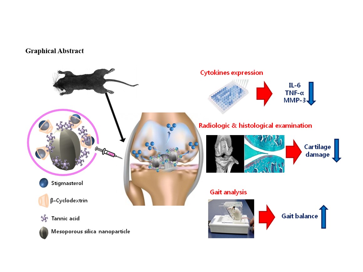
MSNs anchored by TA and stigmasterol
The surface modification of MSNs was accomplished by anchoring stigmasterol using TA as a surface modifying agents following the method previously reported by Noh et al. [18]. For this purpose, TA (30 µg/mL) was dissolved in PBS (5 mL) contained in 10 mL vials, followed by the addition of MSNs (10 mg). The resultant TA-MSN solution was incubated in the dark for 24 hours. The subsequent TA-MSNs were subjected to washing with distilled water (DW) and were lyophilized for two days. Henceforth, these TA-modified MSNs are referred to as TA-MSNs. The PBS employed in the reaction was preserved for the quantification of TA on the MSN surface. To anchor stigmasterol onto the TA-MSN surface, a solution of 10 or 50 µg of stigmasterol in DMSO was incrementally added to β-CD dissolved in DW while maintaining constant stirring for four hours, thereafter, TA-MSNs were introduced and incubated overnight. The final samples were separately washed with PBS and subsequently lyophilized for two days. The DW utilized for anchoring stigmasterol with β-CD onto the TA-MSN surface was collected for estimation of the stigmasterol loading amount. The resulting stigmasterol-loaded β-CD-MSNs with anchored TA were composed of stigmasterol(10 µg)/β-CD-MSNs and stigmasterol(50 µg)/β-CD-MSNs.
Characterization
The morphologies of bare MSNs, TA-MSNs, stigmasterol(10 µg)/β-CD-MSNs, and stigmasterol(50 µg)/β-CD-MSNs were analyzed using a JEM-F200 field-emission transmission electron microscope (FE-TEM; JEOL Ltd., Tokyo, Japan). Prior to microscopic examination, samples (100 µg/mL) were distributed in 5 mL ethanol (C2H5OH) in a test tube, sonicated utilizing an ultra sonicator (KFS-250N; 20 kHz, power: 45 W; Korea Process Technology Co., Ltd. Seoul, Korea) emplying a pulse function (pulse on/off = 10/2), for a duration of 10 minutes in an ice bath. Following this, they were relocated onto a Cu-TEM dish (CF200; Electron Microscopy Sciences, Hatfield, PA, USA) for imaging.
The FE-TEM was utilized at 200 kV for each specimen’s detailed characterization. A SZ-100V2 Nano Particle Analyzer (HORIVA, Kyoto, Japan), operating with a He-Ne laser (633 nm) and leveraging dynamic light scattering (DLS), was employed for assessing the particle size and zeta potential of all MSNs variants. The zeta-potential (mV) was also gauged via a laser Doppler electrophoretic method with a Zetasizer.
To characterize the C1s, O1s, and Si2p of each MSN variant before and/or after surface modification, an ESCALAB250 X-ray Photoelectron Spectrometer (Thermo Fisher Scientific Inc., Waltham, MA, USA), fitted with a monochromatic Al Ka X-ray source (1486.6 eV photons) and operating under super-high vacuum conditions (1 × 10− 9 bar), was deployed. The Brunauer-Emmett-Teller (BET) and Barrett-Joyner-Halenda (BJH) method were used in conjunction with an N2 adsorption instrument (Autosorb-iQ 2ST/MP; Quantachrome Instruments Co., Boynton Beach, FL, USA) to estimate the surface area, pore volume, and pore size of all MSN samples. The quantity of TA on the MSN surface was evaluated using the bicinchoninic acid (BCA; Thermo Fisher Scientific Inc., Waltham, MA, USA) method. A 50 µL supernatant, derived from PBS solution, was transferred into an E-tube and combined with the BCA working reagent (250 µL) under vigorous mixing for 10 s. This solution was incubated at 37°C for one hour, subsequently transferred to 96-well plates, and its absorbance measured at 562 nm via a multimode microplate reader (Thermo Fisher Scientific Inc., Waltham, MA, USA).
Stigmasterol release from stigmasterol/β-CD-MSNs
The stigmasterol release from stigmasterol(10 µg)/β-CD-MSNs and stigmasterol(50 µg)/β-CD-MSNs was examined by enclosing 10 µg of each sample in a dialysis bag (MWCO 3500 Da; Spectrum Laboratories, Inc., Rancho Dominguez, CA, USA), filled with 1 mL of PBS (pH 7.4). Each bag was immersed individually in a 50 mL test tube containing 20 mL of PBS (pH 7.4) and gently agitated at a frequency of 100 times/min in a water bath maintained 37°C. The PBS solution was refreshed at specified time intervals (1, 3, and 10 h, and 1, 5, 7, 14, 21, and 28 days), succeeded by the introduction of an equivalent volume of fresh PBS for additional incubation. The released stigmasterol level was quantified at 645 nm utilizing a multimode microplate reader.
Cytotoxicity evaluation
RAW 264.7 macrophages, procured from the Korean Cell Line Bank (Seoul, Korea), were employed for assessing the cytotoxicity of each MSN sample. The cells (5 × 10⁴ cells/well) were cultured in 96-well plates using DMEM (Thermo Fisher Scientific Inc., Waltham, MA, USA) supplemented with 10% FBS and 1% antibiotics (100 U/mL penicillin and 0.1 mg/mL streptomycin), and incubated at 37°C for 24 hours. Subsequent to the 24 hour incubation, the cells were exposed to the MSN samples (100 µg/mL). After 24 and 48 hours, the DMEM was substituted with PBS, CCK-8 reagent was added, and the cells were incubated for an additional one hour at 37°C in the dark. The absorbance was gauged at 450 nm using a multimode microplate reader, and the cell viability (%) was determined by comparison with control cells (no exposure).
Cytokine analysis
Cells (1 × 10⁵ cells/well) were cultured in 24-well plates and rested overnight. Following the rest, cells were treated with LPS (100 ng/mL) or the individual samples (100 µg/mL), or left untreated (control). Following induction for three days, the supernatant was collected to analyze M1-associated cytokines, specifically IL-6 and tumor necrosis factor-α (TNF-α), using enzyme-linked immunosorbent assay (ELISA) kits (Elabscience®, Houston, Texas, USA). The absorbance was observed at 450 nm employing a Multimode Reader.
Real-time quantitative polymerase chain reaction (PCR)
Macrophages were stimulated with LPS or each MSN sample for 72 hours. Subsequently, total RNA was isolated utilizing the TRIzol reagent kit (Life Technologies, Grand. Island, NY, USA) and served as a template for the synthesis of complementary DNA (cDNA) via AccuPower RT PreMix (Bioneer, Daejeon, Republic of Korea). PCR amplification was performed employing AccuPower HotStart PCR PreMix (Bioneer Daejeon, Republic of Korea). The primer sequences were as follows: IL-6 (F) 5ʹ-CCG TTT CTA CCT GGA GTT TG-3ʹ; (R) 5ʹ-GTT TGC CGA GTA GAC CTC AT-3ʹ; TNF-α (F) 5ʹ-CTC CCA GAA AAG CAA GCA AC-3ʹ, (R) 5ʹ-CGA GCA GGA ATG AGA AGA GG-3ʹ; matrix metalloproteinase-3 (MMP-3) (F) 5ʹ-ACC TGT CCC TCC AGA ACC TG-3ʹ; (R) 5ʹ-AAC TTC ATA TGC GGC ATC CA-3ʹ. Amplification and detection procedures were executed on the ABI7300 Real-Time Thermal Cycler (Applied Biosystems, Foster, CA, USA). Expression levels of IL-6, MMP-3, and TNF-α were normalized against glyceraldehyde 3-phosphate dehydrogenase (GAPDH) and reported as relative values.
Establishment of OA rat model and treatment
All animal-related procedures adhered to the Guidelines of the Laboratory Animal Research Center at the Korea University College of Medicine and were approved by the Institutional Animal Care and Use Committee at the Korea University Medical Center (KOREA-2021-0036). Eight-week-old male Sprague-Dawley (SD) rats (DooYeol Biotech, Seoul, Republic of Korea) were employed for the creation of the OA rat model. For a seven-day acclimation period, rats were housed in an environment maintained at 20–24°C with a 12 hour light/dark cycle and were provided with a standard diet and water. Monosodium iodoacetate (MIA; Sigma-Aldrich, St. Louis, MO, USA), dissolved in 0.9% normal saline at a concentration of 10 mg/mL, was used for model establishment. Post-acclimation, the rats were anesthetized with isoflurane and received a single 50 µL intra-articular injection of MIA solution in the right knee. Rats were then allowed a week for OA development. Beginning one week post-MIA administration, the rats were subjected to intra-articular injections in the right knee joint with the following MSNs: bare MSNs, stigmasterol(10 µg)/β-CD-MSNs, and stigmasterol(50 µg)/β-CD-MSNs. The injection volume for each MSN sample was 100 µL. The stigmasterol dose was 70.1 ng/rat for the stigmasterol(10 µg)/β-CD-MSNs and 350.9 ng/rat for the stigmasterol(50 µg)/β-CD-MSNs. Rats were randomly allocated into five groups: untreated controls; negative control (MIA induction); MIA plus MSNs; MIA plus stigmasterol(10 µg)/β-CD-MSNs; MIA plus stigmasterol(50 µg)/β-CD-MSNs. The in vivo experiment involved a total of 25 rats.
Weight-bearing on the right hind paw
The estimation of pain control was determined by quantifying the change in weight distribution between the experimental right and control left hind paws. This was conducted initially (prior to treatment), and subsequently at 1st (post-MIA induction), 2nd, 4th, 8th weeks (post-stigmasterol/β-CD-MSN treatment) using an IITC Incapacitance Test Meter (IITC Life Science Inc., Woodland Hills, CA, USA). Briefly, the rats were positioned in an angled plexiglass enclosure, and the force exerted by each hind limb was recorded an averaged over a duration of three seconds. The distribution of weight-bearing between the right and left limbs was calculated as follows: weight on the right leg (%) = [(weight on right leg) / (weight on left leg + weight on right leg)] × 100. For each rat at every time point, an aggregate of three readings was acquired, and their mean was calculated for subsequent analyses.
In vivo micro-computed tomography (CT)
Quantitative assessment of the hind knee joints was performed using a SKYSCAN 1172 micro-CT system (Bruker, Kontich, Belgium). The X-ray source for each smaple was set at 60 kV with 250 µV. The nominal resolution was established at 35 µm/pixel and the rotation increment was fixed at 0.5°. Cross-sectional images were reconstructed employing a filtered back-projection algorithm through NRecon software (Bruker micro-CT; Bruker, Kontich, Belgium). For each scan, series of 286 cross-sections was reconstructed at a resolution of 2,000 × 1,336 pixels.
Histological examination
Hind knee joint samples were first fixed in 10% phosphate-buffered formalin for 24 hours, followed by decalcification in a 14% EDTA-glycerol solution for two weeks at ambient conditions. The samples then underwent routine processing, paraffin embedding, and sectioning into 7 µm thick slices. These slices were stained with hematoxylin and eosin (H&E) for evaluation, and to examine cartilage health, safranin-O-fast green staining was performed. Based on the stained tissues, the degenerative grade and stage of cartilage were assessed utilizing the Osteoarthritis Research Society International (OARSI) Scoring System. The histological grade of cartilage degeneration was established according to the structural attributes as follows: Grade 0, smooth surface and healthy morphology; Grade 1, intact surface; Grade 2, surface discontinuity; Grade 3, vertical fissures (clefts); Grade 4, erosion; Grade 5, denudation; Grade 6, deformation.
Measurement of pro-inflammatory component levels in the serum
Blood samples were procured for analysis of pro-inflammatory cytokines, including IL-6, TNF-α, and MMP-3. The samples were then processed to collect serum through centrifugation at 2,500 rpm for a period of 10 minutes. The resultant serum was transferred into fresh tubes and stored at -80°C until further analysis. Quantitative estimation of serum IL-6, TNF-α, and MMP-3 were performed employing specific ELISA kits as per the instructions provided by the manufacturer. The absorbance values were at 450 nm utilizing a multimode microplate reader.
Statistical analysis
All experimental results are expressed as means ± standard deviation (SD). One-way ANOVA with Tukey’s post-hoc test for multiple comparisons was executed for statistical analysis using SigmaPlot software (Systat Software, Inc., San Jose, CA, USA). Intergroup comparisons were made based on p-values with values less less than 0.05 or less than 0.01 considered statistically significant.