Physical characterizations:
XRD pattern of the calcined Fe3O4, ZnO and Fe3O4-ZnO nanocomposites exhibited identical well-resolved X-ray diffraction peaks. The X-ray diffraction (XRD) analysis does not exhibit any apparent diffraction peaks corresponding to the existence of Fe3O4 in Fe3O4-ZnO nanocomposites. This observation provides evidence of the successful encapsulation of Fe3O4 nanoparticles by ZnO. The X-ray diffraction (XRD) pattern of core Fe3O4 is also shown in Fig. 1a. The average size of the precursor Fe3O4 crystals (referred to as the mean crystallite size, denoted as D) has been determined by utilising Scherrer's formula. This formula calculates D by considering the full width at half-maximum (FWHM) of the most prominent peak, with additional parameters including the shape factor (k), the wavelength (λ) of the X-ray used, and the diffraction angle (θ).
Debye–equation Scherrer's was used to calculating average crystallite sizes. (Xia et al. 2011; Atla et al. 2018)
$$D= \frac{K\lambda }{\beta cos\theta }$$
1
It is important to note that the FWHM value should be expressed in radians. The dimensionless form factor exhibits variability depending on the specific shape of the crystallite, with spherical crystals often possessing a characteristic value of about 0.94. The determined average size of Fe3O4, ZnO and Fe3O4-ZnO crystallites are 39, 42 and 29 nm. Figure 1a displays the X-ray diffraction (XRD) pattern of virgin ZnO, The X-ray diffraction (XRD) pattern seen corresponds to the known XRD pattern of zincite ZnO, as documented in the standard reference database JCPDS 36-1451. (Hong et al. 2008; Xia et al. 2011; Atla et al. 2018)
Figure 1b depicts the differential reflectance spectra (DRS) of interlink particles, such as Fe3O4/ZnO nanocomposites, that were synthesised using sol-gel techniques and pure ZnO. The Diffuse Reflectance Spectra (DRS) are represented by the function F(R), which is obtained by the application of the Kubelka-Munk approach to the observed reflectance (R). The absorption edge of the Fe3O4/ZnO nanocomposites that were synthesised has been determined to be 392 nm, a number that roughly corresponds to the absorption edge of pure ZnO, which is measured at 389 nm. This discovery offers empirical support for the existence of ZnO nanostructures inside the synthesised network-like architecture. The observed phenomenon of visible light being absorbed in a distributed manner in the Fe3O4/ZnO nanocomposites may be explained by the presence of Fe3O4 as the core ingredient. The Fe3O4 nanocrystals possess the capacity to absorb electromagnetic radiation within the visible light spectrum. The above phenomena is well supported by the previously reported literature (Atla et al. 2018; Elshypany et al. 2021; Astuti et al. 2023)
Figure S1a shows the Raman spectra of the Fe3O4/ZnO nanocomposites of the pristine ZnO.. The presence of two prominent peaks seen at 100 and 438 cm− 1 may be ascribed to E2(low) and E2(high), respectively, which are suggestive of the wurtzite lattice structure. The above statement may be reformulated as a subtraction operation, whereby the quantity denoted as E2(high) mode represents the oscillation of oxygen atoms occurring at a relatively high frequency. Conversely, the E2(low) mode corresponds to the vibration of the zinc sublattice, manifesting at a lower frequency. The observation of the prominent E2(high) mode indicates a substantial level of crystalline structure. In addition, the observed phenomena might be attributed to the existence of intrinsic flaws associated with oxygen, namely oxygen vacancies (VO), since it only refers to the oscillation of oxygen atoms. The user's material is insufficient in length to be rewritten in an academic way. The presence of a small peak seen at 380/382 cm− 1 suggests the presence of the A1(TO) mode.(Hong et al. 2008; Atla et al. 2018) The frequencies observed at 534/535 and 578/580 cm− 1 are attributed to the A1(LO) and E1(LO) modes, respectively. The observed peaks at 207 and 330 cm− 1 may be ascribed to the second-order Raman spectra arising from 2TA(M) and E2(high) − E2(low), respectively. The observed values are in accordance with these modes.(Zou et al. 2010; Xu et al. 2014) The occurrence of a broad peak seen at around 666 cm− 1 may be ascribed to the occurrence of surface phonon scattering that arises from zone-boundary phonons. Furthermore, a significant asymmetrical peak is seen at around 1150/1149 cm− 1. The spectra demonstrate resemblances to the previously reported research (Długosz et al. 2021; ASLAN 2022)
After XRD analysis, FTIR spectra has been used to check the functional group present in the synthesized material. Fourier Transform Infrared Spectroscopy (FT-IR) is a very efficient technique for elucidating the chemical makeup of various items. In Figure S1b, it was shown the freshly prepared Fe3O4-ZnO nanocomposites and also it was shown the FTIR spectra of Fe3O4-ZnO nanocomposites, there was also no significant changes in the functional groups which support the stability of the optimized nanostructured materials under 1 M KOH solution. The vibrational spectra of Fe3O4–ZnO nanocomposites and their modified counterparts, respectively. The FT-IR findings indicate that a distinctive peak corresponding to Fe-O was detected at a wavenumber of 543 cm− 1, whereas the band found at 565 cm− 1 was attributed to the stretching frequency of M–O in Fe3O4.(Abazari et al. 2016) The stretching vibration seen at wavenumbers 3370, 3513, and 3449 cm− 1 was assigned to the presence of the hydroxyl (-OH) functional group. The vibrational mode seen at a wavenumber of 1618 cm− 1 was attributed to the bending vibrations of the hydroxyl (-OH) group. Likewise, the occurrence of a stretching vibration at a wavenumber of 1119 cm− 1 may be attributed to the existence of iron and carboxyl groups. Primarily, FT-IR spectroscopy has high sensitivity towards metal-oxygen bond vibrations. Typically, the stretching vibration band associated with M-O bonds is often seen at frequencies below 1001 cm− 1, providing substantial evidence for the successful production of metal oxides. Consequently, the identification of stretching vibration bands associated with the Fe-O bond was determined by the existence of two peaks seen at 432 and 608 cm− 1 in the Fe3O4-ZnO nanocomposite. Furthermore, prior research has shown that the stretching vibration of M-O bonds, namely Fe-O and Zn-O bonds, may be identified by the presence of peaks at 428, 625, and 527 cm− 1, respectively. Furthermore, in a study conducted by researchers, it was observed that certain peaks were detected at 3438, 1631, and 1381 cm− 1, which were shown to be linked to the adsorption of water molecules on the surface of magnetic nanocomposites. (Zou et al. 2010; Xu et al. 2014; Abazari et al. 2016)
Morphology of the synthesized Fe3O4-ZnO nanocomposites catalysts was analysed by FESEM which are indicates that the presence of highly aggregated particles with uniform morphology towards gradually evolved into agglomerated form (Fig. 2a-c) with minor fragments of particles (Fig. 2c). The morphology on the surface of Fe3O4-ZnO nanocomposites particle agglomeration is shown in more detail in Fig. 2c. there are no significant changes in the overall morphology. Overall, the diameter was found to be 800 nm to 2 µm in size and their shapes appeared varied from elongated too somewhat curved or circular. As seen, the Fe3O4-ZnO nanocomposites catalyst shows spherical agglomerates with rough surfaces (Fig. 2c) which also probably support the deposition of Fe3O4-ZnO nanocomposites. The overall morphology does not have any noteworthy changes after the heat treatment along with loading of Fe3O4-ZnO nanocomposites.
As shown in Fig. 3 the nitrogen adsorption-desorption isotherms of the Fe3O4-ZnO nanocomposites demonstrated typical type II curves with type-H3 hysteresis loop (according to the IUPAC classification), in the relative pressure range of 0.1 ~ 1.2, suggesting the typically pores between the particles. Nitrogen adsorption-desorption isotherm patterns of all the samples are almost identical and correspond to type II indicating that the synthesized Fe3O4-ZnO nanocomposites have typically pores between the particles. The BET surface area of the synthesized Fe3O4-ZnO nanocomposites was 98 m2 g− 1 with a total pore volume of 0.112 cm3 g− 1. The pore size distribution analysis using BJH (Barrett-Joyner-Halenda) method from desorption part of the isotherm of Fe3O4-ZnO nanocomposites (inset Fig. 3) reveals a very narrow pore size distribution. (Hong et al. 2008; Astuti et al. 2023)
X-ray photoelectron spectroscopy (XPS) in the range from 0 to 1200 eV (Figure S2) indicated the presence of carbon, oxygen, and iron arising from the Fe3O4-ZnO nanocomposites. The Fe3O4/ZnO nanocomposite's composition was subsequently evaluated using X-ray photoelectron spectroscopy (XPS). Figure 4a-b depicts the X-ray photoelectron spectroscopy (XPS) spectra pertaining to the Fe 2p and Zn 2p areas of the Fe3O4-ZnO nanocomposite. As a result of spin-orbital coupling, the Fe 2p energy levels undergo a division into Fe 2p3/2 and Fe 2p1/2 components. The spin-orbit splitting occurs at energies of 725.60 eV and 710.29 eV for the Fe 2p3/2 and Fe 2p1/2 components, respectively. The presence of Fe2+ with Fe3+ ions is indicated by the widening of the Fe 2p3/2 and Fe 2p1/2 peaks.(Yamashita and Hayes 2008) Fig. 5b shows the presence of Zn in the nanocomposite by the observation of two distinctive peaks positioned at binding energies of 1044.80 and 1023.01 eV, corresponding to Zn 2p1/2 and Zn 2p3/2, respectively. The reference spectrum displays the peak associated with the C 1s orbital (Figure S2). Furthermore, the X-ray photoelectron spectroscopy (XPS) analysis of the Zn 2p and O 1s energy levels provided further confirmation that Zn is present in the form of Zn2+ ions and that oxygen is present in the Fe3O4/ZnO nanocomposite's.(Yang et al. 2005; Yamashita and Hayes 2008) The O1s spectra seen in Fig. 5c has a peak at a location of 529.78 eV, which has been designated as oxygen in the Fe-O bonds of Fe3O4/ZnO nanocomposite's, indicating the presence of ferro-ferric oxide.(Yamashita and Hayes 2008) The analysis of the O1s signals using deconvolution demonstrated the existence of Zn–O bonds in ZnO, with a binding energy measured at 530 eV. The observed peak at 531.75 eV might perhaps be attributed to the presence of the hydroxyl group from the biomolecular capping process, as well as the chemisorption of oxygen onto the surface of Fe3O4/ZnO nanocomposites. Additionally, Under the influence of electrochemical environment in 1 M KOH electrolytic solution, the active oxygen species present on the surface of the Fe3O4/ZnO nanocomposite's may play a role in the acceleration of alkaline water oxidation by the generation of oxygen ions, namely O− 1 and O− 2. This, in turn, enhances the electrocatalytic performance of the Fe3O4/ZnO nanocomposite's.(Yang et al. 2005; Yamashita and Hayes 2008; Zhan et al. 2015). This strategy has shown a high level of concordance with the findings obtained from the X-ray diffraction (XRD) investigation. This approach is used to investigate nanostructured samples.(Zhan et al. 2015; Dwivedi et al. 2021)
Electrochemical Oxygen Evolution Reactions
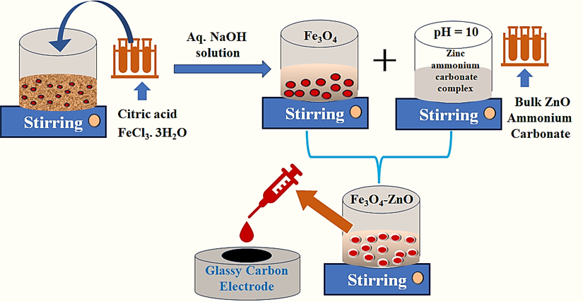
The synthesized Fe3O4-ZnO nanocomposites has been used for the electrocatalytic Oxygen Evolution Reaction (OER) performance. The synthesized material has been loaded on the clean surface of glassy carbon electrode (GCE). The OER test was performed in a nitrogen (N2) gas saturated 1M KOH solution in three-electrode system. Later, the iR corrected linear seep voltammetry (LSV) has been done at 5mV/s scan rate in three electrodes set up.(Galani et al. 2020) The Linear Sweep Voltammetry data has shown in Fig. 5.
Yet, all the synthesized materials of Fe3O4-ZnO nanocomposites shown the lower onset potential towards the OER. The overpotential has been prerequisite to attain the current density at 10 mA/cm2 (ɳ10) which was correspond to 10% efficient solar water-splitting system which was shown in Fig. 5(a-d). All electrochemical results have been shown in the Table 1, which is also indicating the presence of potential electrochemical properties of the nanostructures Fe3O4-ZnO. The data on Figure S4, has shown that at two different current density (10 and 40 mA/cm2), Fe3O4-ZnO nanocomposites has shown good electrochemical performances compared to other ZnO and Fe3O4 materials due to their multi-porosity and high geometrical surface areas of the materials. (Li et al. 2020; Zhang et al. 2021; Wang et al. 2022)
In Fig. 5a, it was shown that the electrocatalytic activity among, whereas Fe3O4-ZnO nanocomposites show the superior electrocatalytic activity among all synthesized samples. For the Fe3O4-ZnO nanocomposites, 10 mA/cm2 current density has been reached at the overpotential of 350 mV for OER. However, Fe3O4 and ZnO nanocomposites has shown the overpotential value around 580 mV and 460 mV respectively. (Zhang et al. 2021)
The Tafel slope was calculated from the recorded linear sweep voltammetry (LSV) curves of the synthesised materials in order to ascertain the steady state of the electrocatalytic process. In general, a decrease in the Tafel slope corresponds to an increase in catalytic activity. In Fig. 5b, it was shown the corresponding Tafel slope which indicate the excellent electrocatalytic activity of Fe3O4-ZnO nanocomposites -based catalyst. Whereas, Fe3O4-ZnO nanocomposites has shown the lowest Tafel slope value of 74 mVdec− 1 towards the OER activity compared to the other Fe3O4-ZnO nanocomposites. The enhanced electrocatalytic activity of the nanostructures loaded on the surface of GCE owing to their mesoporous nature and enhanced surface area. Owing to their porous nature, it helped to electrolytes onto their active surface of the Fe3O4-ZnO. Moreover, due to the development of dual structure, multi porosity of the Fe3O4-ZnO nanocomposites which was further facilitate the overall electrocatalysis process. (Wang et al. 2022)
For the long-term stability test, Chronoamperometry test has been performed in 1 M KOH solution. Due to that, our optimized electrocatalyst Fe3O4-ZnO nanocomposites as a working electrode at 1.5 V (vs RHE) was applied for 24 hrs for the stability test and it was shown in Fig. 5c.
For additional stability test, we performed the cyclic voltammetry test before and after LSV test in 1M KOH at 5 mV s− 1 of Fe3O4-ZnO nanocomposites. To check the stability of 1 Fe3O4-ZnO nanocomposites, LSV of the fresh catalyst and after the 3000 cycles in cyclic voltammetry. The corresponding result has shown in Fig. 5d. Following the comprehensive cyclic voltammetry examination, it was observed that the polarisation curve of the Fe3O4-ZnO nanocomposites exhibited a very high degree of similarity, with a 98% resemblance. This finding serves as strong evidence for the exceptional stability of the aforementioned nanocomposites. There is very little change of the overpotential values of 15 mV at 100 mA/cm2 with 98% retention.
Furthermore, EIS has been done to check the charge transfer resistance (Rct) of Fe3O4-ZnO nanocomposites to check their kinetics towards the OER motion. In Fig. 6, it was clearly seen that the corresponding Nyquist plot, clearly indicating the Fe3O4-ZnO nanocomposites loaded on the surface of Carbon has shown the minimum curvature which tell the low charge transfer resistance. Owing to their unique physicochemical properties Fe3O4-ZnO nanocomposites than other samples which shows low Rct values which was helped to pass the charge easily. This characteristic of the synthesized material facilitates the overall OER process. (Matsumoto and Sato 1986; Li et al. 2020)
Table 1
Summary of the electrochemical information for all the catalysts.
|
Catalyst
|
Tafel slope (mV/dec)
|
Overpotential (mV) η10
|
Electrochemically active surface area (ECSA (cm2))
|
Roughness factor (Rf)
|
Rct
|
|
Fe3O4
|
78
|
460
|
92
|
55
|
48
|
|
ZnO
|
93
|
530
|
45
|
30
|
63
|
|
Fe3O4-ZnO
|
62
|
350
|
115
|
93
|
23
|
Overall, Cyclic voltammetry (CV) has been used to calculate the capacitive current of the Fe3O4-ZnO nanocomposites along with the other synthesized samples. CV has been done of the different samples such as Fe3O4-ZnO nanocomposites on modified glassy carbon electrode (GCE) in 1 M KOH solution. The expected result was shown in Figure S5 (Supporting Information). The corresponding scan rate were varied from 10 mV/s to 60 mV/s in the potential range of 1.10 V to 1.30 V to enumerate their double layer capacitance. The double layer capacitance can be obtained due to their mesoporous nature and maximum accumulation of electrolyte molecules on the surface of heterojunction materials such as Fe3O4-ZnO nanocomposites. Whereas, maximum result has shown in 1% loaded RuO2 on the surface of Carbon. The electrochemical active surface area (ECSA) has been calculated from the corresponding formulae(Zhu et al. 2023) (see Figure S5 Supporting Information).