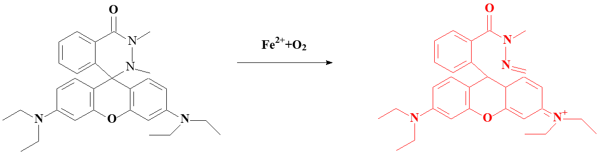
2.2 Synthesis of probe 1
Phosphorus oxychloride (459 mg, 3 mmol) was added slowly to a 1,2-dichloroethane solution (10 mL) of rhodamine B (479 mg, 1 mmol) and the mixture was stirred at 80°C for 4 h. When the solution was cooled to room temperature, 1,2-dichloroethane was removed by a rotary evaporate. The residue was added dropwise a dicloromethane solution (15 mL) of rhodamine B (479 mg, 1 mmol), 1,2-dimethylhydrazine dihydrochloride (133 mg, 1 mmol) and N,N-diisopropylethylamine (1.349 g, 10 mmol). The mixture was stirred at room temperature for 12 h. Then the product was purified by silica column to get the white solid (reaction yield 25%). [45] 1H (NMR, 400 MHz, CDCl3) δ (ppm): 1.16 (t, J = 7.2 Hz, 12H), 2.31 (s, 3H), 2.79 (s, 3H), 3.28–3.42 (m, 8H), 6.28–6.31 (m, 2H), 6.51 (d, J = 2.4 Hz, 2H), 6.71 (d, J = 8.4 Hz, 2H), 7.30 (d, J = 6.4 Hz, 1H), 7.48–7.56 (m, 2H), 8.24–8.26 (m, 1H). 13C NMR (100 MHz, CDCl3) δ (ppm): 12.6, 34.6, 38.9, 44.4, 63.3, 99.1, 106.3, 110.0, 127.4, 127.7, 129.0, 130.1, 130.3, 132.2, 138.0, 148.6, 154.2, 164.1. (HRMS) (ESI): calculated for C30H37N4O2 [M + H]+, 485.2911; found, 485.2915.
2.3 Cytotoxicity of probe and Cell culture
The MTT assay standard was used to test the cytotoxicity of the probe and to determine whether the probe is toxic to cells. RAW 264.7 macrophage cells (1 × 10⁵ cells/mL) were selected and were plated in a microwell plate with medium (100 µL) (CO2 incubator, 37 ℃, 24 h). Then the cells were incubated with different concentrations of probe (0, 5, 10, 20, 30 µM) for another 24 h and washed with PBS buffer solution (0.01 M, pH = 7.4) three times. Next, MTT solution (10 µL) was added to each well for 4 h and DMSO solution (150 µL) was added to each well to dissolve the Formazan crystals in the cells after the medium was removed. Finally, the cell survival rate was calculated by the data obtained from the absorbance of each well at 570 nm.
RAW 264.7 macrophage cells were seeded in DMEM medium which contained 10% FBS in incubator (5% CO2/95% air, 37 ℃) for 24 h. After washed with PBS three times, the cells were treated in turn with the probe (5 µM) and Fe3+ (100 µM) for 30 min. Then washing with PBS three times, the fluorescence images of cells was gathered with a laser confocal scanning microscope.