1 Ethics statement
The use of human aqueous humour (AH) samples from cataract eyes during surgery was approved by the Institutional Review Board of Eye and ENT hospital of Fudan University. This study was performed in accordance with the tenets of the Declaration of Helsinki for research involving human subjects. Written informed consent was obtained from every enrolled participant.
2 Collection of AH and human lens epithelium
We collected AH and human lens epithelium from 36 patients with diabetes and cataracts (age from 45-76 years old, free of other ocular diseases, and lenticular opacity ranging from C3-4, NO2-3, NC2-3, and P1-3 by LOCSIII) and 43 patients with age-related cataracts (age from 62-89 years old, free of other ocular diseases, C3-4, NO2-3, NC2-3, and P1-2) before cataract surgery at Eye and ENT Hospital of Fudan University. AH samples were obtained before the collection of lens epithelium samples. The lens epithelium samples were acquired by intact continuous curvilinear capsulorhexis during cataract surgery for ARC patients by the same experienced surgeon (Yi Luo). All AH and human lens epithelium samples were stored in a freezer at -80℃ until the next step.
3 Isolation of exosomes
The AH samples from 36 patients with diabetes and cataracts were pooled together as the DMC group, and the AH samples from 43 patients with age-related cataracts were pooled together as the ARC group. Exosomes were isolated using ultracentrifugation. Procedures were as followed: a) Take AH samples out and thaw in 27℃ water bath. b) 4℃, 2000 g, 10 min, and remove supernatant. c) 4℃, 10000 g, 30 min, and take supernatant. d) 4℃, 110000 g, 75 min, and discard supernatant. e) Resuspend pellet and filter with 0.22 um membrane. f) 4℃, 110000 g, 75 min, and abandon supernatant.
4 Transmission electron microscopy (TEM) and nanoparticle tracking analysis (NTA) of AH exosomes
Purified exosomes were diluted in PBS. 5 μL samples were absorbed onto copper grids and dried for 5 minutes at room temperature. After that, a drop of 2% uranyl acetate solution was added for 20 minutes, and the sample was air-dried and examined by TEM (Tecnai G2 Spirit BioTwin, FEI, USA). Particle size, concentration, and distribution of exosomes were determined by NTA (ZetaView, Particle Matrix, Germany).
5 Exosomal RNA extraction and miRNA sequencing analysis
Exosomal RNA was extracted from the DMC group and the ARC group using the miRNeasy Micro Kit (217084, QIAGEN, Germany) according to the manufacturer’s guidelines. RNA libraries were prepared and sequenced on an Illumina HiSeq 2500 platform. Read counts were obtained by FeatureCounts software. FastQC software was used for quality control. Additionally, we used Cutadapt software to remove low-quality reads and high-quality reads were used to analyse miRNAs by mapping to the human reference genome using Bowtie software. A fold change>1.2 or <0.83 was considered to indicate differentially expressed miRNAs by DESeq2.
6 RNA extraction of epithelium samples and quantitative real-time PCR (qRT-PCR)
Epithelium samples from 43 patients with age-related cataracts were classified as ARC group. Epithelium samples from 36 patients with diabetes and cataracts were classified as DMC group. In each group, 4 to 5 epithelium samples were pooled together to obtain enough RNA. Total RNA from all epithelium samples was extracted using TRIzol reagent (Invitrogen, Carlsbad, CA, USA) and reverse transcribed with the RT reagent Kit (Takara Bio, Inc, Japan) according to the manufacturer’s protocol. Expression of mRNAs was detected using the SYBR Green detection kit (Takara, Japan) on the LightCycler 480II Real-Time PCR System (Roche, Switzerland). GAPDH was detected as the internal control. RNA expression was determined by the 2-ΔΔCT method.
7 Human lens epithelial cell culture and transfection
HLECs were cultured in 35-mm culture Petri dishes with growth medium containing DMEM (Gibco, USA) with 10% foetal bovine serum (FBS, Gibco, USA). MiR-29b mimics (50 nM) and inhibitors (100 nM) were transfected into HLECs when the cells covered 70-80% of the entire dish.
8 Examination of Ca2+ of AH samples and cell culture supernatant
Concentration of Ca2+ of AH samples and cell culture supernatant was detected by using Calcium Assay Kit (Colorimetric) from Abcam (Cambridge, MA, USA) according to the manufacturer’s protocol. We added 50 μL of AH samples and cell culture supernatant to each well of the 96-well plates. Then 90 μL of the Chromogenic Reagent and 60 μL of Calcium Assay Buffer were added into each well for 10 minutes at room temperature protected from light. Absorbance was measured at a wavelength of 575 nm using an automatic microplate reader (Tecan, Switzerland). Concentration of Ca2+ was equivalent to Abs/Vol (ug/μL). (Abs referred to absorbance of AH samples and cell culture supernatant, Vol referred to the volume of AH samples and cell culture supernatant added to each well)
9 Cell Counting Kit-8 (CCK-8) cell proliferation and cytotoxicity assay
Cell viability was determined by using CCK-8 kit (Dojindo, Japan) according to the manufacturer’s protocol. Transfected cells were plated onto 96-well plates and cultured for 24 hours. CCK-8 (10 μL) was then added for 1 hour at 37℃. We used Tert-butyl hydroperoxide solution (TBHP) as an oxidative stimulus. Absorbance was measured at a wavelength of 450 nm using an automatic microplate reader (Tecan, Switzerland). The cell viability was equivalent to (At-Ab)/(Ac-Ab). (At referred to absorbance of transfected cell groups, Ac referred to absorbance of controlled groups, Ab referred to absorbance of blank groups)
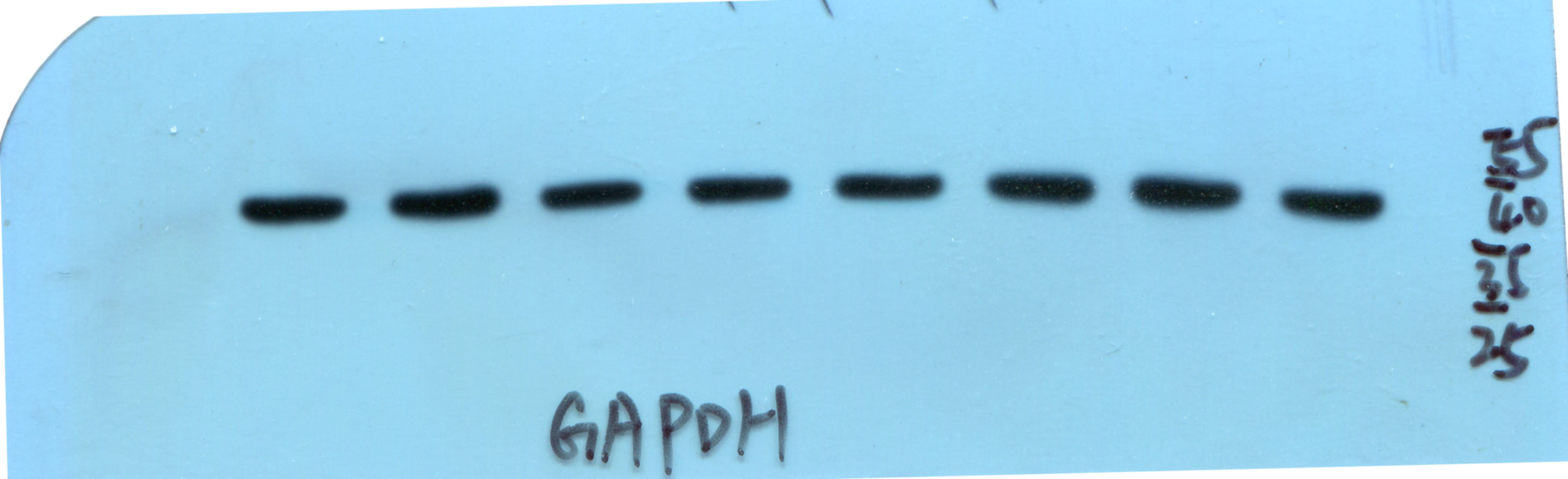
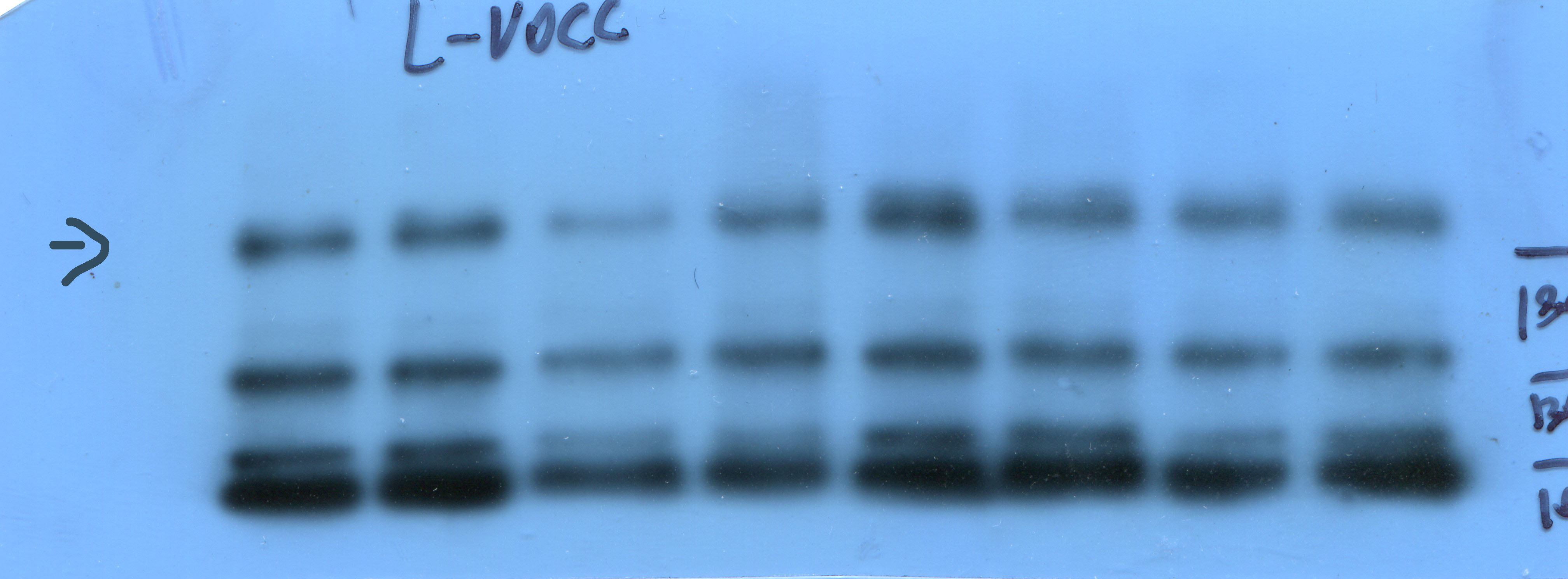
10 Western blot
Protein was extracted by RIPA lysis buffer (Biotech Well, Shanghai, China). Equal amounts of proteins were resolved by SDS-PAGE using 5% acrylamide-containing gels, followed by electrophoretic transfer to PVDF membranes. The membranes were blocked with transfer buffer (Biotech Well, Shanghai, China) and incubated overnight with the monoclonal primary antibodies at a 1:200 dilution, followed by secondary antibodies at a 1:2000 dilution. The signaling of western blotting was then observed using ECL prime reagents (Biotech Well, Shanghai, China) and scanned using a Peiqing automatic gel imaging analysis system (Shanghai, China). The L-VOCC polyclonal antibody (21774-1-AP) was purchased from Proteintech Group (USA). The anti-GAPDH antibody and the goat anti-rabbit IgG (H+L) secondary antibody were purchased from Biotech Well (Shanghai, China)
12 MicroRNA target prediction
Potential targets of miR-29b were predicted by the Targetscan database (http://www.targetscan.org)[33].
13 Statistical analysis
All data are shown as the mean±SD, and experiments were repeated three times. Statistical significance was determined by two-tailed Student’s t-test, One-way ANOVA or chi-square test using IBM SPSS 21.0(USA). P-value<0.05 was considered statistically significant.