In the present study, we investigated the possibility of using CPC cells, collagen, fibroin-based substrates, two 3D porous matrices, and an electrospun net, to create a vascularized and functional in vivo model of a cardiac organoid. We tested first the viability and differentiation of CPC cell-collagen organoids in vivo, second the host response to biomaterials, and then the host response to the CPC cell-scaffold-collagen combination in immunocompromised animals.
Scaffolds with three different geometries were prepared using a fibroin-water solution, including two sponges with different pore sizes and pore distributions and an electrospun net with randomly distributed fibers. Characterization of the morphological properties of the fibroin-based constructs showed that the pore size and pore distribution of the sponges were tuned according to the different protocols used to fabricate the scaffolds, which included freeze-drying and a temperature gradient in the quenching step. The results of the morphological analysis showed a homogeneous pore distribution and bipolar pore size with a well-oriented lamellar structure for the sponges. The electrospun net was found to contain randomly distributed fibers with a constant diameter. The pore size ranges and water absorption capacity of the scaffolds were previously measured and published [15–17].
All porous scaffolds induced a foreign body reaction with giant cells and capsule formation. The electrospun net (F-scaffold) did not induce an evident foreign body reaction, but a small capsule was still visible. The foreign body reaction was confirmed by the presence of CD11b + giant cells and the expression of the inflammatory interleukins, IL -4 and IL -13.
Silk fibroin can be isolated in sufficient quantities from the cocoons of the silkworm Bombyx mori and is considered an adjustable and versatile commercially available biomaterial. The biocompatibility of silk fibroin can be improved by separating the immunogenic protein family, sericins, as we did in the present study, so that this biomaterial elicited only a moderate inflammatory response during subsequent implantation in rodents [18]. The biocompatibility of scaffolds has been shown to depend on a number of different parameters, including microstructure and architecture, both of which influence the inflammatory response. In addition, implantation of hybrid constructs, i.e. scaffolds pre-seeded with cells, can trigger an adaptive immune response towards the biological component and thus influence the host response to the implanted device [19].
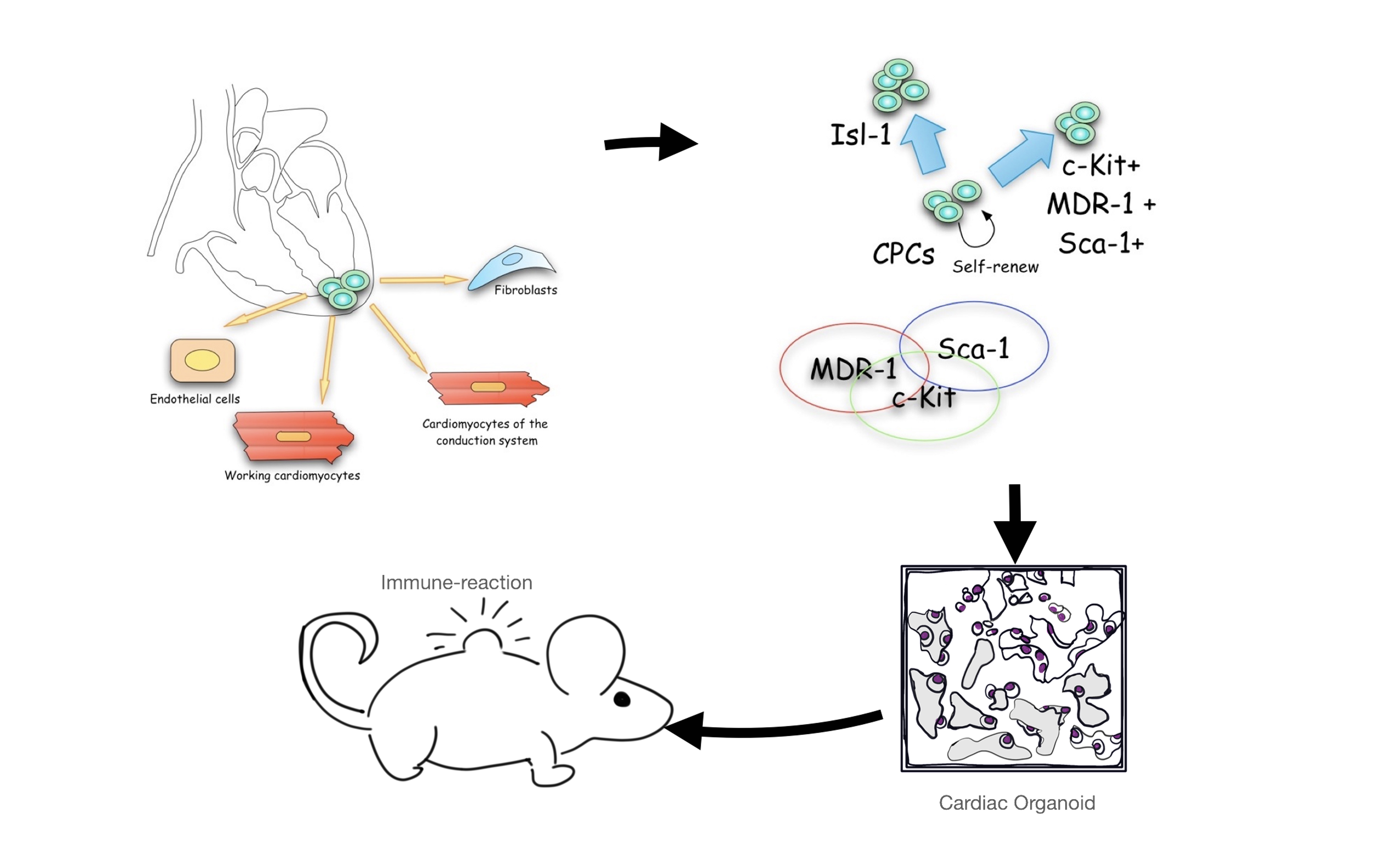
The CPC cells that we were able to isolate as primary cultures from adult rat hearts after a long collagenase digestion [8] did not induce tumor masses in the several organs analyzed, nor did they induce CFUs in vitro. We verified that the cells expressed the usual stem cell markers (c-Kit, Sca-1 and MDR-1) and some structural proteins typical of the heart in both 2D and 3D cultures ( Titin and cardiac Troponin T2), as studied by Bernstein and Srivastava [20]. In fact, we had already shown in a previous work that c-Kit + CPC cells isolated by repeated treatments with collagenase and cultured in 3D under different conditions can differentiate and express many cardiac proteins in vivo [8–9]. However, even though several research groups have isolated similar cardiac progenitor cells, no one has yet investigated their applicability as the main component of an in vivo model of a cardiac organoid. Because their immunogenicity has not been studied in vivo, we also examined the immune response induced by injection of the CPC cell-collagen organoid, the host response to the CPC cell-scaffold-collagen combination in immunosuppressed animals.
Unfortunately, injection of the CPC cell-collagen organoids induced a cell-mediated immune response with the formation of a capsule. The implanted cells were completely destroyed by the immune reaction.
Because the F-fibers were the least reactive scaffolds studied, we implanted them together with the CPC cells and collagen (Sc + cell) into the subcutaneous area of nude mice, but even though the F-scaffolds alone did not induce a foreign body reaction with giant cells, the CPC cells were destroyed by the lymphocyte infiltrate.
A foreign body reaction usually occurs when biomaterials alone are implanted for therapeutic clinical applications in tissue engineering. This reaction is the starting point for tissue regeneration in orthopedics because the foreign body reaction stimulates the regeneration of germinal/progenitor cells of the host tissue while degrading the scaffolds in a reasonable time frame [21]. However, this phenomenon is not useful for stem cell therapy or in vivo modeling of vascular and functional organoids because, as shown here, the foreign body response destroys the cells very quickly after implantation, even when injected into immunosuppressed animals.
Organoids are structures that arise from the cells of a particular organ, or from a combination of different, partially differentiated tissues, or from progenitor cells of the organ itself that are capable of giving rise to the various tissues of the organ. They have gained importance in scientific research because they represent a valid alternative to animal experiments, and because they can be a valid tool for testing drugs, new molecules or even simulating the environment of a particular pathology. The use of organoids reduces the number of animals used for experiments.
Many organoids arise from mesodermal cells and are formed from a single mass of progenitor cells. When cells in the center of the mass are deprived of nutrients and oxygen, necrosis occurs, even in vitro. Our progenitor cells (CPC cells) are naturally capable of giving rise to cells of endothelial origin, as we have shown previously [8], and are capable of forming vessels even in vitro within the collagen I gel. For this reason, we hoped that the same mass of cells could develop an adequate network of vessels in a more complex environment such as the subcutaneous region of immunocompromised mice. Vascular organoids are important both to ensure the supply of nutrients and oxygen to all cells in the mass and to provide a mesenchymal nice useful for maintaining organoid structure [22]. Previous studies have shown that induced human pluripotent stem cells (hPSCs) are able to differentiate into a microvascular network and this was integrated with the host vasculature to form a functional blood system when implanted into immunodeficient mice [23].
Here, we used FISH to show that the vasculature present in the implanted organoids was derived from the host and not from the donor cells.
In conclusion, CPC cells are capable of expressing cardiac structural markers and organizing a vascular network in vitro, but when implanted (with or without fibroin nets ) into the subcutaneous region of immunocompromised mice, they are likely to be destroyed by CD3 + lymphocytes, which are still active in this type of animals.