Ethics statement
All animal experiments were performed according to the recommendations outlined in the Guide for the Care and Use of Laboratory Animals. The Institutional Animal Care and Use Committee approved all research animal protocols at the Southwest Medical University, Lu Zhou.
Mice
The RA reporter Tg (RARE-Hspa1b/lacZ) 12Jrt/J mouse strain with a C57BL/6 genetic background was obtained from Jackson Laboratories. C57BL/6 was purchased from GemPharmatech. Mice were kept in a room with a 12 h light-dark cycle at 22◦C, fed by an OVA-free diet sterilized by irradiation. Water was sterilized by autoclaving, and aspen wood shaving was used for bedding.
Reagents
The following reagents were purchased: DDAOG (Invitrogen or Setareh Biotech); SiO2 and urate crystals (Invivogen); AM80 and AGN193109 (MedChemExpress); UAMC-3203 and erastin (Selleck); liproxstatin-1, deferoxamine mesylate and ferrostatin-1 (MedChemExpress); BMS493 and CD2665 (APExBIO).
FACS and antibody
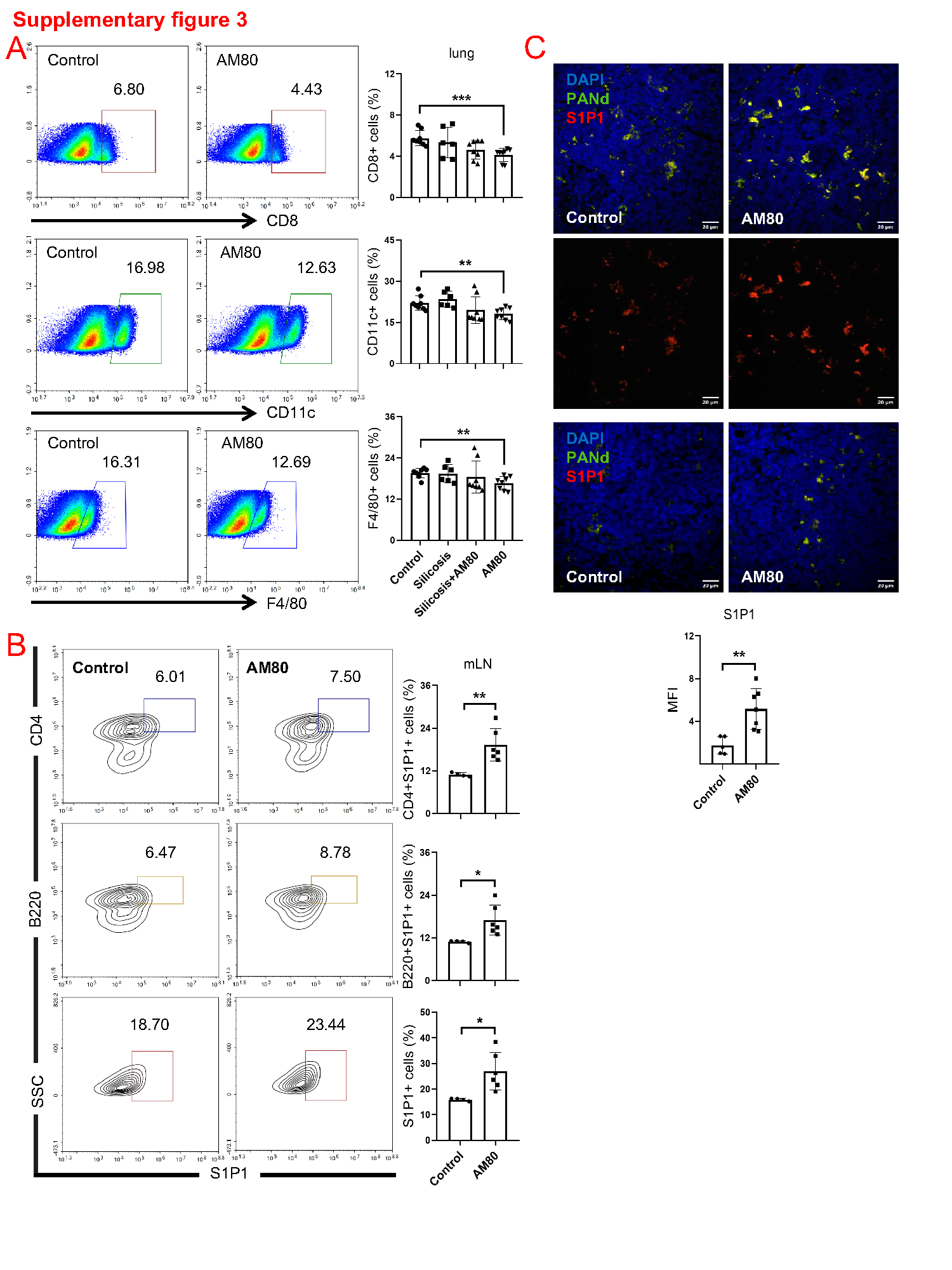
BMDCs and RAW264.7 were washed and suspended in FACS buffer (PBS, 2% FBS), incubated with fluorochrome-coupled agents for 30 min at 4°C, and then washed in FACS buffer. Data were obtained with the Acea NovoCyte series flow cytometer and analyzed by FlowJo software v9.6 (FlowJo, Ashland, USA). The following anti-murine antibodies were used for flow cytometry: CD11c (N418), CD4 (RM4-5), CD8 (53 − 6.7), B220 (RA3-6B2), CD3 (17A2), F4/80 (BM8) (all purchased from eBioscience); Ly6G (1A8) (purchased from Biolegend); S1P1 (T4-H28) (purchased from R&D Systems).
β-galactosidase activity
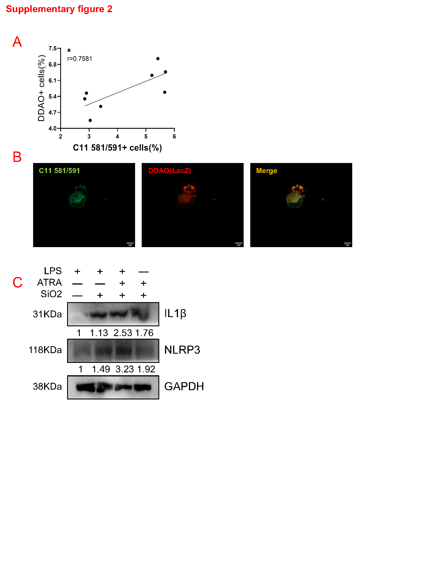
BMDCs from RA reporter mice were incubated with the galactosidase substrate DDAO galactoside (DDAOG) (10 µM) in serum-free Hank's buffer for 0.5 − 2 h at 37°C. The cells were washed three times with Hank's buffer before flow cytometry. DDAOG yields the hydrolytic product DDAO in DCs, which can be excited with the 633 nm laser (excitation/emission maxima ~ 645/660).
BMDC culture
DCs were generated from bone marrow (BM) cells obtained from 5–7-week-old RA reporter mice or C57/B6 mice. Briefly, BM cells were flushed out from the femurs and tibias. BM cells were cultured in a complete culture medium (RPMI 1640 supplemented with 10% FBS, 25 mM HEPES, 5 mM 2-ME, and antibiotics; Hyclone) containing 10 ng/ml GM-CSF and 10 ng/ml IL-4 (Peprotech).
Western blots
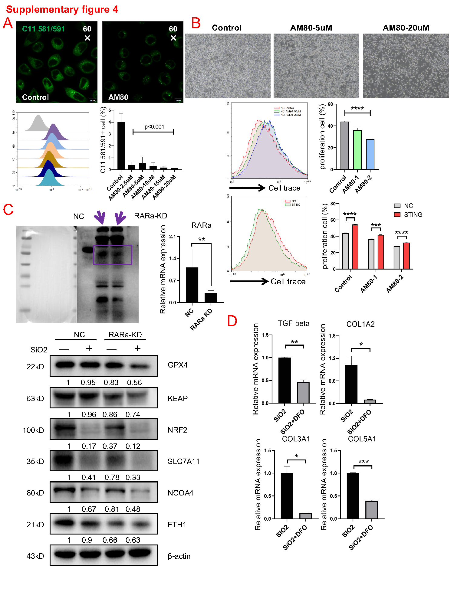
DCs and lungs were collected and lysed on ice with RIPA Buffer (CST) containing protease/phosphatase inhibitor cocktail (CST) and 1 mM PMSF (Beyotime). Cell and tissue extracts were loaded onto Biofuraw™ Precast Gel (Tanon), separated by electrophoresis, and transferred onto NC membranes (Millipore). Signals were generated with ECL western blotting substrate (ChemiDoc Touch system). GPX4, NCOA4 (E8H8Z), KEAP1 (D6B12), NRF2 (D1Z9C), FTH1 (D1D4), and xCT/SLC7A11 (D2M7A) (purchased from CST); RARa (purchased from Santa Cruz or Abcam); RARa-S77 and RARa-S96 (purchased from Invitrogen); AKT, AKT-S473, and AKT-T308 (purchased from CST); GAPDH and β-actin (purchased from Beyotime); Goat anti-Rabbit IgG H&L (HRP) (purchased from Abcam).
Detection of cell apoptosis
BMDCs were cultured in 96-well plates and then treated with a different concentration of SiO2 for 14 h. 200 ul Annexin V binding solution, 5 ul Annexin V, and 10 ul propidium iodide staining solution were added and mixed gently in suspension cells, then incubated at room temperature in the dark for 20 min. Data were obtained with the Acea NovoCyte series flow cytometer and analyzed by FlowJo software v9.6 (FlowJo, Ashland, USA), Apoptosis detection kit (purchased from Beyotime or US Everbright).
Intracellular ROS
BMDCs were collected and suspended in the diluted DCFH-DA after SiO2 exposure for 14 h, incubated for 20 minutes at 37°C, and then washed with serum-free cell culture medium three times. ROS level was detected by the Acea NovoCyte series flow cytometer and analyzed by FlowJo software v9.6 (FlowJo, Ashland, USA), ROS detection kit (purchased from Beyotime).
Silicosis model mice
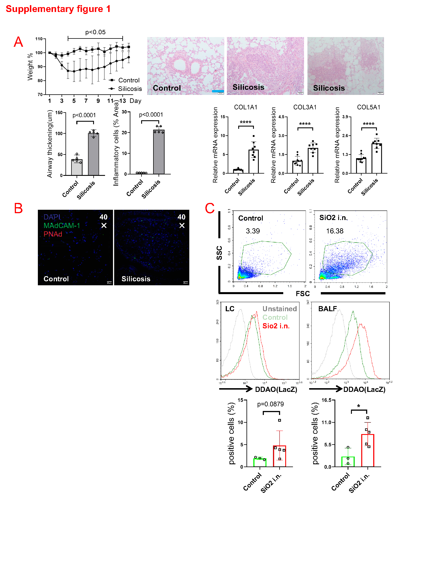
6 weeks C57BL/6 mice were randomly divided into sham operation and silicosis model groups. These mice were given 50 ul SiO2 suspension (100 mg/mL) or 50µl ddH2O intratracheally (i.t.) under the anesthetic of ketamine-xylazine, then shook the mice from side to side to make sure that the suspension spread over in the lungs uniformly. Mice were sutured and kept under warm conditions until awakened.
AM80 treatment
Am80 was mixed in feeding pellets to make a rodent maintenance diet (supported by Xietong Biotechnology). Am80 (1 mg/kg/day) administered to mice started 1 week before SiO2 exposure intratracheally (i.t.).
RAW264.7 and DC2.4 culture
The cells were cultured in DMEM or RPMI 1640 cell culture medium with 10% FBS and 100 IU/mL penicillin/streptomycin (Hyclone) and incubated at 37°C with 5% CO2. Subculture or subsequent experiments were administrated when cells were at 80% confluence.
Retroviral knockdown
Scrambled sequence (5′-TTCTCCGAACGTGTCACGT-3′) and RARa target sequences (5′-GCAGTTCCGAAGAGATAGT-3′) were cloned into the retrovirus-based vector GV493 (Genechem) containing puromycin resistance gene and GFP fluorescent reporter. 293T cells were transfected with packaging plasmids in a transfection mixture for 6 hours, replaced with 10% FBS medium, and collected virus particles after 48 hours. DC2.4s were cultured in 12-well plates and treated with Retroviral transfection for 14 h at 37 ℃ when the cells were grown to 30% confluence; the mixed clone stable strains were screened by puromycin (2ug/ml).
Quantitative real-time PCR
RNA (BMDCs or lung) was extracted with the total RNA isolation kit (Vazyme), and cDNA was synthesized from total RNA by the Hiscript III reverse transcription system (Vazyme). qPCR was performed with the ChemQ Universal SYBR qPCR master mix and LightCycler 480 (Roche). PCR primers of mouse genes were purchased from OriGene. GAPDH or β-actin was used as an internal control.
Immunofluorescence staining
BMDCs, RAW264.7 cells, mLN, hLN, and lung tissue sections from SiO2 exposure were fixed with 4% paraformaldehyde at room temperature for 0.5 h. After blocking with 10% FBS, the samples were incubated with the primary antibody (1:1000) at 4°C overnight (or fluorochrome-coupled agents at 4°C for 0.5 h). The next day, these samples were incubated with the secondary fluorochrome-coupled antibody, and the nucleus was localized by 4',6-Diamidino-2-phenylindole (DAPI). CD11c (N418), F4/80 (BM8), MAdCAM-1 (MECA-367), and PNAd (MECA-79) antibodies (purchased from eBioscience); S1P1 antibodies (T4-H28) (purchased from R&D systems); RARa antibodies (purchased from Santa Cruz); RARa-S77 and RARa-S96 antibodies (purchased from Invitrogen); ALDH1A2 antibodies (purchased from Abcam); All secondary antibodies (AlexaFluor488 and 555) were purchased from Invitrogen.
Ferrostatin-1 treatment
The day before SiO2 exposure, 6 weeks of C57BL/6 mice were injected intraperitoneally (i.p.) with Ferrostatin-1 (dissolved in 10% DMSO and 90% corn oil). The administration of Ferrostatin-1 (1 mg/kg/2 days) to mice lasted for 10 days.