2.1. Materials and Reagents.
All reactants were commercially obtained and used without further purification. Iron(III) chloride hexahydrate (97%) and ethyl ester 1,3,5-benzenetricarboxylic (97%) were obtained from Alfa Aesar. Doxorubicin hydrochloride (Dox, 98%) was purchased from Merck. All aqueous solutions were prepared using ultrapure water from a Millipore Milli-Q Academic pure-water system.
2.2. Experimental techniques.
Routine X-ray powder diffraction (XRPD) patterns were collected using a conventional PANalytical Empyrean powder diffractometer (PANalytical Lelyweg, Netherlands, θ-2θ) using λCu Kα1, and Kα2 radiation (λ = 1.54051 and 1.54433 Å). The XRPD patterns were carried out with a 2θ scan between 3–35° with a step size of 0.013° and a scanning speed of 0.1°·s-1. Fourier transform infrared (FTIR) spectroscopic analyses were performed in a Nicolet 6700 (Thermo Scientific, USA) infrared spectrometer with the help of an attenuated total reflectance (ATR) diamond accessory. High-resolution transmission electron microscopy (HRTEM) images were obtained, and energy dispersive X-ray spectroscopy (EDX) elemental mapping was performed using a high-resolution transmission electron microscope (Centre of Scientific Instrumentation of the University of Granada). 25 µL of each sample were incubated on carbon-coated grids for 5 minutes before being washed off with ultra-pure water. Uranyl acetate was employed for negative stained samples. Grids were observed in a High-Resolution TEM (HRTEM) TITAN from FEI Company operated at 300 kV. Doxorubicin and Furazan fluorescence measurements were performed with a Cary Eclipse Fluorescence Spectrometer and a PerkinElmer Spectrum FL 1.4.0.
2.3. Synthesis of MIL-100(Fe) nanoparticles (NPs).
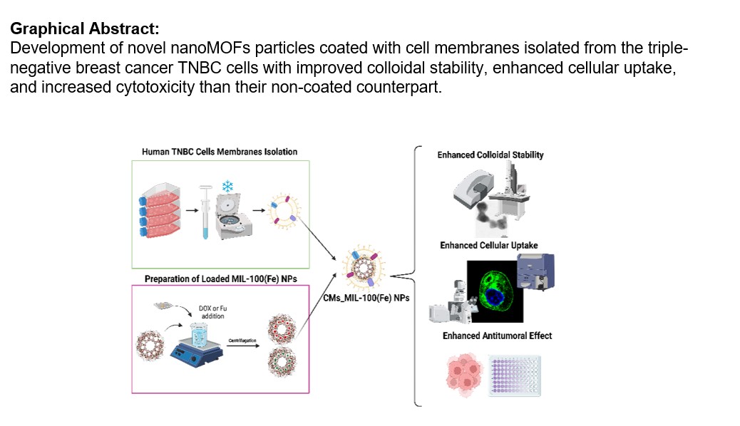
MIL-100(Fe) NPs or [Fe3O(H2O)2OH(C9H3O6)2]·nH2O were synthesized following a microwave assisted synthesis according to previously described [20].
2.4. Cell membrane extraction.
MDA-MB-468 breast adenocarcinoma CMs were isolated following a previously described method [21, 22]. Briefly, cells were grown in T-175 culture flasks to full confluency and physically detached with a scrapper in PBS. Cells were collected and washed in PBS three times by centrifuging at 500 × g for 5 min. Then, cells were suspended in a hypotonic lysis buffer consisting of 10mM Tris-HCl pH = 7.4, 1 mM KCl, 25 mM sucrose, 1 mM MgCl2, 10ug·mL-1 of DNAse and RNAse and EDTA-free protease inhibitor. Cells were disrupted using a dounce homogenizer with a tight-fitting pestle under ice-cold conditions. The solution was centrifuged at 600 × g for 5 min. The supernatant was saved while the pellet was resuspended in a hypotonic lysis buffer and subjected to further homogenization and centrifugation. The collected supernatant was further centrifuged at 17,000 g for 30 min at 4°C. The pellet was collected and washed with PBS. The final membrane-rich pellet was collected and stored for subsequent experiments. CMs were characterized by western blot analysis. Samples were prepared at the same final protein concentration as measured by a BCA assay (Pierce). Samples were mixed with loading buffer (62.5 mM Tris-HCl (pH = 6.8 at 25°C), 2% (w/v) sodium dodecyl sulfate (SDS), 10% glycerol, 0.01% (w/v) bromophenol blue, 40 mM dithiothreitol (DTT)), boiled for 5 minutes at 100°C. and an equal sample volume was loaded into each well of a 12% gel polyacrylamide gel (12% (Mini-PROTEAN® TGX™). Protein was transferred to nitrocellulose membranes (Whatman) using an XCell II Blot Module (Invitrogen) in transfer buffer (Invitrogen) following the manufacturer’s instructions. Membranes were probed using a antibodies cocktail (ab140365, abcam) against Sodium Potassium ATPase, GRP78, ATP5A, GAPDH and Histone H3 along with a horseradish peroxidase (HRP)-conjugated anti-rabbit IgG (sc-2357, Santa Cruz). Films were developed using ECL western blotting substrate (Pierce) and a Mini-Medical/90 Developer (ImageWorks).
2.5. Cell membrane coating of MIL-100(Fe).
CM coating was carried out by mixing 1 mg of MIL-100(Fe) in 1 mL of MilliQ water previously sonicated using an ultrasound tip (Bandelin Sonoplus, 20% amplitude, 1 min, ice), with 1 mg (protein) of membranes and sonicating the mixture for 3 minutes in a bath sonicator operating at 50/60 Hz and 360 W (JP Selecta™ 3000513) [23]. After each coating procedure, CMs_MIL-100(Fe) NPs were cleaned by centrifugation (7600g x for 10’) to remove not coupled membranes and excess molecules, and were resuspended in phosphate buffer solution (PBS, pH = 7).
2.6. Physicochemical characterization of the prepared NPs.
Hydrodynamic diameter, polydispersity index (PDI) and z-potential were determined by dynamic light scattering (DLS). Measurements were performed with a Zetasizer Nano-S system (Malvern Instruments, UK). The self-optimization routine in the Zetasizer software was used for all measurements, and the z-potential was calculated according to the Smoluchowsky theory. Samples were diluted with a low ionic strength phosphate buffer (1.13 mM KH2PO4, pH = 7) and measured at 25°C in triplicate. Results appeared as the mean value ± standard deviation (SD). During the colloidal stability studies, the pH effect was analyzed using buffers with identical ionic strength (0.002 M), while the ionic strength effect was analyzed at stable neutral pH of 7. The colloidal stability of the prepared systems in PBS (pH = 7.4 and 150 mM) was also determined.
2.7. Doxorubicin loading conditions.
Doxorubicin (DOX) encapsulation was performed following a similar procedure than previously described [24]. 10 mg of MIL-100(Fe) NPs were suspended in 3.0 mL of DOX aqueous solution (10 mg·mL-1) for 24 h at room temperature (RT). The drug-loaded NPs (MIL-100(Fe)@DOX) were recovered by centrifugation (5600 g, 15 min, RT) and kept at 4 ºC in the dark in a refrigerator. The amount of encapsulated DOX was determined by fluorescence spectroscopy (emission maximum at 551 nm when excited at 472 nm). After the DOX loading, DOX@MIL-100(Fe) NPs were coated using the same procedure as previously described for empty MIL-100(Fe). It should be noted that the amount of DOX leached during the coating process was estimated to be 0.033 wt%, and considered in subsequent calculations.
2.8. DOX release.
The release of DOX was studied by suspending 1 mg of MIL-100(Fe)@DOX or CMs_MIL-100(Fe)@DOX in 1 mL of PBS (0.153 M, pH = 7.4). These suspensions were kept under bidimensional stirring for different incubation times (from 15 min to 5 days). At each point, an aliquot of 0.5 mL of supernatant was recovered by centrifugation (7600g x for 5 min) and replaced with the same volume of fresh PBS. Released DOX was quantified by fluorescence spectroscopy and the potential linker release was also determined by HPLC.
2.9. Quantification of trimesic acid by high performance liquid chromatography (HPLC).
Quantification of trimesic acid (H3BTC) was performed using HPLC using a reversed phase Jasco LC-4000 series system, equipped with a PDA detector MD-4015 and a multisampler AS-4150 controlled by ChromNav software (Jasco Inc., Easton, MD, USA). A Purple ODS reverse-phase column (5 µm, 4.6 ✕ 150 mm2 Análisis Vínicos) was employed. The mobile phase consisted of a 50:50 solution (v/v) of buffer (0.04 M, pH = 2.5) and methanol (MeOH). The injection volume was set at 30 µL with a flow rate of 1 mL·min-1 and the column temperature fixed at 25 ºC. The standards used for the calibration curve consisted of trimesic acid solutions in MilliQ water solution with a concentration range from 9.65 to 0.01 µg·mL-1 (correlation coefficient > 0.99). The chromatogram of the standard solution showed a retention time (rt) of 2.70 min (λmax at 225 nm).
2.10. Cellular uptake.
Cellular uptake of the prepared NPs by MDA-MB-468 cells was assessed by flow cytometry and confocal fluorescence microscopy. For the flow cytometry assay, 1 × 10⁵ cells were seeded into 24-well culture dishes and incubated for 2, 24, and 48 hours with Fu-containing coated and non-coated MIL-100(Fe) NPs at a concentration of 64 µg·cm-2, as previously described [25]. Then, cells were detached, centrifuged at 500 g for 5 min, washed twice with PBS, resuspended in 300 µL of PBS, and analyzed by flow cytometry with a FACS Canto II (FACSCanto II, Becton Dickinson, New Jersey, US) using the software FACSDiva 6.1.2 (Becton Dickinson) for data analysis. Confocal microscopy images were taken with a Leica Sp8 spectral laser confocal microscope. 3.5 × 10⁴ MDA-MB-468 cells were seeded in 35 mm glass bottom IBIDI chambers (81158, INYCOM). After 24 h the culture media was changed and cells were incubated with 64 µg·cm-2 of NPs for 24 h. Cells were washed twice with prewarmed (37 ºC) PBS, and stained with Hoestch for 5 min for nucleus visualization. Samples were washed twice with prewarmed PBS after each step. Fu-containing NPs were visualized with a 480 nm laser. During the cellular uptake assay, IBIDI chambers were placed in a thermostatic chamber, which was kept at 37 ºC.
2.11. Cytotoxic assays.
MDA-MB-468 cells were seeded on 96 well/plates (5 ×10³ cells/well) and allowed to grow overnight. Free DOX, MIL-100(Fe)@DOX and CMs_MIL-100(Fe)@DOX samples were prepared on RPMI culture medium. Cells with the corresponding incubation medium were used as positive (100%) inhibition controls, while wells with only incubation medium and no cells were used as positive (0%) growth controls. Cells were incubated for 48 h. Then, the incubation medium was removed, cells were washed with PBS, and 100 µl of 3-(4,5-dimethylthiazol-2-yl)-2,5-diphenyltetrazolium bromide (MTT, 0.6 mM) were added per well. Plates were incubated again at 37 ºC, with an atmosphere of 5% of CO2, and 95% humidity, for 3 h. MTT was removed, cells were washed with PBS, and 100 µl of dimethyl sulfoxide (DMSO) were added per well. Absorbance was recorded at 570 nm (HEALES MB-580 microplate reader, Bangladesh). Each sample was tested in triplicate.
2.12. Statistical analysis and representation.
The obtained data were analyzed using Origin ® software (OriginLab Corporation, Northampton, Massachusetts, USA). Data appears as the mean value ± standard deviation. Data pairs were analyzed with one-way ANOVA with Tukey mean comparison method (p < 0.05).