Materials and reagents
Unless otherwise specified, all cell culture reagents were obtained from Invitrogen-Gibco (Inchinnan, UK). TRIzol® reagent (Invitrogen, USA) was utilized for RNA extraction, whereas LunaScript RT SuperMix and qPCR Master mix (New England Biolabs, UK) were utilized for cDNA synthesis and quantitative polymerase chain reaction (qPCR). All the primers used are listed in Table S1 in a supplemental document.
Adipose tissue collection
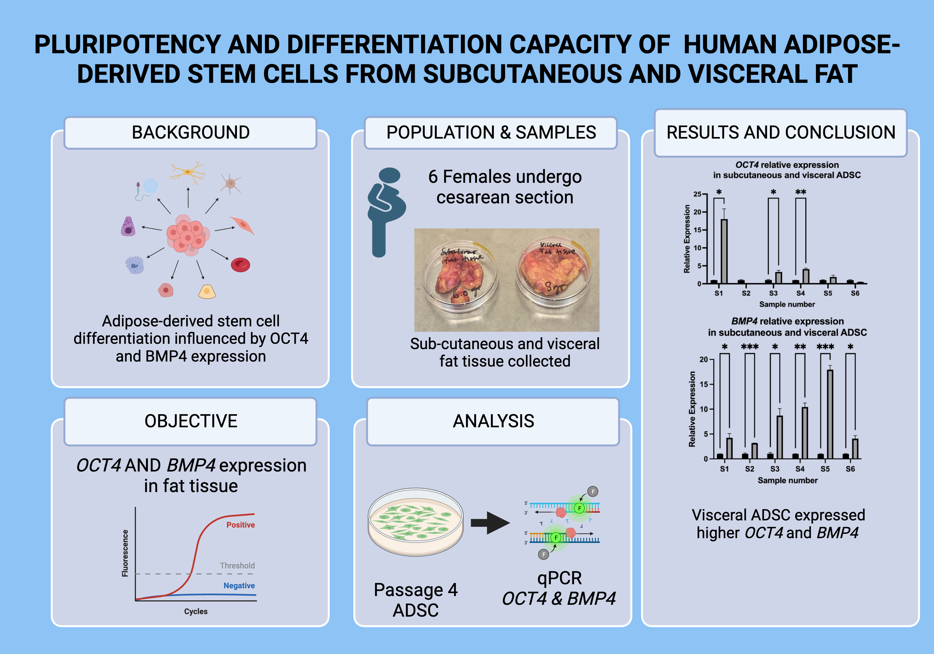
Subcutaneous and visceral adipose tissue were collected from consented 6 females (mean age 34.3 ± 7.4 years, range 27–47 years) undergo caesarean section or laparotomy at Obstetrics and Gynecology department of Hospital Canselor Tuanku Muhriz, Universiti Kebangsaan Malaysia (UKM). The procedure was reviewed and approved by the Universiti Kebangsaan Malaysia Medical Research and Ethics committee (JEP-2019-705) and completed with signed patient’s informed consent. Inclusion criteria were female gender, age below 50 years. Exclusion criteria were having cancer, on chemotherapy treatment and having peritoneal dialysis. Approximately 2–5 mg of subcutaneous and visceral adipose tissue samples were collected from each patient. The samples were kept in cold 50 ml of Dulbecco's Modified Eagle Medium: Nutrient Mixture F-12 (DMEM:F12) supplemented with 1% of antibiotic-antimitotic and processed immediately.
Isolation, culture and expansion of human adipose-derived stem cells
The subcutaneous and visceral adipose tissue were processed and isolated using stromal vascular fraction (SVF) isolation method as previously described with some modification [22, 23]. In brief, the adipose tissue samples were minced and washed with sterile PBS to remove blood. Sample were then digested using 0.3% collagenase type I solution and incubated for 4 hours at 36°C. Tissue samples were then centrifuged at 700 g for 10 minutes to isolate the SVF. The SVF cells were cultured in DMEM:F12 supplemented with 10% foetal bovine serum (FBS), 1% vitamin C, 1% ascorbic acid, 1% of antibiotic-antimitotic and 1% glutamax in 6-well plates. The cells were seeded at 10,000 cells/cm2 until confluence and expanded up to passage 4 (P4).
ADSC cell line purchased from the Centre for Tissue Engineering and Regenerative Medicine (CTERM), UKM was used as a positive control. The cells were cultured in DMEM:F12 supplemented with 10% foetal bovine serum (FBS), 1% vitamin C, 1% ascorbic acid, 1% of antibiotic-antimitotic and 1% glutamax until confluence and multiplied up to P6 and used as a positive control of pluripotent cells.
RNA Isolation and RT-PCR
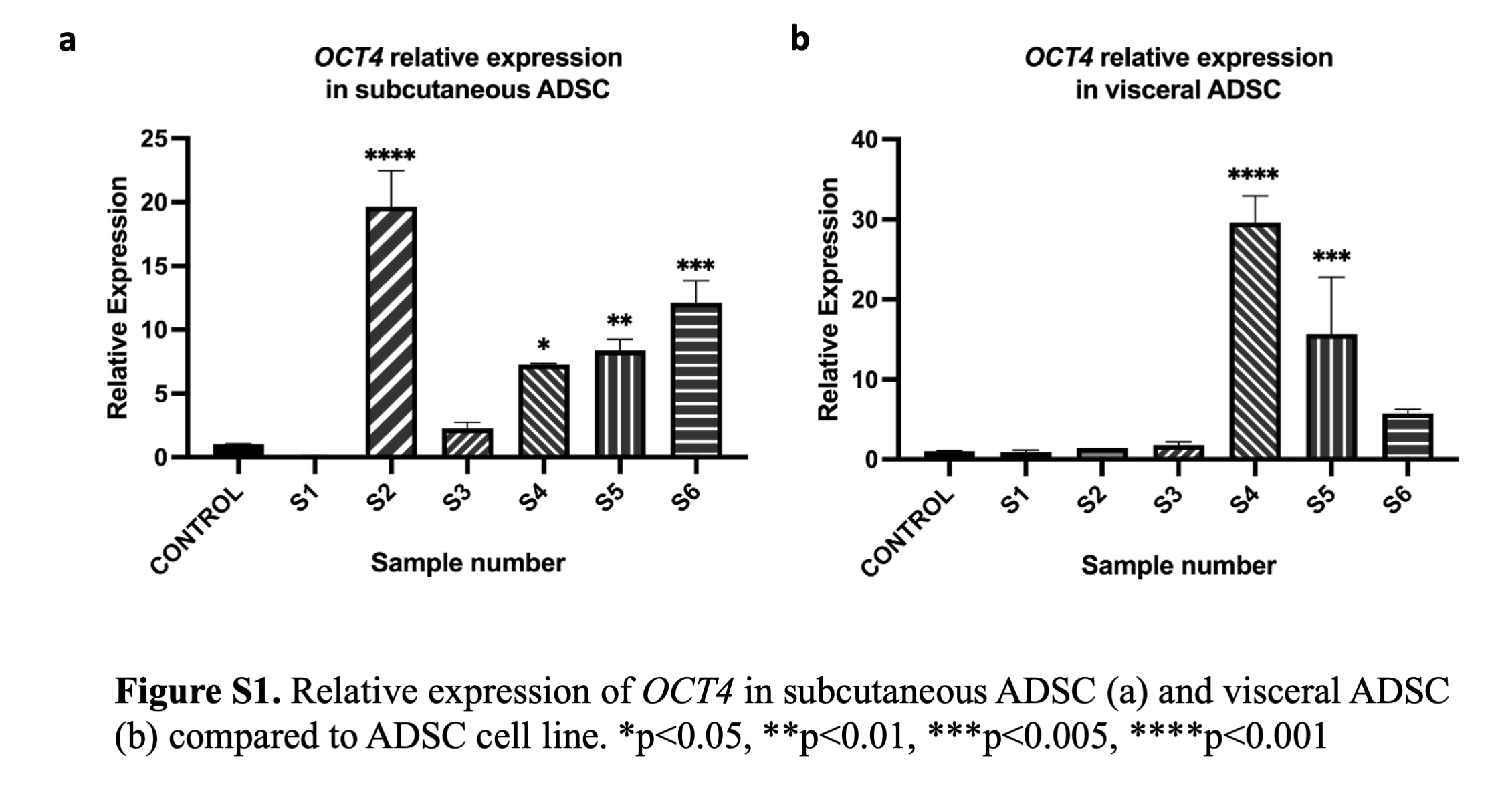
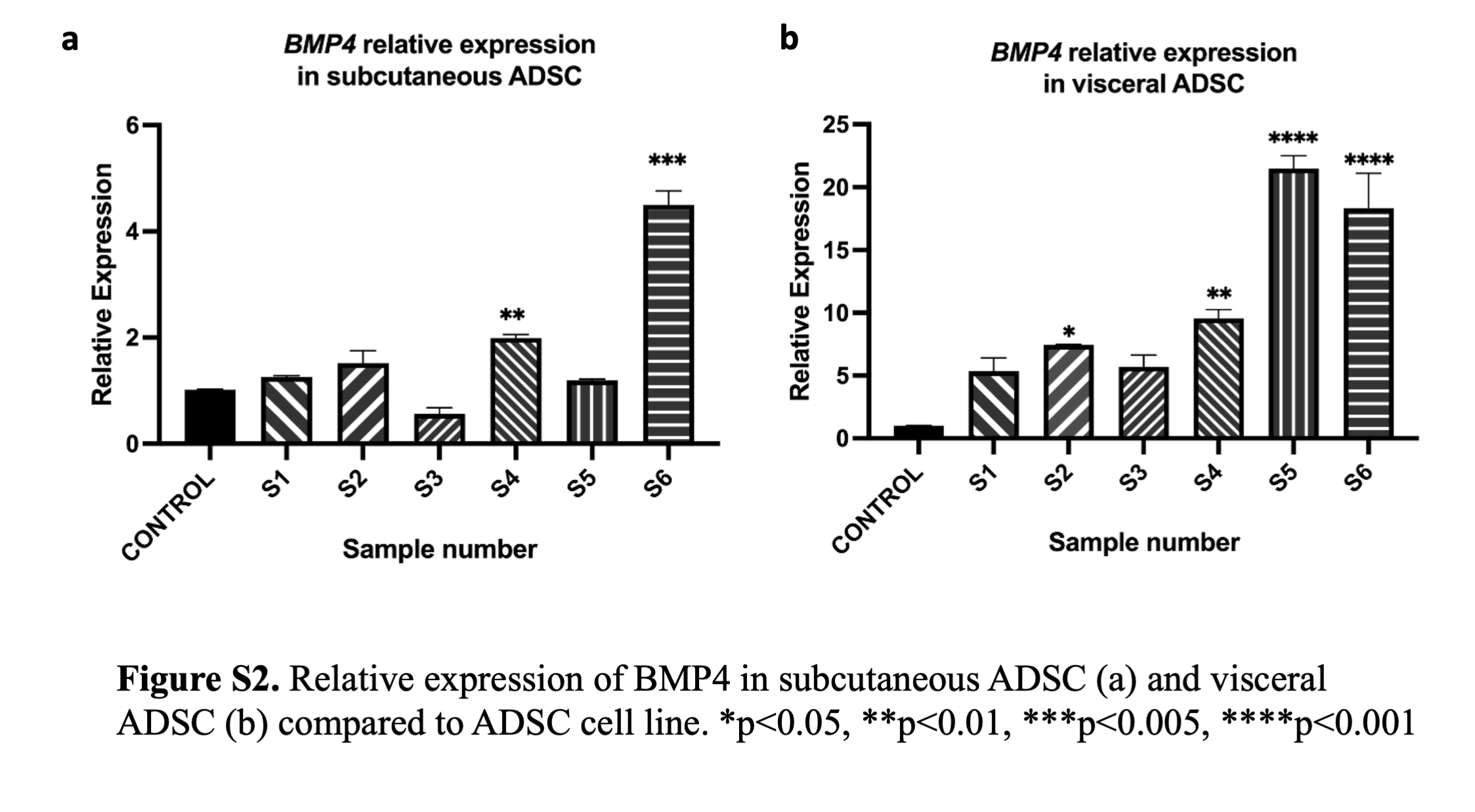
RNA was extracted from the cultured cells using the Trizol method according to the company guideline. The concentration and quality of RNA were determined using a Nanodrop spectrophotometer. The expression levels of OCT4 and BMP4 genes were evaluated by quantitative real-time PCR (qPCR) using the glyceraldehyde 3-phosphate dehydrogenase (GAPDH) gene as a reference gene. Results were analysed using the 2–ΔΔCt method and expressed as the fold change in gene expression relative to the ADSC cell line [24, 25].
Statistical Analysis
Standard error of the mean (SEM) was used to present the gene expression results. GraphPad Prism version 9.0.0 for Windows, (GraphPad Software, San Diego, California, USA, www.graphpad.com) was used to analyse the data. One-way analysis of variance (ANOVA) was used to determine the statistical significance for comparisons made within the groups while student t-test was used to evaluate the statistical significance between subcutaneous and visceral ADSC groups. P values of 0.05 or less were considered as statistically significant.