An eco-friendly green route is employed for the successful synthesis of heterostructured ZnO-CuO nanocomposites using Calotropis gigantea plant and the investigation of their antimicrobial properties against skin ulcer pathogens. Binary ZnO-CuO nanocomposites prepared at calcination temperature of 300 °C exhibited superior antimicrobial effect on S. aureus, whereas the negative control sample did not show any antibacterial activities. High ZnO nanoparticles of 75 wt.% ZnO-CuO nanocomposites showed zero count of Staphylococcus aureus at a minimum inhibitory concentration of 0.625 mg/mL and minimum bactericidal concentration (MBC) of 2.5 mg/mL. Interestingly, the 75 wt.% ZnO-CuO nanocomposites exhibited strong antimicrobial activity against multi-drug resistant pathogens, with MBC ranging from 0.3125 mg/mL to 1.25 mg/mL. A time-kill assay captured a reduction in viable count from 4.3 log 10 to 1.3 log 10 after 12 h of incubation for S. aureus . Elucidating the antimicrobial activities could be useful for identifying novel ways to incorporate ZnO-CuO nanocomposites in polymers for applications in biocide materials, such as for wound dressing. Further, molecular studies are needed to explain the underlying biocidal mechanism of ZnO-CuO nanocomposites especially in the presence of Cu 2+ and Zn 2+.
Research
Calcination Temperatures, Compositions and Antimicrobial Properties of Heterostructural ZnO–CuO Nanocomposites from Calotropis Gigantea Targeted for Skin Ulcer Pathogens
https://doi.org/10.21203/rs.3.rs-37393/v1
This work is licensed under a CC BY 4.0 License
Journal Publication
published 08 Jan, 2021
Version 1
posted
You are reading this latest preprint version
heterostructural
green nanoparticles
binary nanoparticles
nanocomposites
copper oxide nanoparticles
zinc oxide nanoparticles
skin ulcer pathogens
biocidal agent
pressure ulcers (PUs)
Ulcerative skin infections arising from colonisation and development of gram-positive bacteria, gram-negative bacteria and multidrug-resistant bacteria are significant health care problems that seriously affect human skin. A prospective quantitative study reported on the prevalence rates of skin pressure ulcers (PUs) at 15.5% in Kuala Lumpur, Malaysia (2013) [1], 33% in Palestine (2017) [2] and 16% in Bandung, Indonesia (2017) [3]. Skin infection was found in 60 (74.0%) of the collected samples from the PUs of hospitalised patients and was mainly constituted with Enterobacteriaceae strains (49.0%) such as Escherichia coli (E. coli), Klebsiella pneumonia (K. pneumonia), Enterobacter spp. and Proteus spp.; followed by Staphylococcus aureus (S. aureus) (28.0%) and non-fermenting GNB (23.0%), mostly Pseudomonas aeruginosa (P. aeruginosa), Acinetobacter spp. and Methicillin-resistant S. aureus (MRSA) [4, 5, 6, 7]. PUs are open infected wounds that develop on the skin as result of pressure on one spot of the body for too long or from friction on the skin. Findings of new inorganic oxide antimicrobial agents synthesised from natural plant became one of the remarkable alternatives for infectious skin treatments of PUs as such options are rich in numerous varieties of metal oxides that release ions and in reactive oxygen species (ROS), such as hydroxyl radical (•OH-) and superoxide (•O2−) which cause increased permeability, rupture and cell death of microorganisms [8, 9].
Recently, the incorporation of inorganic metal and metal oxides in sponges [10], hydrogels [11, 12] and bandages [13, 14] have gained priority in research because of their advantages as antimicrobial agents for treating locally infected skin ulcers. Mixed inorganic metal and metal oxides are effective disinfectants because of their relatively non-toxic profile, chemical stability and efficient antibacterial activity (Table 1). Binary antimicrobial agents (e.g. CuO and ZnO and Ag and ZnO) have been highlighted over single antimicrobial agents given the strong synergic effect of the former in eliminating bacterial colonies at low concentration [10, 24, 38], more pronounced wound healing [10], lower cytotoxicity [10], better biocompatibility [24] and improved cell viability which is safe for human application [24]. Therefore, this paper focuses on the preparation of green synthesised binary ZnO-CuO nanocomposites using Calotropis gigantea (C. gigantea) leaf extract and investigates the microbial activity of these nanocomposites upon culturing with skin ulcer pathogens such as E. coli, K. pneumonia, S. aureus, P. aeruginosa and MRSA. Further, the effect of different compositions and calcination temperatures on ZnO-CuO nanocomposites were explored with respect to their prospective antimicrobial application.
Preparation of the leaves extract and binary inorganic oxides
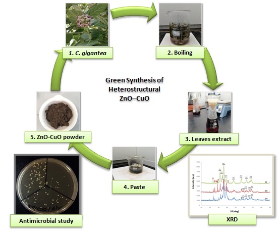
For this present investigation, the whole plant of C. gigantea was collected from Perai Pulau Pinang, Malaysia and was identified by the expert of Unit Herbarium, Pusat Pengajian Sains Kajihayat USM Pulau Pinang. (Herbarium No.: 11843). In this experiment work, 5 g of C. gigantea leaves were added to 100 mL of deionized water and boiled for 60 min at temperature of 90-100°C using hot plate [39, 40]. Then, 50 mL of filtered leaves extracts were taken and boiled to 60-80°C using a stirrer-heater. Binary inorganic oxides ZnO-CuO nanocomposites were prepared by adding Copper (II) Nitrate Trihydrate and Zinc Nitrate Hexahydrate into the extract solutions simultaneously and then boiled until it reduced to paste. These pastes were calcinated in an air heated furnace for 2 h [39, 40]. In 1st stage, ZnO-CuO nanocomposites were prepared at fixed rotation speed and composition of 50 wt.% of ZnO and 50 wt.% of CuO by varying the calcinations temperature (300°C, 400°C and 500°C). In 2nd stage, the weight percentage of binary oxides ZnO-CuO (25 wt. %, 50 wt. % and 75 wt. % of ZnO) was varied with constant rotation speed and calcinations temperature of 300°C.
XRD analysis
The crystal phases of ZnO-CuO nanocomposites were studied using X-ray diffraction (XRD), Bruker D8 powder diffractometer operating in reflection mode with a Cu Kα radiation (40 kV, 30 mA) diffracted beam monochromator, using a step scan mode with step size of 0.030° in the range of 10° to 90°. The crystallite size was estimated from the XRD pattern using the Scherer’s Equation [1]:

where K= 0.9 is the shape factor, λ is the X-ray wavelength of Cu Kα radiation (0.1541 nm), θ is the Bragg diffraction angle, and β is the FWHM of the respective diffraction peak.
Minimum inhibitory concentration/minimum bactericidal concentration determination and tolerance level
Antibacterial activity of ZnO-CuO nanocomposites against S. aureus 29213, E. coli 25922, P. aeruginosa 27853, K. pneumonia 700603 and MRSA 38591 were assessed using broth dilution method on 96-well plates as described by NH Harun et al. (2020) with slight modifications [41]. The bactericidal and bacteriostatic capacity of the samples was determined by the tolerance level [41].
Time-kill assay
The antibacterial activity of ZnO-CuO nanocomposites against time was carried out using time-kill assay as illustrated in protocol before [41]. The adjusted S. aureus bacterial suspension to 0.5 McFarland standard turbidity was used and diluted with samples solution with final concentration of 2.5 mg/mL.
Calcination temperatures and composition of heterostructural ZnO–CuO nanocomposites
Seven characteristic peaks of ZnO and five characteristic peaks of CuO were found in green ZnO-CuO samples prepared at different calcination temperatures (300, 400 and 500 °C) as shown in Figure 1. Diffraction peaks at 31.87°, 34.54°, 36.37°, 47.59°, 56.59°, 62.92° and 65.89° which correspond to crystal surfaces (100), (002), (101), (102), (110), (103) and (200) belonged to ZnO. Conversely, CuO was detected at 35.62°, 38.83°, 57.49°, 61.42° and 67.96°, which correspond to crystal surfaces (–111), (111), (202), (–113) and (220). The XRD pattern of ZnO-CuO nanocomposites confirms the presence of pure ZnO and CuO. Furthermore, few additional peaks were observed at 23.65°, 25.69°, 27.73°, 29.47° and 40.78° from Figures 1 and 2. This outcome is possibly due to the presence of the phytochemical element of C. gigantea leaves as a capping and reducing agent [39]. The additional peaks detected at 29.47° and 40.78° are attributed to the natural graphene-like carbon present in the ZnO-CuO nanocomposites [42] as carbon is a main phytochemical element in the leaves of the C. gigantea medicinal plant [43, 44]. Natural carbon in binary ZnO-CuO nanocomposites could further enhance the synergic effect on antimicrobial activity [45, 46]. However, these peaks lessened at higher calcination temperatures.
Prominent diffractive peaks on the differential ratio of binary ZnO and CuO nanocomposites are indexed in Figure 2. Six characteristic peaks of ZnO for sample 75ZnO25CuO-300C were identified at 31.72°, 34.45°, 36.25°, 47.35°, 56.41° and 62.71° and corresponded to crystal surfaces (100), (002), (101), (102), (110) and (103). Another two characteristic peaks of CuO at 38.62° and 67.78° corresponded to crystal surfaces (111) and (220). For sample 25ZnO75CuO-300C, 31.72°, 34.45°, 36.25°, 47.35°, 56.41°, 62.71° and 68.05° peaks, respectively belonged to the (100), (002), (101), (102), (110), (103) and (201) indices of ZnO nanoparticles. Conversely, the diffractive peaks of CuO detected at 35.68°, 38.62°, 58.33°, 61.27° and 65.80° corresponded to crystal surfaces (–111), (111), (202), (–113) and (–311). The peak intensity is drastically increased with higher amounts of ZnO or CuO in the binary ZnO-CuO nanocomposites (Figure 2), thereby indicating the variation in composition (25, 50 and 75 wt. % of ZnO) during green synthesis.
Superior antimicrobial properties of heterostructural ZnO–CuO nanocomposites
The antimicrobial efficacy of binary 50ZnO/50CuO nanocomposites at three different calcination temperatures (300°C, 400°C and 500°C) were initially characterised by S. aureus minimum inhibitory concentration (MIC)/minimum bactericidal concentration (MBC) as presented in Table 2. The MIC of 50ZnO/50CuO-300C and 50ZnO/50CuO-400C for S. aureus were 2.5 mg/mL, except for 50ZnO/50CuO-500C at 5 mg/mL. Moreover, the MBC of all green synthesised 50ZnO/50CuO samples for S. aureus was at 20 mg/mL. S. aureus colonies were counted less at a concentration of 5 mg/mL for the sample 50ZnO/50CuO-300C prepared at low calcination temperature (300°C) relative to the 50ZnO/50CuO-400C and 50ZnO/50CuO-500C samples (Figure S1). The effect of smaller sized nanoparticles generated at low calcination temperature is suggested to possibly enhance surface reactivity in killing the microbes [47, 48, 49]. Particle size is crucial in antimicrobial activity effectiveness. Azam et al. (2012) and Salah et al. (2011) verified that smaller particle sizes mean greater efficacy in inhibiting bacterial growth, a feature that is possibly associated with the larger surface areas of nanoparticles [50, 51].
Next, further characterisation of the differential ratios of binary ZnO and CuO nanocomposites at a calcination temperature of 300 °C were presented in Figure S2 and Table 2. The MIC of 25ZnO/75CuO-300C, 50ZnO/50CuO-300C and 75ZnO/25CuO-300C were 5 mg/mL, 2.5 mg/mL and 0.625 mg/mL for S. aureus, respectively. Similar to the MIC values, the 25ZnO/75CuO-300C and 50ZnO/50CuO-300C green samples had MBC values of 20 mg/mL and the counterpart for the 75ZnO/25CuO-300C sample was 2.5 mg/mL for S. aureus. The 75ZnO/25CuO-300C sample exerted a higher bactericidal effect against the S. aureus strain at the lowest MIC/MBC values (0.625 mg/mL/2.5 mg/mL). The antimicrobial activity was further enhanced by increasing the amount of ZnO nanoparticles in the binary compound (ZnO-CuO). The phenomenon observed can be explained by the fact that the binary 75ZnO/25CuO-300C nanocomposites are highly diffusible and generate more Zn2+ [19]. Moreover, Cu2+ ions bind the cell wall of host cells through surface proteins and enter the cell [19]. Subsequently, the change in the metabolism of cells leads to the microbe’s cell death [19].
Further antimicrobial analysis of 75ZnO/25CuO-300C on selected skin ulcer pathogens are shown in Table 3. These pathogens are commonly associated with skin ulcer disease [4, 5, 6, 7]. The MIC values of the green synthesised ZnO-400C for E. coli, P. aeruginosa, K. pneumonia and MRSA were at 0.3125, 0.15625, 0.625 and 0.15625 mg/mL, respectively. By contrast, the MBC values were 2.5, 0.3125, 1.25 and 0.3125 mg/mL, respectively. Furthermore, the MIC amounts for the 75ZnO/25CuO-300C sample were 0.625, 0.15625, 0.625 and 0.15625 mg/mL for E. coli, P. aeruginosa, K. pneumonia and MRSA, respectively. MBC values with 2.5, 0.3125, 1.25 and 0.3125 mg/mL were also observed for this green binary inorganic oxide sample. The tolerance level according to the MBC/MIC ratio showed that all tested microbes are sensitive to bactericidal agents except for the CuO-500C sample against E. coli, P. aeruginosa and MRSA and the ZnO-400C sample towards E. coli. Table 3 indicates that for all tested microbes, only the tolerance levels for 75ZnO/25CuO-300C sample were less than 4, and these values identifies the sample as a strong bactericidal agent relative to other samples (ZnO-400C and CuO-500C).
Moreover, higher MBC values of the CuO-500C sample against all tested microbes possibly transpire from the slow Cu2+ ion release from CuO nanoparticles [52]. Clearly, the ZnO-400C and 75ZnO/25CuO-300C samples show very promising results against all tested microbes. That outcome may arise from the ZnO nanoparticle’s larger surface to volume ratio and the penetration of the cell membrane of the bacteria by its ions. Furthermore, the ZnO-400C sample showed better antimicrobial activity relative to the CuO-500C counterpart at a similar concentration. Some studies reported that the antimicrobial effectiveness of green synthesised inorganic oxide nanoparticles depends on particle dosage, size and treatment condition, such as calcination temperatures. This situation could be the one of the reasons for the higher antimicrobial activities of ZnO and ZnO-CuO over that of CuO particles.
Additionally, the antimicrobial activities of the ZnO-400C and 75ZnO/25CuO-300C samples are much better than that of the CuO-500C sample alone towards multi-drug resistant strains P. aeruginosa, K. pneumonia and MRSA. That outcome is obviously due to the high diffusion of Zn2+ ions in the medium. The poor activity of CuO particles within a shorter duration suggested that the time requirement for water diffusion and subsequent Cu2+ release influence efficacy. The attacking delay was also associated with the cell walls of gram-negative strains. Studies have reported that gram-negative bacterial strains exhibit higher resistance or tolerance against nanomaterials compared with gram-positive bacteria [53] because of the lipopolysaccharide situated in the outer membrane of the former [54]. The cytoplasmic membrane, which is inherent to gram-negative bacteria, significantly maintains cellular viability. Hence, gram-negative microbes are not readily attacked by free radicals or Cu2+. More time and concentrated Cu2+ ions are thus required to effectively decompose the cell membrane of the bacteria. The antimicrobial activity of ZnO, CuO and ZnO-CuO nanoparticles is due to the electrostatic interaction between positively charged zinc and copper ions (Zn2+ and Cu2+) and negatively charged microbial cell membranes [21]. In addition, the antimicrobial activity of inorganic oxide nanoparticles relies on the generation of ROS as well [17, 19].
In the time-kill assay results were presented in terms of the changes in the log10 CFU/mL of viable S. aureus colonies and indicated that the green synthesised binary 75ZnO/25CuO-300C sample exhibited significant bactericidal activity. The outcomes of the time-kill assay were captured in Figure S3. Figure 3 presents the time-kill curve graph for the strain. A reduction in viable count from 4.3 log10 to 3.4 log10 was captured after 6 h of incubation for S. aureus. By 12 h, only 1.3 log10 of bacterial colonies were seen. At 24 h, the bacteria were completely killed. Therefore, the effective control of gram-positive S. aureus bacteria was achieved by the synergistic combination of 75 wt.% of ZnO and 25 wt.% of CuO nanoparticles with the presence of phytochemical constituents such as cardiac glycosides, tannins, saponins, terpenes, flavonoids and phenolics in the leaf extract of the C. gigantea medicinal plant [55, 56, 57, 58].
In summary, heterostructural ZnO-CuO nanocomposites prepared at a calcination temperature of 300 °C and with a composition of 75 wt.% of ZnO and 25 wt.% of CuO demonstrated significant antimicrobial property against skin ulcer pathogens. The mechanisms that underlie the biocidal activity of ZnO-CuO nanocomposites were reflected by the presence of Cu2+ and Zn2+ ions and ROS. This finding could reduce the environmental bio-burden in a hospital atmosphere, specifically in relation to pressure ulcerative skin infections.
C. gigantea: Calotropis gigantea, E. coli: Escherichia coli, K. pneumonia: Klebsiella pneumonia, S. aureus: Staphylococcus aureus, P. aeruginosa: Pseudomonas aeruginosa and MRSA: Methicillin-resistant S. aureus.
Ethics approval and consent to participate
Not applicable.
Consent for publication
Not applicable.
Availability of data and materials
The datasets generated and/or analysed during the current study are not publicly available due to the patent application for methods of making and using and compositions of binary nanocomposites formed by green synthesis but are available from the corresponding author on reasonable request.
Competing interests
The authors declare no conflict of interest.
Funding
This research was funded by the research university grant 2019/2020.
Authors’ contributions
G Ambarasan Govindasamy carried out the green sample preparation, sample characterization and the antibacterial assays, included bacterial preparation, MIC, MBC and time kill-assay. Nor Hazliana Harun and Srimala Sreekantan assist in the experimental procedures. Rabiatul Basria S. M. N. Mydin contributes in the experimental design, writing process and gave final approval of this paper for publication. All authors have given approval to the final version of the manuscript.
Acknowledgements
The support of all the technical staff of Advanced Medical and Dental Institute, Universiti Sains Malaysia, Pulau Pinang, Malaysia, in the characterization of the sample is acknowledged.
Authors’ information
G Ambarasan Govindasamy − 1Oncological and Radiological Sciences Cluster, Advanced Medical and Dental Institute, Universiti Sains Malaysia, 13200 Bertam, Kepala Batas, Pulau Pinang, Malaysia; 2Ann Joo Integrated Steel Sdn Bhd, Lot 1236, Prai Industrial Estate, 13600 Prai, Penang, Malaysia; Email: [email protected]; ORCID iD: 0000-0002-9263-5083
Rabiatul Basria S. M. N. Mydin − 1Oncological and Radiological Sciences Cluster, Advanced Medical and Dental Institute, Universiti Sains Malaysia, 13200 Bertam, Kepala Batas, Pulau Pinang, Malaysia; 3Department of Biological Sciences, National University of Singapore, 14 Science Drive 4, 117543 Singapore; Email: [email protected]; ORCID iD: 0000-0001-8971-8809
Nor Hazliana Harun − 1Oncological and Radiological Sciences Cluster, Advanced Medical and Dental Institute, Universiti Sains Malaysia, 13200 Bertam, Kepala Batas, Pulau Pinang, Malaysia; Email: [email protected]; ORCID iD: 0000-0002-5563-4145
Srimala Sreekantan − 4School of Materials and Mineral Resources Engineering, Universiti Sains Malaysia, Engineering Campus, 14300, Nibong Tebal, Pulau Pinang, Malaysia; Email: [email protected]; ORCID iD: 0000-0001-5125-8683
- Khor, H. M., Tan, J., Saedon, N. I., Kamaruzzaman, S. B., Chin, A. V., Philip J.H. Poi, P. J. H. and Tan, M. P. (2014). Determinants of mortality among older adults with pressure ulcers. Archives of Gerontology and Geriatrics. 59. p.536-541.
- Qaddumi, J. A. S. and Almahmoud, O. (2019). Pressure Ulcers Prevalence and Potential Risk Factors Among Intensive Care Unit Patients in Governmental Hospitals in Palestine: A Cross-sectional Study. The Open Public Health Journal.12. p.121-126.
- Sari, S. P., Everink, I. H., Sari, E. A., Afriandi, I., Amir, Y., Lohrmann, C., Halfens, R. J. and Schols, J. M. (2019). The prevalence of pressure ulcers in community-dwelling older adults: A study in an Indonesian city. Int Wound J. 16. p.534-541.
- Park-Lee, E. and Caffrey, C. (2009). Pressure Ulcers Among Nursing Home Residents: United States, 2004. NCHS Data Brief. 14.
- Braga, I. A., Brito, C. S., Filho, A. D., Filho, P. P. G. and Ribas, R. M. (2016). Pressure ulcer as a reservoir of multiresistant Gram-negative bacilli: risk factors for colonization and development of bacteremia. The Brazilian Journal of Infectious Diseases. p.1.
- Dana, A., N. and Bauman, W. A. (2014). Review Bacteriology of pressure ulcers in individuals with spinal cord injury: What we know and what we should know. The Journal of Spinal Cord Medicine. p.1.
- EL-TORAEI, M.D., Ch.M., F.LC. S, AND B. CHUNG, M.D. (1977). The Management of Pressure Sores.J. Dermatol. Surg. Oncol. 3 (5). p.507.
- Sumbal, Nadeem, A., Naz, S., Ali, J. S., Mannan, A. and Zia, M. (2019). Synthesis, characterization and biological activities of monometallic and bimetallic nanoparticles using Mirabilis jalapa leaf extract. Biotechnology Reports 24.
- Alavi, M. and Karim, N. (2018). Antiplanktonic, antibiofilm, antiswarming motility and antiquorum sensing activities of green synthesized Ag–TiO2, TiO2–Ag, Ag–Cu and Cu–Ag nanocomposites against multi-drug-resistant bacteria. ARTIFICIAL CELLS, NANOMEDICINE, AND BIOTECHNOLOGY. 46 (3).
- Lu, Z., Gao, J., He, Q., Wu, J., Liang, D., Yang, H., & Chen, R. (2017). Enhanced antibacterial and wound healing activities of microporous chitosan-Ag/ZnO composite dressing. Carbohydrate Polymers 156. p.460-469.
- Nguyen, T. D., Nguyen, T. T., Ly, K. L., Tran, A. H., Nguyen, T. T. N., Vo, M. T., Ho, H. M., Dang, N. T. N., Vo, V. T., Nguyen, D. H., Nguyen, T. T. H. and Nguyen, T. H. (2019). In Vivo Study of the Antibacterial Chitosan/Polyvinyl Alcohol Loaded with Silver Nanoparticle Hydrogel for Wound Healing Applications, Hindawi International Journal of Polymer Science.
- Elbadawy A. Kamoun, E., Kenawy, E. S., Tamer, T. M., El-Meligy, M. A. and Eldin, M. S. M. (2015). Poly (vinyl alcohol)-alginate physically crosslinked hydrogel membranes for wound dressing applications: Characterization and bio-evaluation. Arabian Journal of Chemistry. 8. p.38-47.
- Kumar, P. T. S., Lakshmanan, V., Anilkumar, T. V., Ramya, C., Reshmi, P., Unnikrishnan, A. G., Nair, S. V., & Jayakumar, R. (2012). Flexible and Microporous Chitosan Hydrogel/Nano ZnO Composite Bandages for Wound Dressing: In Vitro and In Vivo Evaluation. ACS Appl. Mater. Interfaces. 4. p.2618-2629.
- Arshad, R., Sohail, M. F., Sarwar, H. S., Saeed, H., Ali, I., Akhtar, S., Hussain, S. Z., Afzal, I., Jahan, S., Rehman, A. U., & Shahnaz, G. (2019). ZnO-NPs embedded biodegradable thiolated bandage for postoperative surgical site infection: In vitro and in vivo evaluation.
- Yulizar, Y., Bakri, R., Apriandanu, D. O. B. and Taufik Hidayat, T. (2018). ZnO/CuO nanocomposite prepared in one-pot green synthesis using seed bark extract of Theobroma cacao. Nano-Structures & Nano-Objects. 16. p.300-305.
- Dobrucka, R., Kaczmarek, M., Lagiedo, M., Kielan, A. and Dlugaszewska, J. (2019). Evaluation of biologically synthesized Au-CuO and CuO-ZnO nanoparticles against glioma cells and microorganisms. Saudi Pharmaceutical Journal. 27. p.373-383.
- Khan, S. A., Noreen, F., Kanwal, S., Iqbal, A. and Hussain, G. (2017). Green Synthesis of ZnO and Cu-doped ZnO Nanoparticles from Leaf Extracts of Abutilon indicum, Clerodendrum infortunatum, Clerodendruminerme and Investigation of their Biological and photocatalytic activities. Materials Science & Engineering C.
- Aloucheh, R. M., Yangjeh, A. H., Bayrami, A., Navid, S. L. and Asadi, A. (2018). Green synthesis of ZnO and ZnO/CuO nanocomposites in Mentha longifolia leaf extract: characterization and their application as antibacterial agents. Journal of Materials Science: Materials in Electronics.
- Widiarti, N., Sae, J. K. and Wahyuni., S. (2017). Synthesis CuO-ZnO nanocomposite and its application as an antibacterial agent. IOP Conf. Series: Materials Science and Engineering. 172. p.012036.
- Hassan, I. A., Sathasivam, S., Sean P. Nair, S. P. and Carmalt, C. J. (2017). Antimicrobial Properties of Copper-Doped ZnO Coatings under Darkness and White Light Illumination. ACS Omega. 2. p.4556-4562.
- Alswat, A. A., Ahmad, M. B. and Tawfik A. Saleh, T. A. (2017). Preparation and Characterization of Zeolite\Zinc Oxide-Copper Oxide Nanocomposite: Antibacterial Activities Colloid and Interface Science Communications. 16. p.19-24.
- Qiu, S., Zhou, H., Shen, Z., Hao, L., Chen, H. and Xinhua Zhou, X. (2020). Synthesis, characterization, and comparison of antibacterial effects and elucidating the mechanism of ZnO, CuO and CuZnO nanoparticles supported on mesoporous silica SBA-3†. RSC Adv.10. p.2767-2785.
- Al-Dhabaan, F. A., Shoala, T., Ali, A. A. M., Alaa, M. and Abd-Elsalam, K. (2017). Chemically-Produced Copper, Zinc Nanoparticles and Chitosan–Bimetallic Nanocomposites and Their Antifungal Activity against Three Phytopathogenic Fungi. International Journal of Agricultural Technology. 13 (5). p.753-769.
- Hu, M., Li, C., Li, X., Zhou, M., Sun, J., Sheng, F., Shi, S., & Lu, L. (2018). Zinc oxide/silver bimetallic nanoencapsulated in PVP/PCL nanofibres for improved antibacterial activity ARTIFICIAL CELLS, NANOMEDICINE, AND BIOTECHNOLOGY. 46 (6). p.1248-1257.
- Basavalingiah, K. R., Harishkumar, S., Udayabhanu, Nagaraju, G., Rangappa, D. and Chikkahanumantharayappa (2019). Highly porous, honeycomb like Ag–ZnO nanomaterials for enhanced photocatalytic and photoluminescence studies: green synthesis using Azadirachta indica gum. SN Applied Sciences. 1 (935).
- Bazant, P., Kuritka, I., Munster, L., Machovsky, M., Kozakova, Z. and Saha, P. (2014). Hybrid nanostructured Ag/ZnO decorated powder cellulose fillers for medical plastics with enhanced surface antibacterial activity. J Mater Sci: Mater Med.
- Motshekga, S. C., Ray, S. S., Onyango, M. S. and Momba, M. N. B. (2015). Preparation and antibacterial activity of chitosan-based nanocomposites containing bentonite-supported silver and zinc oxide nanoparticles for water disinfection. Applied Clay Science. 114. p.330-339.
- Yang, S., Zhang, Y.,Yu, J., Zhen, Z., Huang, T., Tang, Q., Chu, P. K., Qi, L. and Lv, H. (2014). Antibacterial and mechanical properties of honeycomb ceramic materials incorporated with silver and zinc. Materials and Design. 59. p.461-465.
- Sharma, M., Hazra, S. and Basu, S. (2017). Synthesis of heterogeneous Ag-Cu bimetallic monolith with different mass ratios and their performances for catalysis and antibacterial activity. Advanced Powder Technology. 28. p.3085-3094.
- Paszkiewicz, M., Galabiehs, A., Rajski, L., Kowal, E., Sajdak, A. and Zaleska-Medynska, A. (2016). The Antibacterial and Antifungal Textile Properties Functionalized by Bimetallic Nanoparticles of Ag/Cu with Different Structures. Journal of Nanomaterials.
- Joshi, B., Regmi,C., Dhakal, D., Gyawali, G. and Lee, S. W. (2018). Efficient inactivation of Staphylococcus aureus by silver and copper loaded photocatalytic titanate nanotubes. Progress in Natural Science: Materials International.
- Azizi-Lalabadi, M., Ehsani, A., Divband, B. & Alizadeh-Sani, M. (2019). Antimicrobial activity of Titanium dioxide and Zinc oxide nanoparticles supported in 4A zeolite and evaluation the morphological characteristic. Scientific Reports. 9. p.1743.
- Daou, I., Moukrad, N., Zegaoui, O. and Filali, F. R. (2017). Antimicrobial activity of ZnO-TiO2 nanomaterials synthesized from three different precursors of ZnO:influence of ZnO/TiO2 weight ratio.Water Science & Technology.
- Shadmehri, A. A., Namvar, F., Miri, H., Yaghmaei, P. and Mahboobeh Nakhaei Moghaddam, M. N. (2019). Assessment of antioxidant and antibacterial activities of Zinc Oxide nanoparticles, Graphene and Graphene decorated by Zinc Oxide nanoparticles. Int. J. Nano Dimens. 10 (4). p.350-358.
- Jaiswal, A. K., Gangwar, M., Nath, G. and Yadav, RR. (2016). Antimicrobial Activity of Bimetallic Cu/Pd Nanofluids. J Adv Chem Eng. 6 (2).
- Al-Asfar, A., Zaheer, Z. and Aazam, E. S. (2017). Eco-friendly green synthesis of Ag@Fe bimetallic nanoparticles: Antioxidant, antimicrobial and photocatalytic degradation of bromothymol blue. Journal of Photochemistry & Photobiology, B: Biology.
- Vasile, B. S., Oprea, O., Voicu, G., Ficai, A., Andronescu, E., Teodorescu, A. and Alina Holban, A. (2013). Synthesis and characterization of a novel controlled release zinc oxide/gentamicin–chitosan composite with potential applications in wounds care. International Journal of Pharmaceutics.
- Chabala, L. F. G., Cuartas, C. E. E., & Lopez, M. E. L. (2017). Release Behavior and Antibacterial Activity of Chitosan/Alginate Blends with Aloe vera and Silver Nanoparticles. Mar. Drugs. 15. 328.
- Sharma, J. K., Akhtar, M., S., Ameen, S., Srivastava, P. and Singh, G. (2015). Green synthesis of CuO nanoparticles with leaf extract of Calotropis gigantea and its dye-sensitized solar cells applications. Journal of Alloys and Compounds. 632. p. 321-325.
- Gawade, V. V., Gavade, N. L., Shinde, H. M., Babar, S. B., Kadam A. N. and Garadkar, K. M. (2017). Green synthesis of ZnO nanoparticles by using Calotropis procera leaves for the photodegradation of methyl orange.J Mater Sci: Mater Electron.
- Harun, N. H., S. M. N. Mydin, R. B., Sreekantan, S., Saharudin, K. A., Basiron, N., Aris, F., Wan Mohd Zain, W. N. and Seeni, A (2020). Bactericidal Capacity of a Heterogeneous TiO2/ZnO Nanocomposite against Multidrug-Resistant and Non-Multidrug-Resistant Bacterial Strains Associated with Nosocomial Infections. ACS Omega. 5. 12027-12034.
- Bhavyasree, P. G. and Xavier, T. S. (2020). Green synthesis of Copper Oxide/Carbon nanocomposites using the leaf extract of Adhatoda vasica Nees, their characterization and antimicrobial activity. Heliyon. 6. e03323.
- Kumar, P. S., Chezhian, A., Raja, P. S. and Sathiyapriya, J. (2012). Computational selections of terpenes present in the plant Calotropis gigantea as mosquito larvicide’s by blocking the sterol carrying protein, AeSCP-2. Bangladesh J Pharmacol. 7. p.1-5.
- Varghese, S., Kuriakose, S. and Jose, S. (2013). Antimicrobial Activity of Carbon Nanoparticles Isolated from Natural Sources against Pathogenic Gram-Negative and Gram-Positive Bacteria. Journal of Nanoscience.
- Kumar, S. R. K., Mamatha, G. P., Muralidhara, H. B., Anantha, M. S., Yallappa, S., Hungund, B. S. and Kumar, K. Y. (2017). Highly efficient multipurpose graphene oxide embedded with copper oxide nanohybrid for electrochemical sensors and biomedical applications. Journal of Science: Advanced Materials and Devices.
- Elumalai, K., Velmurugan, S., Ravi, S., Kathiravan, V. and Ashokkumar, S. (2015). Bio-fabrication of zinc oxide nanoparticles using leaf extract of curry leaf (Murraya koenigii) and its antimicrobial activities. Materials Science in Semiconductor Processing. 34. p.365-372.
- Azam, A., Ahmed, A. S., Oves, M., Khan, MS. and Adnan Memic, A. (2012). Size-dependent antimicrobial properties of CuO nanoparticles against Gram-positive and Gram-negative bacterial strains. International Journal of Nanomedicine. 7. p.3527-3535.
- Nasihatsheno, N. (2019). Synthesis and Characterization of Nano-Structure Copper Oxide from Two Different Copper (II) Metal-Organic Framework Precursors. Nanochem Res. 4(1). p.94-100
- Oh, S. W., Bang, H. J., Bae, Y. C. and Sun, Y. K. (2007). Effect of calcination temperature on morphology, crystallinity and electrochemical properties of nano-crystalline metal oxides (Co3O4, CuO, and NiO) prepared via ultrasonic spray pyrolysis. Journal of Power Sources. 173. p.502-509.
- Azam, A., Ahmed, A. S., Oves, M., Khan, M. S., Habib, S. S., & Memic, A. (2012). Antimicrobial activity of metal oxide nanoparticles against Gram-positive and Gram-negative bacteria: a comparative study. International Journal of Nanomedicine. 7. p.6003-6009.
- Salah, N., Habib, S. S., Khan, Z. H., Memic, A., Azam, A., Alarfaj, E., . . . Al-Hamedi, S. (2011). High-energy ball milling technique for ZnO nanoparticles as antibacterial material. International Journal of Nanomedicine. 6. p.863-869.
- Ren, G., Hu, D., Cheng, E. W. C., Vargas-Reus, M. A., Reip, P., Allaker, R. P. (2009). Characterisation of copper oxide nanoparticles for antimicrobial applications. International Journal of Antimicrobial Agents. 33. p.587-590.
- Azam A, Ahmed AS, Oves M, Khan MS, Habib SS, Memic A. (2012). Antimicrobial activity of metal oxide nanoparticles against Gram-positive and Gram-negative bacteria: a comparative study. International Journal of Nanomedicine. 7.
- Silhavy TJ, Kahne D, Walker S. (2010). The bacterial cell envelope. In: Shapiro L, Losick R, editors. The GRam-negative cell envelope: Cold Spring Harbor Pesrspective in Biology. p.1-16.
- Ahmad, W. (2020). Preliminary phytochemical, antimicrobial and photochemical study of Calotropis gigantea leaf extract. Current Chemistry Letters. 9. p.105-112
- Patil, S. M. and Saini, R. (2012). Antimicrobial Activity of Flower Extracts of Calotropis Gigantea Int.J.Pharm.Phytopharmacol.Res. 1 (4). p.142-145
- Alam, M. A., Habib, M. R., Nikkon, F., Rahman, M. and Karim, M. R. (2008). Antimicrobial Activity of Akanda (Calotropis gigantea L.) on Some Pathogenic Bacteria. Bangladesh J. Sci. Ind. Res. 43 (3). p.397-404
- Kumar, G., Karthik, L. and Kokati Venkata Bhaskara Rao, K. V. B. (2010). ANTIBACTERIAL ACTIVITY OF AQUEOUS EXTRACT OF Calotropis gigantea LEAVES – AN IN VITRO STUDY. International Journal of Pharmaceutical Sciences Review and Research. 4 (2).
|
Mixed oxides |
Route of synthesis |
Size (nm) |
Shape |
Calcination temperature |
Killing mechanism |
Antimicrobial activity |
Refs |
|
ZnO/CuO |
Green route-Theobroma cacao seed bark extract |
20-50 |
Spherical and rice grains |
400°C |
Nil |
Nil |
[15] |
|
CuO-ZnO |
Biological route-Cnici benedicti |
28 |
Spherical |
Nil |
Nil |
S. aureus, E. coli, P. aeruginosa and C. albicans |
[16] |
|
Cu-doped ZnO |
Solution combustion-Clerodendrum infortunatum extract |
17.49 |
Rod |
200°C |
Generation of reactive oxygen species |
Klebsiella, B. subtilis and T. harzianum |
[17] |
|
Cu-doped ZnO |
Solution combustion-Clerodendrum inerme |
20.73 |
Rod |
200°C |
Generation of reactive oxygen species |
E. coli, S. aureus, Klebsiella, B. subtilis, A. niger and T. harzianum |
[17] |
|
ZnO/CuO |
Green route-Mentha longifolia leaf extract |
At 10 wt.% CuO: 10, ZnO: 14 |
Spherical |
60°C |
Nil |
S. aureus and E. coli |
[18] |
|
CuO-ZnO |
Sol-gel |
15.99 |
Uniform particle |
500°C |
Production of Zn2+ ions and reactive oxygen species |
S. aureus and E. coli |
[19] |
|
Copper-Doped ZnO |
Depositions |
50 and 100 & 100 and 600 |
Globular structure consisting |
Nil |
Oxidative stress |
E. coli |
[20] |
|
Zeolite\ZnO-CuO |
Facile method |
ZnO: 25.9, CuO: 56.2 |
CuO and ZnO formed on surface of zeolite cubic structure |
450°C |
Release of Cu2+ and Zn+2 ions |
B. subtilis and E. coli |
[21] |
|
Mesoporous silica SBA/CuZnO |
Impregnation |
2 µm |
2D hexagonal and honeycomb structure |
550°C |
Release of dissociated metal ions and the release of reactive oxygen species |
E. coli and S. aureus |
[22] |
|
CS/Zn-Cu |
Physico-chemical |
1.7-23.7 |
Nil |
60°C |
Nil |
B. cinerea |
[23] |
|
ZnO/Ag |
Green route-Mirabilis jalapa leaf extract |
19.3-67.4 |
Plates, sheets, and spherical |
Nil |
Activation of electrons; ions release and particle penetration |
K. pneumonia and S. aureus |
[8] |
|
Zinc oxide/silver |
Oxalate decomposition |
ZnO: 40.07 ± 9.70, Ag: 37.46 ± 12.02 |
Spherical |
ZnO: 500°C, Ag: 40°C |
Ag+ ions release & ZnO produces ROS |
S. aureus and E. coli |
[24] |
|
Ag/ZnO |
Deposition-precipitation |
Length: 100-400 and width: 50-200 |
Rod-like structures |
60°C |
Nil |
E. coli, S. aureus, P. aeruginosa, DREC and MRSA |
[10] |
|
Ag-ZnO |
Green route- |
15, pore diameter: 70-500 |
Spherical, porous and honeycomb structure |
500°C |
Nil |
Nil |
[25] |
|
Ag/ZnO |
Stepwise microwave |
ZnO: 1 µm; Ag: 100 |
ZnO: hollow and |
40°C |
Silver ion and generation of reactive oxygen |
E. coli and |
[26] |
|
Ag-ZnO Bent |
Microwave-assisted synthesis |
Ag: 9-30 and ZnO: 15-70 |
Aggregated particle |
70°C |
Nil |
E. coli and E. faecalis |
[27] |
|
Honeycomb doped silver and zinc |
Wet ceramic powder process in combination with co-firing |
Nil |
Honeycomb structure with a porous surface |
Nil |
Nil |
E. coli |
[28] |
|
Ag-Cu |
Green route-flower aqueous extract of A. haussknechtii |
24.82 ± 4.85 |
Berries like |
Nil |
Electrostatic interaction and production of reactive oxygen species |
E. coli, S. aureus and P. aeruginosa |
[9] |
|
Ag-Cu |
Nanocasting |
Core diameter: 25, Cu shell: 3.7 |
Rough pores |
80°C |
Silver ions generate ROS and copper induces hydroxyl radicals |
E. coli and B. subtilis |
[29] |
|
Ag/Cu |
Chemical reduction & impregnation |
1-30 & 100-200 |
Spherical |
200°C |
Penetration of Ag NPs, Ag+ and Cu2+ ions release |
C. albicans, E. coli and S. aureus |
[30] |
|
Ag-Cu/TNTs |
Microwave assisted alkaline hydrothermal |
TNTs: 7.5-10 thickness and ~5 inner diameter |
Bundle |
80°C |
Reactive oxygen species & superoxide radical anion |
S. aureus |
[31] |
|
Cu-Ag |
Green route-flower aqueous extract of A. haussknechtii |
33.79 ± 18.73 |
Needle |
Nil |
Electrostatic interaction and production of reactive oxygen species |
E. coli, S. aureus and P. aeruginosa |
[9] |
|
Ag-TiO2 |
Green route-flower aqueous extract of A. haussknechtii |
36.99 ± 12.03 |
Spherical |
Nil |
Electrostatic interaction and production of reactive oxygen species |
E. coli, S. aureus and P. aeruginosa |
[9] |
|
TiO2-Ag |
Green route-flower aqueous extract of A. haussknechtii |
35.55 ± 9.88 |
Cubic |
Nil |
Electrostatic interaction and production of reactive oxygen species |
E. coli, S. aureus and P. aeruginosa |
[9] |
|
TiO2/ZnO-4A zeolite |
Hydrothermal method & ion exchange process |
10-50 |
Equiaxed |
500°C |
Production of ROS; Zn2+ release and particle’s penetration |
S. aureus, P. |
[32] |
|
ZnO/TiO2 |
Precipitation method & sol-gel |
100 |
No defined shape |
500°C |
Zn2+ ions release |
S.aureus, |
[33] |
|
Au-CuO |
Biological synthesis using using Cnici benedicti |
13 |
Spherical |
Nil |
Nil |
S. aureus, E. coli, P. aeruginosa and C. albicans |
[16] |
|
Graphene-ZnO |
Green route-Crocus sativus petal extract |
25 |
Spherical |
100°C |
Ion release & production of reactive oxygen species |
S. aureus and E. coli |
[34] |
|
Cu/Pd |
Facile method |
3 |
Hexagonal |
Nil |
Metal ions release |
E. coli, P. aeruginosa, E. faecalis and S. aureus |
[35] |
|
Ag/Fe |
Green route-palm dates fruit |
5-40 |
Irregular-truncated triangular polyhedral nano-disks and spherical |
50°C |
Electrostatic interaction of ions |
S. aureus and E. coli |
[36] |
|
Zinc oxide/gentamicin-CS |
Forced hydrolysis & coating |
15 |
Polyhedral |
80°C |
Nil |
S. aureus and P. aeruginosa |
[37] |
|
Samples |
MIC (mg/mL) |
MBC (mg/mL) |
MBC/MIC |
|
50ZnO/50CuO-300C |
2.5 |
20 |
8 |
|
50ZnO/50CuO-400C |
2.5 |
20 |
8 |
|
50ZnO/50CuO-500C |
5 |
20 |
4 |
|
75ZnO/25CuO-300C |
0.625 |
2.5 |
4 |
|
25ZnO/75CuO-300C |
5 |
20 |
4 |
|
Strain |
Samples |
MIC |
MBC |
MBC/MIC |
|
mg/mL |
||||
|
S. aureus 29213 |
CuO-500C |
5 |
20 |
4 |
|
ZnO-400C |
0.625 |
1.25 |
2 |
|
|
75ZnO/25CuO-300C |
0.625 |
2.5 |
4 |
|
|
E. coli 25922 |
CuO-500C |
0.625 |
5 |
8 |
|
ZnO-400C |
0.3125 |
2.5 |
8 |
|
|
75ZnO/25CuO-300C |
0.625 |
2.5 |
4 |
|
|
P. aeruginosa 27853 |
CuO-500C |
0.15625 |
10 |
64 |
|
ZnO-400C |
0.15625 |
0.3125 |
2 |
|
|
75ZnO/25CuO-300C |
0.15625 |
0.3125 |
2 |
|
|
K. pneumonia 700603 |
CuO-500C |
1.25 |
5 |
4 |
|
ZnO-400C |
0.625 |
1.25 |
2 |
|
|
75ZnO/25CuO-300C |
0.625 |
1.25 |
2 |
|
|
MRSA 38591 |
CuO-500C |
0.3125 |
2.5 |
8 |
|
ZnO-400C |
0.15625 |
0.3125 |
2 |
|
|
75ZnO/25CuO-300C |
0.15625 |
0.3125 |
2 |
|