Study participants
PIM2 expression in NK cells from MM patients were analyzed using flow cytometry (FCM) and quantitative real-time PCR (qRT-PCR). The study subjects included 44 newly diagnosed MM patients (NDMM) and 37 healthy donors who were admitted to the Department of Hematology, Tianjin Medical University General Hospital in China, from 2019 to 2022 (Table 1). To determine the effect of PIM2 inhibition on NK cell function and immune checkpoints in MM, clinical samples were collected from 44 NDMM. All participants provided informed consent. The study protocol was approved by the ethics committee (Ethical NO.IRB2023-WZ-120).
Table 1
Information on study participants
| Clinical information | NDMM(n = 44) | Healthy donnors (n = 37) |
| Median age (range) | 64.5(43–85) | 46(24–74) |
| Sex | | |
| Male | 18(40.91) | 28(75.68) |
| Female | 26(59.09) | 9(24.32) |
| M protein type | | |
| IgA | 15 | |
| IgG | 16 | |
| Light chain type | 11 | |
| Non-secretory type | 2 | |
| DS stage | | |
| I | 1 | |
| II | 6 | |
| III | 37 | |
| ISS stage | | |
| I | 4 | |
| II | 17 | |
| III | 23 | |
Reagents and antibodies
The PIM2 kinase inhibitor Smi-16a was purchased from Med Chem Express (MCE). The natural flavonoids, kaempferol and quercetin dihydrate were obtained from TOPSCIENCE. Antibodies against PIM1, PIM2, PIM3, NF-KB, and ETS-1 were purchased from Cell Signaling Technology (CST). Flow antibodies against BTLA-APC-CY7, VISTA-PC7, LAG-3-BV421, CD226-FITC, TIM-3-APC-CY7, CD96-PC7, CD96-PE, PD-1-BV421, TIGIT-FITC, NKG2D-PC7, CD107a-PE, TNF-PE, IFN-γ-PE, granzyme-PE, perforin-PC7, CD138-APC, CD138-BV421, CD56-APC, CD3-Percp, CD3-PE, CD56-FITC, CD69-FITC, PD-1-PE, and TIM-3-BV421 were purchased from BD or Biolegend.
Single-cell RNA sequencing (scRNA-seq)
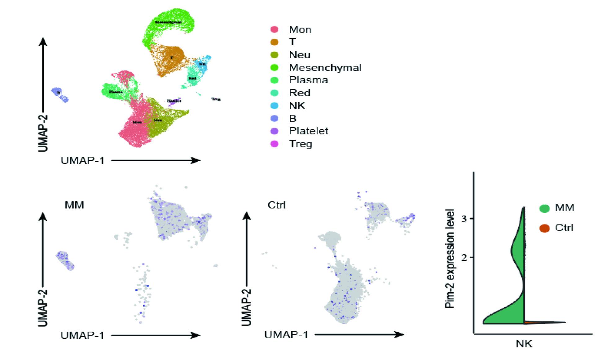
ScRNA-seq data were obtained from the GEO database (https://www.ncbi.nlm.nih.gov/geo/). Data from the GSE124310(MM samples and normal bone marrow(NBM) samples). These results provided a comprehensive map of immunologic changes and PIM2 expression on NK cells from MM patients. We subsequently explored the expression of inhibitory TIGIT receptors and PIM2 expression on NK cells.
Meanwhile the GSE166902 (peripheral blood of one healthy human) and GSE188632 (peripheral blood of one end-stage MM patient) datasets were cleaned to avoid the batch effect using the gord package in R software version 4.0.3. Cells were clustered using the UMAP method and manually annotated using the TOP10 marker genes to obtain the NK cell population. PIM2 expression was compared between the NK cell populations in each dataset (GSE166902 and GSE188632). Cell clustering and gene expression were analyzed using an online analysis tool (https://www.aclbi.com/static/index.html).
FCM analysis of PIM2 kinase expression in NK cells
Bone marrow from each MM patient and healthy donor (2 mL) was placed into an anticoagulant tube with 5 µL of CD3-PerCP (BD Biosciences) and CD56-APC (BD Biosciences) monoclonal antibodies, mixed well with 100µL bone marrow blood, and incubated for 15 min at 25 ℃ in the dark. Samples were then incubated with diluted hemolysin (2 mL) in the dark for 10 min at 25 ℃ and centrifuged at 1500 rpm for 5 min. After hemolysis and centrifugation, the supernatant was removed, mixed with 2 mL of cell cleaning solution (PBS), and centrifuged again at 1500 rpm for 5 min. After the supernatant had been discarded, 100 µL of fixing solution A was added to each test tube, shaken using an oscillator, and incubated for 5 min at 25 ℃ in the dark. The cells were then fixed, resuspended in 2mL PBS, and centrifuged at 1500 rpm for 5 min before the supernatant was discarded. After the cells had been washed, 40 µL of membrane breaker B was added to each centrifuge tube with 5µL of isotype control mouse IgG1-FITC monoclonal antibody (BD Biosciences) or 5µL of PIM2-FITC antibody (BD Biosciences; dilution ratio 1:20). The mixture was oscillated using a vortex oscillator and incubated at 25℃ under light for 15 min. Cells were washed again with PBS and resuspended in 300 µL PBS, and the number of NK cells (defined as CD3−CD56+) with PIM2 kinase expression was detected using FCM (Beckman CytoFLEX). At least 10^5 cells were collected in each test tube. Data were analyzed using Cell QuestTMPro 4·0·2 software.
qRT-PCR analysis of PIM2 kinase expression in NK cell
Total RNA was isolated from NK cells using TRIzol reagent (Invitrogen). First-strand cDNA was synthesized using an iScript cDNA Synthesis Kit (Bio-Rad). qRT-PCR was performed using the following primers: PIM2 forward, GCA CTG CTA TGG AAA GTG GGT; PIM2 reverse, ATG GAC AAC TCC ACG GGA ATG; GAPDH forward, CTC ACC GGA TGC ACC AAT GTT; GAPDH reverse, CGC GTT GCT CAC AAT GTT CAT. qRT-PCR was performed using SYBR Green (Invitrogen) with 40 amplification cycles at 95℃ for 30s and denaturation at 95℃ for 5s, followed by extension at 57.2℃ for 30s. PIM2 levels were calculated using the 2^-ΔΔCt method [(Ct, target gene Ct, GAPDH) sample - (Ct, target gene Ct, GAPDH) control] after the data was been normalized using GAPDH as a housekeeping gene.
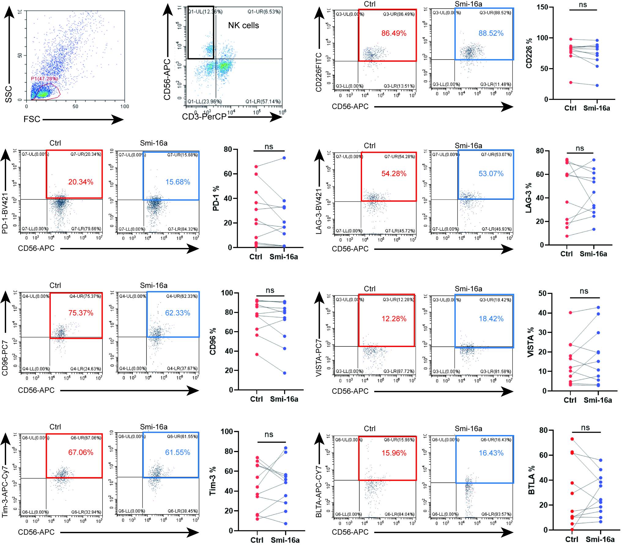
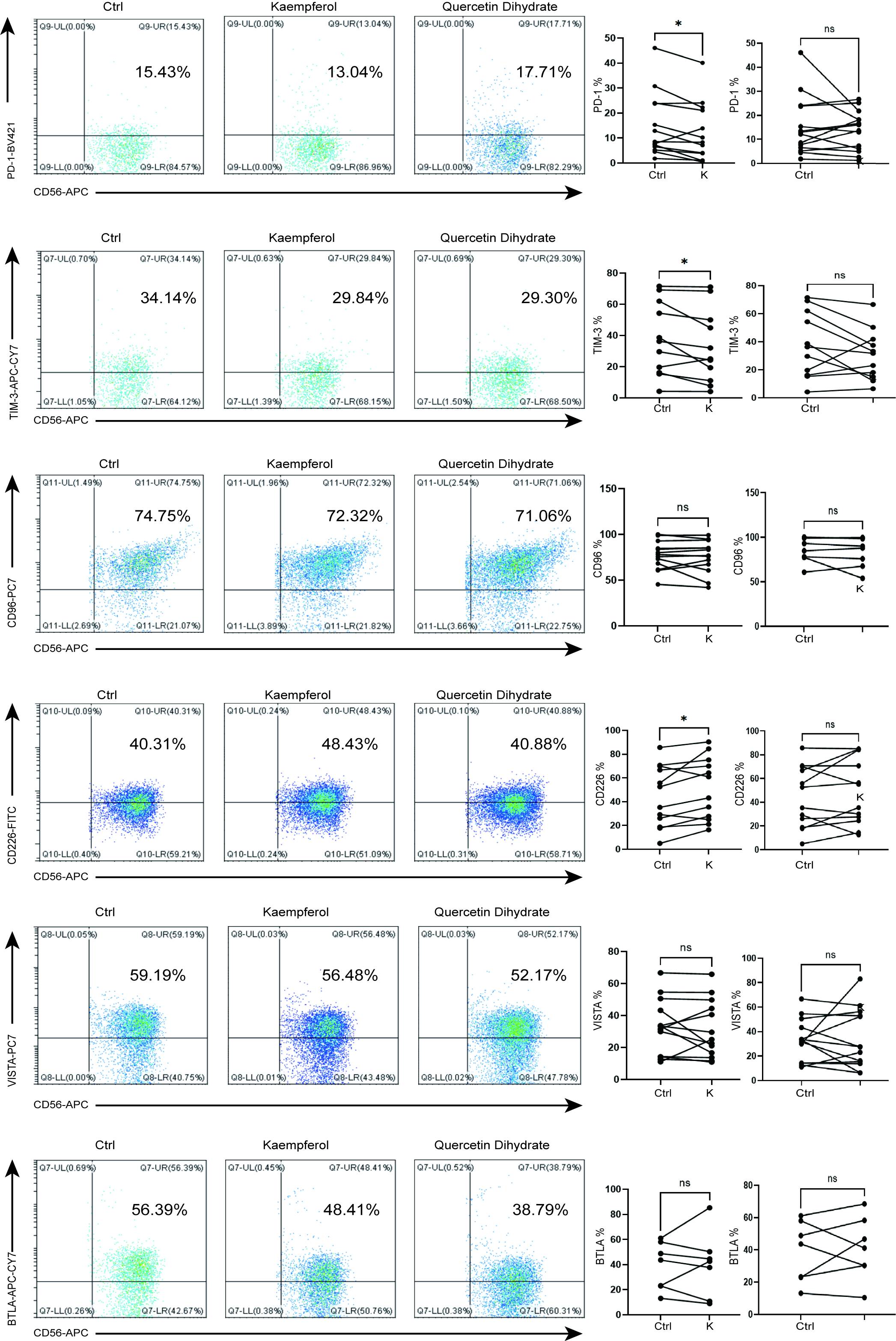
The NK cell function and immune checkpoints expression
Extracted mononuclear cells were isolated using lymphocyte separation solution, added to 50 mL RPMI-1640 medium (Gibco) containing 7·5 mL fetal bovine serum (FBS), 500 µL double antibody, and 50 µL IL-2), and divided into four groups treated with kaempferol (group A; TOPSCIENCE), quercetin dihydrate (group B; TOPSCIENCE), and Smi-16a (group C; Med Chem Express) at their respective IC50 concentrations, or the same volume of DMSO (group D; Solarbio, Beijing, China). After the cells were incubated at 37 ℃ with 5% CO2 for 48 h, groups A, B, and C were stimulated with 2µL Leukocyte Activation Cocktail (BD Biosciences) for 6h and centrifuged at 1500 rpm for 5 min. The supernatant was then discarded and the cells were washed using PBS (Solarbio) and incubated with the following antibodies: BTLA (APC/CY7), VISTA (PE/CY7), LAG3 (BV421), CD226 (FITC), CD107a (PE), Percp-CD3, APC-CD56, Tim3 (APC/CY7), CD96 (PE/CY7), PD1 (BV421), TIGIT (FITC), IgG1 (PE), IFN (PE), and TNF (PE; BD Biosciences). The immune checkpoints expression and function on NK cells were detected using FCM (Beckman CytoFLEX). At least 10000 cells were counted per tube. Data were analyzed using Cell QuestTMPro 4.0.2 software.
Cell culture
NK cells (NK92) were provided by Procell Life Science & Technology and cultured in α-MEM supplemented with 12.5% FBS, 12.5% equine serum, 0.2 mM inositol, 0.1 mM β-mercaptoethanol, 0.02 mM folic acid, and 100–200 U/mL recombinant IL-2 (BD Pharmingen) at 37°C in a 5% CO2 humidified incubator. Human MM cells (U266 and OPM2) were obtained from the American Type Culture Collection and maintained in RPMI 1640 (Welgene, Daegu, Republic of Korea) supplemented with 10% FBS (Gibco) and penicillin/streptomycin at 37°C in a 5% CO2 humidified incubator. Stem cells were derived from the bone marrow blood of patients with MM. Mononuclear cells were extracted from 5mL of bone marrow blood and incubated in basic DMEM/F2 (1:1) containing 15% FBS (Gibco), 100 µg/mL penicillin (Gibco), and 100 U/mL Streptomycin (Gibco) at 37°C in a 5% CO2 humidified incubator for 14 days, and then transcultured for three generations before subsequent experiments.
Lentiviral gene transduction
The lentivirus (customized by GENE) stored at -80 ℃ was thawed slowly on ice and diluted in a complete medium. To determine suitable transfection conditions, the following experiments were carried out according to the manufacturer’s instructions. Cells were suspended in 2 mL of the complete medium at a density of 105 cells/mL in a 12-well plate with the virus diluent (20µL) and the corresponding volume of HiTransGP infection enhancement solution. After 8–12 h, a sufficient volume of exchange medium was added to the cell culture medium for further culture. After 72 h, the transfection effect was observed using fluorescence microscopy, and an appropriate number of cells was collected for follow-up experiments. After protein extraction, PIM2 (CST), NF-KB (CST), and ETS-1 (CST) expressions were detected using western blotting.
Natural compound identification
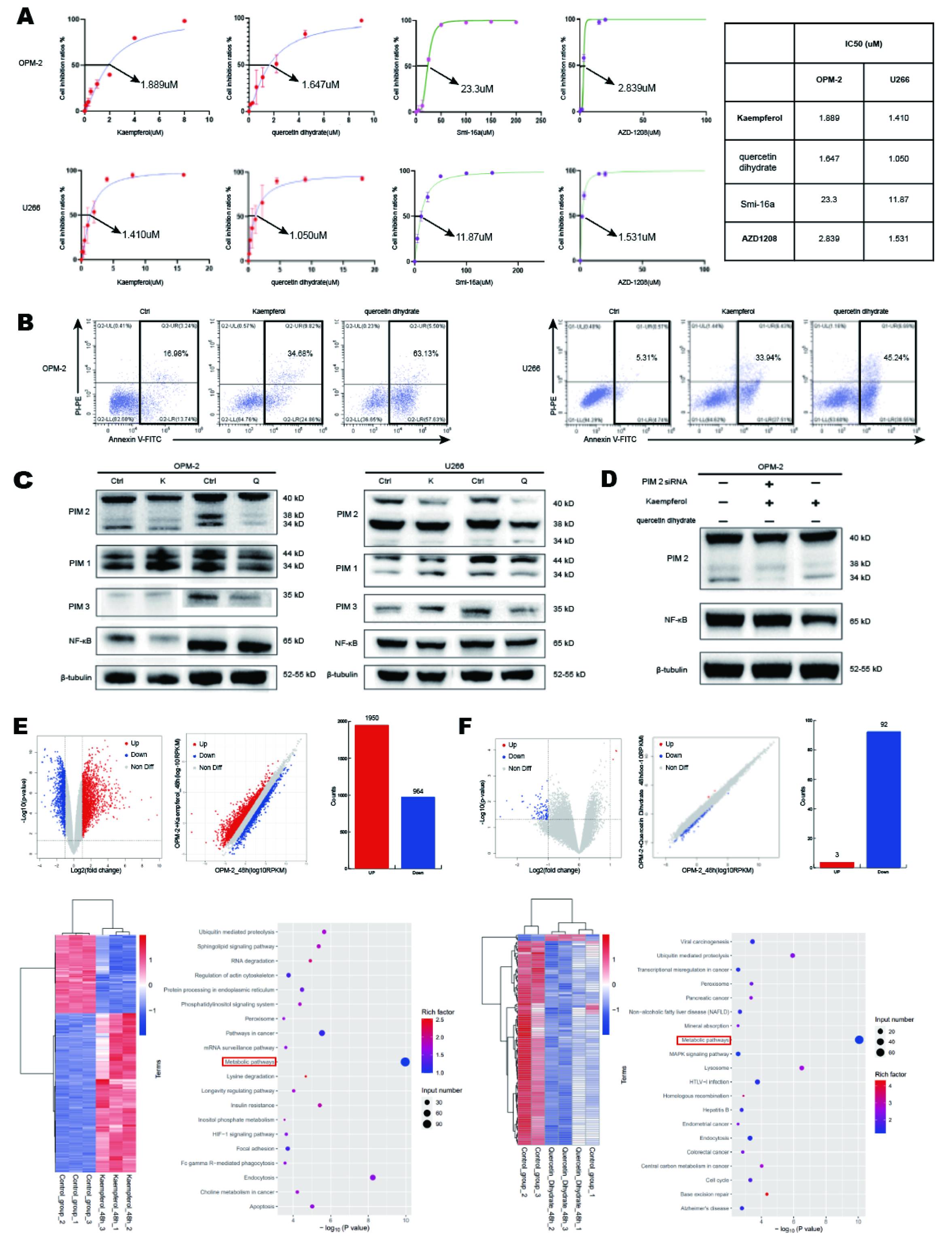
A total of 160 flavonoids (Supplementary Table 1) were screened from a library of 2592 natural compounds (TOPSCIENCE; https://www.tsbiochem.com/index), mostly derived from plants. Using the ADP-GLO method18,19, Two natural compounds kaempferol and quercetin dihydrate that inhibited PIM2 kinase by over 90% were selected.
Molecular docking analysis
The molecular structures of kaempferol and quercetin dihydrate were retrieved from PubChem (https://pubchem.ncbi.nlm.nih.gov/). The ligands were processed using the MM2 procedure with a minimized energy module in Chemdraw 3D and saved as “mol2” files. PIM2 protein structure was downloaded from the PDB database (http://www.rcsb.org), defined as a receptor, and saved in pdbqt format after excluding all water molecules, adding hydrogens, calculating charges, and distributing charges using Mgtools 1.5.6. Molecular docking was performed using AutoDock Vina 1.1.2. A higher-scoring docked conformation model was chosen and the results were analyzed and visualized using PyMOL and Discovery Studio.
Cell proliferation assay
For CCK8 assays, 5×10⁴ MM cells (U266 and OPM2) were inoculated into 96-well plates containing different concentrations of kaempferol (TOPSCIENCE), quercetin dihydrate (TOPSCIENCE), Smi-16a (Med Chem Express), and AZD1208 (Med Chem Express), with a final volume of 100µL, and cultured at 37 ℃ with 5% CO2 for 48 h. After 10µL CCK8 (Bimake) had been added to each well and incubated at 37℃ with 5% CO2 for 3h, absorbance at 490 nm was measured using an enzyme-labelling instrument. Cell inhibition rates and IC50 values were calculated.
Cell apoptosis analysis
For FCM cell apoptosis analysis (Beckman CytoFLEX) MM cells (OPM2, U266, NK92) were treated with kaempferol (TOPSCIENCE), quercetin dihydrate (TOPSCIENCE), and Smi-16a (Med Chem Express) for 48 h, washed twice with PBS and suspended in binding buffer (10mmol/L N-2-hydroxypiperazine-N´-2-ethanesulfonic acid/NaOH, pH7.4140 mmol/L NaC, 12.5 mm/L CaCl2). The cell suspensions (100 mL) were then incubated for 20 min with 5µL Annexin V-FITC (BD Biosciences) and 5µL PI (BD Biosciences) at 25 ℃ in the dark for 3min. Apoptotic cells were counted using FCM (Beckman CytoFLEX).
Western blot analysis
To detect PIM1, PIM2, PIM3, and NF-κB expression after the addition of kaempferol, quercetin dihydrate, or lentiviral knockdown, MM and NK92 cells treated with drugs or the lentivirus were washed with PBS and suspended in a lytic buffer Lysates were centrifuged at 12000 rpm for 15 min to remove cell fragments and total protein was quantified using a BCA kit (Sigma-Aldrich). Proteins (25µg) were separated using SDS-PAGE and transferred onto PVDF membranes that were blocked using 5% milk in Tris-buffered normal saline (TBST) containing 0·05% Tween 20 for 1h and then incubated with primary antibodies overnight at 4℃. Membranes were then incubated with HRP-labelled secondary antibodies at 25 ℃ for 1 h and developed using an ECL substrate. Data represent the findings of at least three independent experiments.
mRNA sequencing analysis
RNA-seq experiments, high-throughput sequencing, and data analysis were conducted by SeqHealth Technology (Wuhan, China).Total RNA was extracted from OPM2 cells treated with kaempferol and quercetin dihydrate for 48h using TRIzol Reagent. After the DNA was digested using DNase I, RNA quality was determined by measuring the absorbance at 260/280 nm using a NanodropTM OneC spectrophotometer (Thermo Fisher Scientific). RNA integrity was confirmed using 1.5% agarose gel electrophoresis. RNA was quantified using a QubitTM RNA Broad Range Assay kit (Life Technologies, Q10210) with Qubit3.0. A stranded RNA sequencing library was prepared from 2 µg total RNA using a KCTM Stranded mRNA Library Prep Kit for Illumina® (Cat # DR08402, Wuhan SeqHealth Technology), according to the manufacturer’s instructions. PCR products of 200–500bp were enriched, quantified, and sequenced on a Novaseq 6000 sequencer (PE150, Illumina).
Raw sequencing data were filtered using Trimmomatic (version 0.36), low-quality reads were discarded, and reads contaminated with adaptor sequences were trimmed. Reads mapped to the exon regions of each gene were counted using featureCounts (Subread-1.5.1; Bioconductor) and RPKMs were calculated. Differentially expressed genes (DEGs) between groups (p < 0.05) were identified using the edgeR package (version 3.12.1). Gene ontology (GO) and Kyoto Encyclopedia of Genes and Genomes (KEGG) enrichment analysis were performed using KOBAS software (version 2.1.1, p < 0.05). Alternative splicing events with a false discovery rate (FDR) < 0.05 and Δψ = 0.05 were detected using rMATS (version 3.2.5).
Co-culture assays
Cultured NK92 and U266 cells were counted using a cell counting board. According to the 1:4 ratio of U266 and NK92 cells, a co-culture system was established and inoculated into a sterile 12-well cell culture plate. U266 cells were cultured alone (group A) or co-cultured with NK92 cells without drug intervention (group B) or with kaempferol (group C), quercetin dihydrate (group D), or Smi-16a inhibitors (group E). Follow-up experiments were carried out according to the IC50 concentration measured or noted in the product manual. After incubation at 37℃ in a sterile 5% CO2 cell incubator for 48 h, U266 cell apoptosis, NK92 cell activation, and functional indices were detected. Experiments were repeated three times per group.
U266 cell apoptosis was detected using FCM (Beckman CytoFLEX) after co-cultured cells were washed with PBS, incubated with CD138-APC (BD Biosciences) at 25℃ in the dark for 15min, stained with annexin V(BD Biosciences) and PI(BD Biosciences).
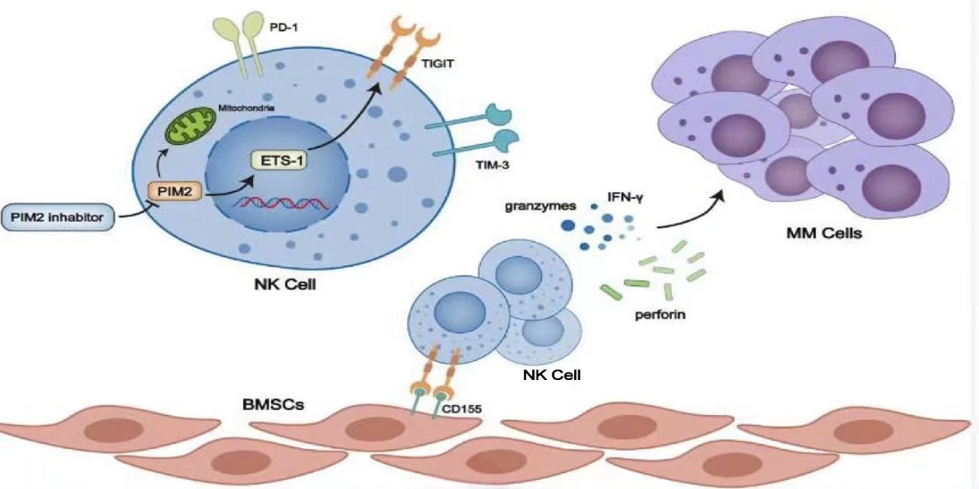
NK92 cell activation, functional indices, and changes in immune checkpoints were detected using FCM (Beckman CytoFLEX). Briefly, stem cells were induced using mononuclear cells from patients with MM. Bone marrow mesenchymal stem cells (BMSCs) in the logarithmic growth stage from passage 3 (P3) to passage 4 (P4) were digested using 0·25% trypsin and cultured in 24-well plates containing DMEM/F-12 at 37°C and 5%CO2 for 12 h. After the supernatant was discarded, 2 × 10⁵ purified NK cells were added at a ratio of 1:4 BMSCs/NK and supplemented with complete RPMI1640 to a final volume of 500µL. Next, PBS was added and cells were cultured with kaempferol, quercetin dihydrate, or Smi-16a inhibitors for six days at 37°C with 5% CO2, before FCM analysis (Beckman CytoFLEX).
Statistical analysis
Statistical analyses were performed using GraphPad Prism software. Gene expression was compared using one-way ANOVA. Paired samples were compared using Student’s t-tests. Differences were considered statistically significant at p < 0.05.