4.1 Morphological characteristics of PDA NPs
PDA NPs have been prepared through the self-polymerization and auto-oxidation of DA in an alkaline environment. The morphologies of the synthesized nanoparticles were observed using SEM and TEM. PDA NPs were found to have a mono-dispersed spherical shape. The sizes of the PDA NPs were determined using SEM and TEM, the average particle sizes are lies in the range of PDA NPs had a calculated size of approximately 120 nm. The obtained results suggested that the respective volume of NaOH during the synthesis of PDA NPs led to a decrease in the average diameter of the nanoparticles. This decrease in size is attributed to the formation of negligible amount of aggregates, possibly caused by an increase in the number of nucleation sites due to the higher volume of NaOH used in the reaction.
4.2 XRD and FT-IR
The XRD pattern for PDA NPs showed a broad reflection peak at 23.4˚, which is shown in the Fig. 2A. This broad peak suggests the presence of amorphous or disordered structures within PDS. Amorphous materials do not have a well-defined crystalline structure, and their XRD patterns typically show broad peaks.[46, 47] The FTIR spectra of the synthesized PDA NPs are represented in the Fig. 2B. The absorption band appeared at around at 3100–3450 cm− 1 and 1550–1700 cm− 1, which are corresponded to the stretching frequency of –OH and N–H groups present in the PDA NPs. The N-H and the –N D species can be produced in the process of self-polymerization of DA. It was found that the photogenerated holes on the catalyst could react with the surface hydrophilic groups and adsorbed water to produce “OH radicals, which were potent oxidants that degraded organic materials in water.
Furthermore, the sharp bands appeared at around 1295 and 1630cm− 1, which are ascribed to the existence of C–O and C = O bonds, broadened and slowly shifted to lower wavenumbers of 1258 and 1605 cm− 1, respectively. In addition, the existence of C = N and C = C almost stays the same is primarily responsible for the band appeared at 1516 cm− 1. Thus, the shifting of the band could be due to the process of polymerization and confirmed the presence of PDA NPs.
4.3 Structures and Properties of Scaffolds
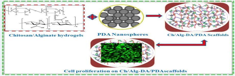
Preparation of Ch/Alg-DA/PDA scaffolds with specific concentrations of PDA NPs with different particle sizes has been illustrated. The scaffolds were synthesized by mixing the aqueous solutions of Ch/Alg-DA with the PDA NPs. Cross-linking of the mixture was achieved by adding 6 wt % of CaCl2. Different concentrations of PDA suspensions (1, 1.5, 2, and 2.5 wt %) were tested when blending with Ch/Alg-DA. At higher concentrations (2 and 2.5 wt %) of PDA, inhomogeneity in the distribution of PDA NPs was observed in the scaffolds. Due to this inhomogeneity, PDA at 2 wt % was preferred to mix together with Ch/Alg-DA for the preparation of composite hydrogel scaffolds. This work also investigated the influence of particle sizes of PDA on the composite scaffolds and concluded that there was no statistically notable difference occurred between the scaffolds with different particle sizes of PDA NPs. The selected conditions involved at 1.5 wt % PDA concentration and PDA NPs with a median size of 150 nm have been selected for further structural and property studies.
4.4 Porosity of various Scaffolds (Ch/Alg, Ch/Alg/PDA, Ch/Alg-DA and Ch/Alg-DA/PDA)
The importance of porosity significantly emphasizes in determining the morphological structure, biological activity and mechanical properties of scaffolds. Porosity refers to the volume of void spaces or pores within a material, and it has a significant impact on the properties and functionality of scaffolds, which is crucial for cell growth and other biological activities within the scaffold. The porosity characteristics of the synthesized Ch/Alg-DA/PDA hydrogel scaffold has been determined using the pycnometer method and was found to range from approximately 72–85% when the Ch/Alg concentration was increased from 2 to 4 wt % (Fig. 3). Moreover, the pore size of the scaffold was affected by the various concentrations (2 to 4 wt %) of Ch/Alg. At higher concentration of Ch/Alg, it increased the cross linking degree in the presence of Ca2+, resulting in smaller pore sizes. The Ch/Alg-DA/PDA hydrogel scaffold exhibited the lowest porosity when compared to the other scaffolds. The order of porosity among the scaffolds was Ch/Alg-DA/PDA < Ch/Alg-DA < Ch/Alg/PDA < Ch/Alg. The results proposed that the smaller pore size of the synthesized hydrogel scaffold promotes high interactions between the catechol groups, contributing to lower porosity. Additionally, the porosity characteristics of the scaffold lowered while increasing the concentration of Ch/Alg. The obtained findings illustrated that both the concentration of Ch/Alg and the components of the scaffolds played a decisive role in controlling pore size and porosity.
Three types of interactions were indicated by the formation of scaffolds: (i) Interconnection between the groups of Ch/Alg and Ca2+ (ii) The interaction of hydrogen between the catechol groups of DA and Ch/Alg. (iii) Covalent and non-covalent bond interactions between the catechol groups in the DA moieties (or PDA NPs). These interactions were believed to synergistically influence the pore sizes in the scaffold. It confirmed that these interactions was challenging due to the complex structures of PDA molecules, as reported earlier.[48] Furthermore, the consequences of various interactions on the pore size and structure in different types of scaffolds, including Ch/Alg, Ch/Alg/PDA complex, DA modified Ch/Alg, and Ch/Alg-DA/PDA complex scaffolds have been demonstrated. The Ch/Alg scaffold showed the largest pore size among the tested scaffolds. This scaffold has the largest pore size and the lowest cross linking degree because there was only one kind of cross linking bond arised between Ch/Alg and Ca2+. The primary interaction involved in this scaffold was between Ch/Alg and Ca2+. The Ch/Alg/PDA scaffold had smaller pores and may be a denser structure compared to the Ch/Alg scaffold.
In this scaffold, there were two more interactions takes place between the catechol groups in addition to the one between Ch/Alg and Ca2+. These extra interactions increased the cross linking degree and reduced the porosity of the scaffold. Like the Ch/Alg-DA/PDA complex scaffold, the Ch/Alg-DA scaffold featured three different kinds of interactions. As a result, the pore size was slightly greater than the Ch/Alg-DA/PDA complex scaffold, but smaller than the Ch/Alg and Ch/Alg/PDA scaffolds. On the other hand, when compared with this scaffold to the Ch/Alg-DA/PDA scaffold, the cross-linking degree was lower because there was no interaction between the PDA NPs. This kind of scaffold may have a denser structure and smaller pore sizes due to the higher cross linking degree created by catechol groups and Ca2+. The attained results suggested that the interactions between Ch/Alg, Ca2+, catechol groups, and PDA NPs played a crucial role in formatting the pore size and structural characteristics of the different scaffolds. These findings high lightened the combination and strength of these interactions can be tailored to control the properties of hydrogel scaffolds for various biomedical applications.
4.5 Mechanical properties of different scaffolds
The incorporation of catechol modification and the existence of PDA NPs in the Ch/Alg-DA/PDA scaffold significantly enhanced its compressive modulus compared to other scaffolds, demonstrating the importance of these interactions in improving mechanical properties for various biomedical applications (Fig. 4). The Ch/Alg-DA/PDA scaffold's compressive modulus was measured, reaching approximately 0.8, 1.2 1.81 MPa at 2, 3 and 4 wt% Ch/Alg concentration respectively. The compressive modulus of the Ch/Alg-DA/PDA scaffold increased, when the Ch/Alg concentration was raised from 2 to 4 wt%. To put things in perspective, tests were conducted on the mechanical characteristics of various scaffolds, including Ch/Alg, Ch/Alg/PDA, and Ch/Alg-DA scaffolds. Among these scaffolds, the Ch/Alg-DA/PDA scaffold exhibited the maximum compressive modulus.
The Ch/Alg scaffold had the lowest cross linking degree and weakest interaction, resulting in lower mechanical strength. Both the Ch/Alg-DA and Ch/Alg/PDA scaffolds have greater interactions and superior cross linking degrees due to the oxidation of catechol groups. Within the Ch/Alg-DA/PDA scaffold, the Ch/Alg-DA chains not only interacted with one another but also established the bonds with inflexible PDA NPs. The occurrence of PDA NPs in the scaffolds had a significant impact on enhancing the mechanical properties and providing additional cross linking points. Consequently, while compared to the other scaffolds, the Ch/Alg-DA/PDA scaffold had a significantly higher compressive modulus. The results concluded that the modification of Ch/Alg hydrogel with catechol groups, either through chemical or physical interactions, efficiently improved the mechanical characteristics of the scaffolds.
4.6 Stress/Strain Behaviour of different scaffolds
It is significant to take into account that the exact composition, processing techniques, and the concentration of the different components in each scaffold (Ch/Alg, Ch/Alg/PDA, Ch/Alg-DA and Ch/Alg-DA/PDA) will determine the precise stress and strain values as well as the shape of the stress-strain curve. Figure 5 represents the stress-strain curves of the various scaffolds, it can be used to evaluate the mechanical integrity and suitability of these scaffolds for the treatment of bone fracture and regeneration of bone tissue applications.
It also determines the material's stiffness, strength, and deformation characteristics under compression. The stress-strain behaviour of Ch/Alg-DA/PDA at 4 wt% exhibited an excellent performance, when compared to the other scaffolds. It also very well correlated with the characteristics of compression modulus and porosity.
4.6 Degradability of various Scaffolds
The results described the importance of scaffold degradation for tissue regeneration and presents findings on the in vitro degradation behaviors of different scaffolds, including those with and without PDA NPs and catechol (DA) moieties.
Scaffolds used for tissue regeneration are typically required to degrade as new tissues grow to achieve the best curative effect for cellular activities. The in vitro degradation behaviors of the scaffolds were investigated using SBF as the degradation solution. An analytical balance was used to measure weight loss in order to track degradation rates (DRs). The Ch/Alg-DA/PDA hydrogel scaffold exhibited the lowest degradation rate, approximately 19, 17 and 15% DR after 35 days at 2, 3 and 4 wt% Ch/Alg concentrations, when compared to Ch/Alg, Ch/Alg-DA, and Ch/Alg/PDA scaffolds (Fig. 6). The lower DR in the Ch/Alg-DA/PDA scaffold was attributed to its high cross linking degree. Similar trends were observed for the porosities of scaffolds with 2 and 3 wt% Ch/Alg concentrations. The DRs of the scaffolds decreased with increasing Ch/Alg concentration. Higher cross linking degrees and concentrations led to denser microstructures with smaller pores, slowing down the penetration rate of the buffer solution and resulting in lower degradation rates. The results suggested that the degradation of the scaffolds was effectively adjusted by the catechol modification., making them suitable for bone or cartilage regeneration. This controlled degradation is important for tissue regeneration applications, where scaffolds need to provide support initially and then gradually degrade as new tissues form.
4.7 In vitro mineralization properties of the synthesized Scaffolds
A cartilage defect involves the necessary for regenerating numerous tissues, including subchondral bone, calcified cartilage and articular cartilage. Therefore, it is essential to stimulate the revival of both subchondral bone and cartilage for effective treatment. HAp is known for its high osteoconductive properties, making it useful in medical implants and the surface of the scaffolds coated by HAp can promote osteointegration. In this study, we have examined the in vitro mineralization of the Ch/Alg-DA/PDA scaffold.
The process of mineralization was achieved by alternating immersion in aqueous solutions of disodium hydrogen phosphate and calcium chloride. The XRD patterns of the mineral layers on the scaffolds were shown in the Fig. 7 and the obtained XRD peaks correlated with the respective peaks of HAp at specific angles, which are observed at 26.5°, 30–34°, and 49.5° indicating that the presence of calcium phosphate apatite (inorganic phase) in the mineral layers. After the modification of the scaffolds by the influence of DA and PDA NPs, the specific regions of characteristic peaks for the mineral layers were decreased slightly. When compared with the Ch/Alg, ChAlg/PDA, Ch/Alg-DA, and Ch/Alg-DA/PDA scaffolds, the Ch/Alg-DA/PDA scaffold exhibited higher peak areas and sharper peaks for HAp. The catechol group, known for its chelating capability with Ca2+, was reported to be beneficial in promoting the formation of HA during mineralization. These alteration in the XRD patterns indicated that the chemical and physical modifications of catechol assisted to enhance mineralization on the scaffold's surface and improving its mineralization performance. The outcome of the results depicted that the mineralization on the surface of the Ch/Alg-DA/PDA scaffold effectively supports on osteointegration. After the scaffolds were filled with Ca2 + to form hydrogel, there was slight interaction takes place between the Ch/Alg and HAp. By chelating Ca2+ with catechol groups, the physical and chemical modifications of catechol (DA or PDA) significantly improved the uniformity of mineral layers. It enriched with Ca2+ on the scaffold's surface and promoted the concentration of Ca2+ to attain a supersaturated state and providing nucleation sites for HAp. Greater numbers of catechol groups in the scaffolds produced more nucleation sites, which in turn produced homogenous HAp particles.
4.8 CFLX release behaviour on various Scaffolds
The primary goal is to prevent infections associated with orthopedic implants by immobilizing antibacterial drugs or anti-inflammatory within the scaffolds. CFLX is used as a model drug to load onto these scaffolds. In the Fig. 8(A), the resulting behaviour indicated that the Ch/Alg-DA/PDA scaffold have the maximum potential to adsorb CFLX compared to other types of scaffolds, including Ch/Alg, Ch/Alg/PDA, and Ch/Alg-DA. The immobilized amount of CFLX on the Ch/Alg-DA/PDA scaffold depends on the concentration of Ch/Alg. Specifically, at the concentration of Ch/Alg was 4 wt%, the maximum immobilized amount of CFLX on the Ch/Alg-DA/PDA scaffold was 1540.4 ± 16.3 ng/mg. In comparison, the immobilized amounts of CFLX on other scaffolds were lower. For instance, on Ch/Alg, Ch/Alg/PDA, and Ch/Alg-DA scaffolds, the immobilized amounts were 672 ± 23.2 ng/mg, 836.3 ± 15.9 ng/mg, and 1320.3 ± 27.5 ng/mg, respectively. The higher immobilization ability of the Ch/Alg-DA/PDA scaffold is attributed to the presence of catechol groups.
These catechol groups provide numerous binding sites for CFLX, like hydrogen bonding and π–π stacking, making them effective for drug immobilization. This study demonstrated that the Ch/Alg-DA/PDA scaffold, with its catechol groups, is highly efficient at immobilizing the anti-inflammatory or antibacterial drug CFLX. This immobilization is concentration-dependent, and the scaffold outperforms other tested scaffolds in terms of drug adsorption capacity. The releasing performance of CFLX from the scaffolds is shown in the Fig. 8(B and C). At the stage of first release at a span of 12 h, the Ch/Alg-DA/PDA scaffolds released approximately 38% of the loaded CFLX. This indicates a relatively slow release rate during the initial phase. On the other hand, the Ch/Alg, Ch/Alg/PDA, and Ch/Alg-DA scaffolds displayed rapid release rates in the initial stage of 12 h, releasing approximately 62%, 56%, and 42% of the loaded CFLX, respectively. These scaffolds released a higher percentage of the drug during the same time frame compared to Ch/Alg-DA/PDA. The release rate decreased as the concentration of Ch/Alg or Ch/Alg-DA increased. It is suggested that at higher concentrations of Ch/Alg or Ch/Alg-DA resulted in slower drug release. The strong interactions between the DA moieties and CFLX, as well as the high cross-linking degree of the scaffold, are responsible for this type of delayed release of CFLX from the Ch/Alg-DA/PDA scaffold. These factors collectively contribute to the sustained and controlled release of the drug over time. The release behavior of CFLX from these scaffolds varies depending on the scaffold type and concentration of Ch/Alg or Ch/Alg-DA. The Ch/Alg-DA/PDA scaffold exhibits a slower initial release, which can be advantageous for controlled drug delivery, and this behavior is attributed to the interactions between DA moieties and CFLX as well as the scaffold's cross linking degree.
4.9 Biocompatibility of various Scaffolds
Cytotoxicity is a critical factor to consider in tissue engineering because it assesses the potential harm that scaffold materials may have on cells, ensuring that the scaffolds are not toxic to cells is essential for their biocompatibility. The cytotoxicity and cell viability of four types of scaffolds, namely Ch/Alg, Ch/Alg/PDA, Ch-Alg-DA and Ch/Alg-DA/PDA were examined in vitro through the MTT assay. According to Fig. 9, the cell viability of osteoblast cells, after incubation with the scaffolds, was examined. The key finding is that the cytotoxicity of the scaffolds did not significantly change after being modified with catechol groups. After the modification of scaffolds with catechol groups, it exhibited tremendous cell viability of oesteoblast cells.
This implied that the scaffolds did not harm the cells and remained biocompatible. The results of this assessment indicate that the scaffolds, even after undergoing physical and chemical modifications involving catechol groups, still maintain good cell compatibility. The cytotoxicity assessment demonstrated that that the scaffolds, with or without catechol group modifications, are non-toxic to cells and exhibited excellent cell viability. This makes them suitable candidates for use in cartilage tissue engineering applications. The suitability of cartilage repairing scaffolds for supporting cell attachment and proliferation, particularly focusing on the morphological observations of cells cultured on different scaffold types. The primary goal for cartilage repairing scaffolds is to provide an environment conducive to cell attachment and proliferation.
4.10 Wound scratch assay of various scaffolds on oesteoblast cells
The impact of the existence of different scaffolds such as Ch/Alg, Ch/Alg/PDA, Ch/Alg-DA and Ch/Alg-DA/PDA, on oesteoblast cells behavior in vitro has been examined by using a scratch wound healing assay, which is an efficient laboratory method for evaluating cell migration. It involves by creating a controlled "wound" in a cell monolayer, and effectively measures the migration of cells close to the wound with respect to the time.[49, 50] In order to scrutinize the influence of different scaffolds on bone fracture, various treatment modalities have been applied to oesteoblast cells (Fig. 10) pursued by evaluating the migration of cells by using the scratch (wound closure) assay. A drastically vigorous inhibition of cell migration has been noticed microscopically in oesteoblast cells treated with a grouping of Ch-Alg, Ch/Alg/PDA, Ch/Alg-DA, Ch/Alg-DA/PDA and the irradiation of light. The percentage of cells that migrated into the scratched area was shown by the respective graphs in Fig. 10B, observed at an appropriate time of 0, 10, 12 and 15 h, respectively.
In oesteoblast cells, the percentages of cells that migrated toward the scratched area in an untreated cells were 68.4% ± 3.8%, 78.9% ± 2.1%, 91.5% ± 2.5%, and 98.9% ± 0.2%, respectively, at 15 h, and 31.3% ± 2.8%, 38.4% ± 3.2%, 42.3% ± 4.3%, 45.4% ± 0.8%, respectively, at 10 h. There is no much differences were observed between 10 and 12 h of the wound closure. As a result, the cells were treated with and without scaffolds (Ch/Alg, Ch/Alg/PDA, Ch/Alg-DA, and Ch/Alg-DA/PDA) in order to observe and quantify the effect of the scaffolds on the rate of cell migration into the denuded area of the culture. Evaluation of cell coverage into the denuded space at predetermined time points allowed for an accurate calculation of migration rates (Fig. 10A). In comparison to untreated control cells, there was a noticeable, dose-dependent increase in culture migration in the existence of scaffolds and displayed ~ 40% increase in wound closure within 15 h (Fig. 10B). Moreover, when compared to the control cells, there is a significant decrease in the scratch wound closure time along with an increase in migration upon scaffold treatment. The Ch/Alg-DA/PDA scaffold-treated cells showed a ~ 2-fold increase in healing rate by 10 h and more quickly filled the wound area than the control cells (Fig. 10C), indicating that the presence of Ch/Alg-DA/PDA affected the rate at which cells migrated in culture. However, at the wound healing process of 15 h, the potential of Ch/Alg-DA/PDA scaffold was significantly higher when compared to the other hydrogel scaffolds. The observed results confirmed that the oesteoblast cells were not only adhering to the surface of the scaffolds but also penetrating into the scaffolds. This study suggested that the seeded cells have been successfully migrated into the inner structure of the scaffolds. The observation of this study demonstrated the cells had moved inside the scaffolds suggested that the' inner structure and pore size of the scaffolds were appropriate for the proliferation of cells and attachment.
This is a positive sign as it shows that the scaffolds provide an environment that supports cell growth. Notably, the number of cells and the area they covered on the Ch/Alg-DA/PDA scaffolds were significantly greater to those on the Ch/Alg, Ch/Alg/PDA, and Ch/Alg-DA scaffolds. The results concluded that the scaffolds modified with catechol groups (Ch/Alg-DA/PDA) were more conducive to adhesion and spreading of cells. The improved performance of catechol-modified scaffolds in terms of cell adhesion and spreading is attributed to the physical and chemical interactions provided by the catechol groups. These interactions create a more favorable environment for cell attachment and growth. The observed cell morphologies on the scaffolds indicated that they support cell attachment and proliferation and the catechol-modified scaffolds (Ch/Alg-DA/PDA) appear to be particularly effective in promoting cell adhesion.
4.11 Cell viability and Proliferation ability assays of various Scaffolds
Furthermore, the cytotoxic effect of different types of scaffolds (Ch/Alg, Ch/Alg/PDA, Ch/Alg-DA and Ch/Alg-DA/PDA) on osteoblast cells was investigated using fluorescence microscopy, employing propidium iodide (red) and calcein-AM (green) as the stain markers for live and dead cells, respectively. Calcein-AM is a fluorescent and highly lipophilic compound that can pass through cell membranes and hydrolyzed by cytosolic esterase enzymes. In viable (live) cells, hydrolysis occurs, resulting in the generation of calcein and emits a strong green fluorescence, which is an indicator of live cells at an excitation wavelength of 488 nm. Propidium iodide is another fluorescent dye, but it is used to stain the nucleus of the cell. It can only cross through the segments of the cell membrane that are distorted or broken. In dead or damaged cells, propidium iodide can enter the cell and intercalate with the DNA double helix, emitting red fluorescence excited at 535 nm, which indicates dead or damaged cells.
All of the Ch/Alg, Ch/Alg/PDA, Ch/Alg-DA, and Ch/Alg-DA/PDA scaffolds showed green fluorescence areas after 1, 2, and 3 days of culture (Fig. 9), indicating the existence of live cells. A higher number of green fluorescence areas were recognized on the Ch/Alg-DA/PDA scaffolds (Figure D, H and L) than on the Ch/Alg, Ch/Alg/PDA, Ch/Alg-DA scaffolds (Fig. 9D-L), indicating that the proliferation of osteoblast cells were highly accelerated on Ch/Alg-DA/PDA and Ch/Alg-DA than on the Ch/Alg and Ch/Alg/PDA. It was suggested that the modified DA/PDA has no cytotoxic effect on osteoblast cells because Ch/Alg-DA/PDA did not exhibited red fluorescence. This type of behaviour revealed that the Ch/Alg-DA/PDA provided a suitable surface for osteoblast proliferation and adhesion. The major cause of higher adhesion and proliferation of osteoblast on Ch/Alg-DA/PDA was attributed to the uniformly distributed surface nanospheres with spaces between neighboring nanospheres. The nanospherical shape reduced the interfacial tensions between the osteoblast surface and spheres, allowing bodily fluid to pass through and supply nutrients for cell growth. Furthermore, monodispersed PDA NPs have much greater surface areas than the other surfaces and contributed to the interlocked configuration of cells.[51–54]