3.1. Expression of CD44 in pancreatic cancer cells
In recent years, nanotechnology has become one of the most important technologies at present, playing an important role in the rapid development of several research fields, especially in the field of precision cancer treatment. HA has good biocompatibility and CD44 receptor targeting32-33, which can accumulate and aggregate in PANC-1 pancreatic cancer tumor tissues overexpressed by CD44 receptor, target the release of chemotherapy drugs, and improve anti-tumor efficiency. Flow cytometry to determine CD44 expression levels in PANC-1 pancreatic cancer cells. The results (Figure 1A) showed that 99.9% of pancreatic cancer cells significantly overexpressed CD44.Pancreatic cancer cells (Figure 1B) and tissue immunohistochemistry(Figure 1C) showed high expression of CD44.
3.2. Constructing MITO@HA
3.2.1 Characterization of MITO@HA
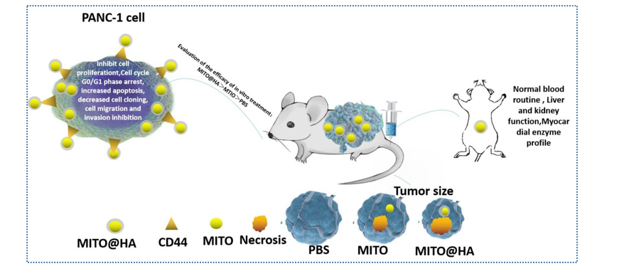
In this study, we successfully created an MITO@HA-targeted nanomedicine through the hydrophilic interaction of HA with MITO, and we evaluated its therapeutic efficacy on pancreatic cancer cells in vitro and on pancreatic cancer animals in vivo via tail vein injection in nude mice to investigate its targeted inhibitory effect on pancreatic cancer. Compared with traditional chemical drugs, polymer nanoparticle-loaded antitumor drugs offer advantages: (1) targeted therapy34 and (2) enhanced tumor accumulation35. MITO@HA nanodrugs can enhance the antitumor efficacy and reduce toxic side effects of these drugs by actively targeting CD44Rs and enhancing the permeability and retention (EPR) effect on the surface of pancreatic cancer tumors.
First, solutions at different concentrations (10 μg/ml, 15 μg/ml, 25 μg/ml, 35 μg/ml, 50 μg/ml, 75 μg/ml) were prepared with a mitoxantrone hydrochloride standard aqueous solution, and the ultraviolet absorption spectrum was determined via an ultraviolet spectrophotometer, with 610 nm as the maximum absorption peak. The linear relationship between the concentration of the drug and the absorbance was determined as a standard curve (Figure 2A and Figure 2B). By measuring the absorbance of MITO@HA at 610 nm, the encapsulation rate and drug loading were calculated to be 45.82±1.72% and 4.15±0.82%, respectively (Figure 2C). The authors demonstrated the successful self-assembly of nanoparticles between hyaluronic acid and mitoxantrone through electrostatic interactions. This study provides a basis for subsequent active targeted therapy for pancreatic cancer. Next, the morphological characteristics and particle size distributions of the nanoions were analyzed. MITO@HA The average hydrated particle size measured by a dynamic light scattering nanolaser was 51.4 ± 2.3 nm, and the particle size in the TEM image was consistent with the particle size measured by particle size analysis. Moreover, the surface of MITO@HA had high electrostatic stability (-26.1 ± 3.2 mV) (Figure 2D).
3.2.2 Screening of the MITO@HA concentration
After treating pancreatic cancer cells with 1 μg/ml, 2.5 μg/mL, 5 μg/mL, 20 μg/mL, and 50 μg/mL MITO, as determined by the CCK-8 method, it was found that the concentration of MITO@HA significantly inhibited the proliferation of cells in the MITO group (Figure 2E). When the concentration was increased to μmol/L, the efficacy and toxicity of seven different concentrations of MITO@HA were evaluated in pancreatic cancer cells after 0, 1, 2 and 3 days. MITO@HA was incubated with pancreatic cancer cell precursors at a concentration of 0.5 μmol/L for 2 days at the most appropriate concentration and time (Figure 2F).
3.3 In vitro study
In the present study, inhibition of pancreatic cancer cells by MITO@HA for 2 days was most suitable for treating pancreatic cancer cells at a concentration of 0.5 μmol/L. Compared with those in the MITO group, the cells in the MITO@HA group exhibited cell cycle arrest at the G0/G1 phase, increased apoptosis, decreased cell replication, cell migration and invasion inhibition.
3.3.1 Cell cycle
The cell cycle results (Figure 3A) showed that the average percentages for each group were 24.91±1.42%、39.37±1.27%和42.67±0.944%, respectively, in the Control group, MITO and MITO@HA groups, indicating that nanoparticles affected the cycling activity of PANC-1 pancreatic cancer cells, resulting in most cells in G0/G1 phase blockade (P<0.0001).
3.3.2 Cell apoptosis
The apoptosis assay results revealed that (Figure 3B) the average percentages of the sum of the total apoptosis rates (early apoptosis rate Q4 and late apoptosis rate Q2) in the control, MITO, and MITO@HA groups were 3.01±0.12%, 5.30±0.49% and 7.28±0.59%, respectively (P<0.0001). MITO@HA had the greatest impact on the apoptosis of PANC-1 pancreatic cancer cells.
3.3.3 Cell migration and invasion
The results of cell migration and invasion are shown in Figure 3C. The average migration and invasion rate of PANC-1 cells in the control group was 280.75±18.51%, which is consistent with the rapid metastasis of pancreatic cancer. The mobility rates of the patients in the MITO@HA and MITO groups were significantly lower than those in the control group (P<0.0001), with 154.50±9.02% and 145.63±7.15%, respectively. Moreover, there was no statistically significant difference between the MITO group and the MITO@HA group (P=0.3539). Similarly, cell invasion was significantly lower in the MITO@HA and MTO groups than in the control group (P<0.0001). The above results showed that MTO has a significant inhibitory effect on tumor growth. In addition, hyaluronic acid (HA) targets the CD44 receptor in pancreatic cancer, and HA-coated MTO can also tightly adhere to the CD44 receptor and target it to inhibit the migration and invasion of tumor cells.
3.3.4 Cell cloning
The cell cloning results showed that (Figure 3 D) for the control, MITO, and MITO@HA groups, the average percentages of cell clones in each group were 100.00±3.56%, 47.02±9.53%, and 45.02±7.38%, respectively. The number of clones of PANC-1 pancreatic cancer cells in the MITO@HA group was the smallest and was significantly lower than that in the control and MITO groups.
Therefore, we believe that hyaluronic acid (HA) targets the CD44 receptor in pancreatic cancer and plays the most important role in killing pancreatic cancer cells. In addition, the constructed MITO@HA nanodrugs target and concentrate in the plasma of pancreatic cancer cells and release MITO, resulting in significant blockade of the G0/G1 cell cycle in pancreatic cancer, promotion of tumor cell apoptosis, significant reduction of cell cloning, and inhibition of cell migration and invasion.
3.4. In vivo study
The results of animal experiments showed that MITO@HA nanodrugs had a significant inhibitory effect on pancreatic tumors. A tumor growth curve was generated for the nude mice with pancreatic cancer (Figure 4A). The growth of the tumors in the control group was the fastest, and the growth of the tumors in the MITO@HA group was slower than that in the control group, indicating that the tumor inhibition effect of the former was significant. The average tumor mass of the nude mouse tumors in each group (Figure 4B) was 0.846±0.016 g in the control group and 0.790±0.0346 g in the MITO@HA group, and the average tumor mass in the MITO@HA group was 0.455±0.529 g (P<0.0001). To further validate the antitumor efficacy of the targeting liposomes, tumor tissue was fixed, sectioned, and HE stained (Figure 4C). Compared with those in the control and MITO groups, the tumor cells in the MITO@HA group exhibited obvious large areas of liquefied necrosis, whereas the control group exhibited no obvious necrosis. The above results showed that MITO@HA had an obvious inhibitory effect on tumors and had good therapeutic effects on tumors. The reason for this difference is that MITO@HA can allow more drugs to reach tumor tissue through EPR effects, and HA can fuse with cell membranes to swallow lipids into cells, increasing tumor cell uptake. In vivo antitumor experiments further proved that surface-modified HA can further promote the accumulation and uptake of MITO@HA at the tumor site through passive and active targeting to better suppress tumor growth.
3.5. In vivo toxicity assessment
The biochemical indices of the nude mice in each preparation group are shown in Figure 5. Important parameters such as white blood cells, red blood cells, PLT, ALT, AST, TBIL, ALB, CK, CK-MB, LDH, LDB1, URES, CREA, and UA in the MITO@HA group were normal, indicating that the MITO@HA treatment had good safety.
In this study, the biosafety of MITO@HA nanoparticles was further confirmed by blood biochemical indices and enhanced antitumor effects compared to those of the MTIO group. In summary, MITO@HA nanoparticles targeting the CD44 receptor can effectively enhance the therapeutic efficacy against pancreatic cancer tumors, and the safety of these nanoparticles is reliable, suggesting the possibility of clinically targeted treatment for pancreatic cancer.