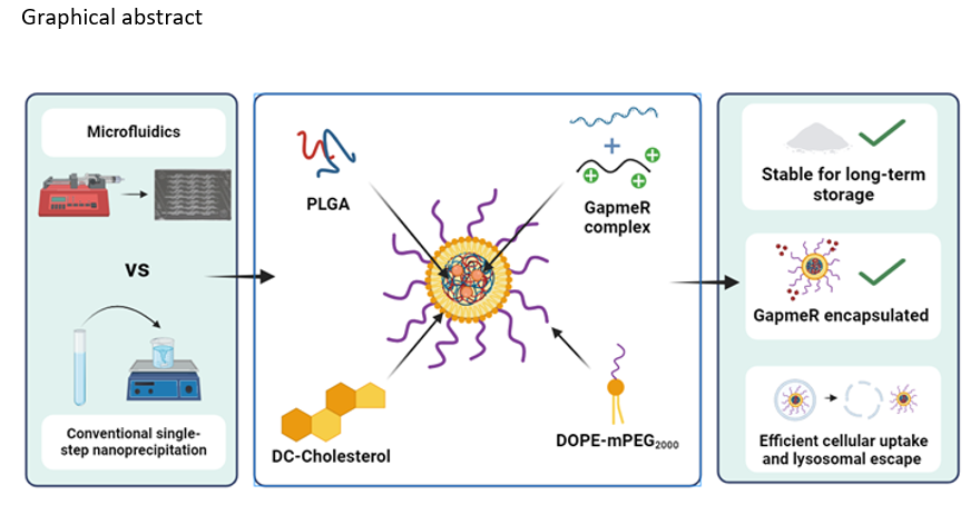
In the present work, we take advantage of the LPHNP characteristics and the two production techniques, conventional SSN and MF, to make a platform for GapmeR delivery. Several variables were evaluated to obtain NPs with suitable physicochemical characteristics.
Regarding the lipid mixture composition, PEGylated lipids provide steric stabilization of the NP suspension during manufacturing and storage. PEGylation also prolongs circulation time and prevents uptake by the mononuclear phagocyte system [36], but has a downstream negative effect by decreasing cellular uptake. In this work, to achieve faster dePEGylation after administration, DOPE-mPEG2000 was chosen instead of the more widely used DSPE-PEG because of its greater dissociation rate from NP surface conferred by its unsaturated chain [37]. Similarly to cholesterol, DC-Chol strongly interacts with polymer, imparting stability to the lipid shell. The positive charge of DC-Chol improves cellular uptake and at the same time acidic pH facilitates endosomal escape [38]. Additionally, it would improve NPs stability by electrostatic repulsion during storage. The effect of lipid/polymer mass ratio was tested in the range of 15–50%. At low lipid/polymer mass ratio (< 25%), NPs with neutral or negative Z-potential by SSN (Table 1) and flluctuating Z-potential or even a clogging of the chip by MF (Table 2) were produced, which indicate insuficient amount of lipids to complete core coating. The NP characteristics were not affected by the lipid/polymer ratio in the range of 25–50%. Thus, to prevent the formation of micelles or liposomes from the lipids not incorporated into the NP surface, the lipid/polymer mass ratio was set at 25%. As expected, an increase in particle size was observed when increasing PLGA concentration. Concentrations higher than 1.25 mg/mL were excessively viscous for the preparation of NPs by MF, producing chip clogging due to the low solvent diffusion rate.
Aditional variables have to be considered for the NPs production by MF. TFR and FRR are the most mentioned parameters that influence the NPs size and PdI. An increase in both TFR and FRR leads to a decrease in size and PdI of lipid, polymeric and lipid-polymeric NPs. However, the micromixer design plays an important role in optimizing the process. Comparison of our results with others previously reported is difficult due to the wide variety of microchips used, which to some extent act as a limiting factor in the TFR and FRR that can be used. It has been reported that increases in FRR between 3 and 10 fold, depending on the geometry of the micromixer (homemade custom-made or commercial) and evidently the composition of the phases, lead to notable reductions in particle size [39–42]. However, the same authors also point out that the FRR has a limit above which further increases have no effect or a negative effect on size and/or PdI [39–41]. With respect to the TFR, some authors also show that the size reduction only occurs up to a certain limit beyond which the effect is reversed [43].
The micromixer chip used in the present study is a commercially available hydrophilic glass device designed to efficiently mix fluids. It combines the hydrodynamic focusing flow (HFF) at the inlet followed by 12 mixing stages consisting of series of alternate paths with different internal cross section (125 µm x 350 µm and 50 µm x 125 µm - depth x width) where a repeated fluid splitting and joining take place. HFF reduces the diffusion length of the organic solvent by compressing the central organic phase with the aqueous phase injected into the two symmetrical side channels. Like other authors,[39–41] the best results were obtained with a FFR of 3 because at higher ratio (tested from 3 to 10) NPs with PdI greater than the maximum established as optimal were obtained. The maximum TFR tolerated by this chip is 5 ml/min to avoid internal overpressure. However, at values greater than 1.5 ml/min (tested from 0.8 to 4 ml/min) the chip is clogging. In the range of 0.8 to 1.5 ml/min, an increase in TFR has a negative effect, leading to NPS with greater size and PdI (Table 2), the same as the behavior mentioned by Li et al. [43].
In any case, LPHNPs with optimal size (150–180 nm), PdI (0.23–0.24) and positive Z-potential (29–32 mV), were obtained by both conventional SSN and MF. It has to be highlighted that it was possible by using the same components and in the same proportion (1.25 mg/ml PLGA, lipid/polymer mas ratio of 25% and DOPE-mPEG2000 at 25% molar ratio of total lipid) and setting FRR 3:1 and TFR of 0.8 ml/min for MF. The TEM images show NPs with a size and PdI consistent with the results obtained by DLS and similar structure which is compatible with that of a polymeric core surronded by a lipid shell.
One of the strategies used to improve oligonucleotides encapsulation in LPHNPs is to form, through electrostatic and hydrophobic interactions, less water-soluble larger-size neutral complexes. The three condensing agents evaluated in the present work could also, improve the endosomal escape. Low molecular weight protamine is one of the most used condensing agents and with less toxicity. In fact, protamines act physiologically as condensers and DNA stabilizers in spermatozoa. However, changes in complexation efficiency have been reported between different protamines due to variations in amino acid composition and therefore in their conformation [44]. It is known that chitosan is also capable of complexing and condensing DNA molecules. The binding efficiency depends on the deacetylation degree and the presence of certain nucleotide sequences [45]. DC-Chol also forms multilaminar condensates, leaving the oligonucleotide chains confined between the lipid bilayers [46]. In our case, greater encapsulation efficiency with protamine, distantly followed by chitosan (56%) and DC-Cholesterol (46%) was obtained, regardless of the preparation technique (Table 1 and 2). In all cases, an excess of net charge was available for GapmeR complexation, therefore, the differences in encapsulation performances should be due to the own condensing agent structure and stability of the formed complex. Acording to the results, protamine sulfate was selected as a condensing agent for subsequent assays.
The stability of NPs during storage plays an important role for translating to the clinic. Despite the steric and electrostatic stabilization (ZP 29–32 mV) provided by DOPE-mPEG2000 and DC-Chol respectively, the NP suspensions did not maintain their physicochemical characteristics beyond 6 hours. On the contrary, they were effectively stabilized for the short and long storage using trehalose. NP suspensions in 2.5% trehalose were stable for at least 24 h at 4ºC, time enough for administration. NPs were also easily reconstituted after freeze-drying using trehalose in the range of 2.5-5% as cryoprotectant, providing suitable storage conditions for long-term use. Trehalose has also been recommended as a cryoprotectant for nanostructured solid lipid nanoparticles at concentrations in the range of 3.75–12.5% [47] and at 1–10% for polymeric particles [48]. However, in this study, concentrations higher than 7.5% do not work. Unfortunately, we have not found previous references to lyophilization of lipid-polymeric nanoparticles similar to those prepared by us. Consequently, the discrepancies with the above authors could be related with the different NP structure and composition but also to the NPs concentration to be freeze-dried.
The physicochemical properties of nanoparticles, size, shape, surface charge and surface chemistry influence the efficiency of cellular uptake [49, 50]. Upon contact with biological fluids or the culture media where they are going to be tested, NPs can change their physicochemical characteristics, which will affect their circulation time, distribution, release profile, interaction with target cells and endosomal escape. These changes are mainly due to the protein corona formation but also to the different pHs during intracellular trafficking. NPs with small sizes (30–50 nm) have greater cell penetration capacity and greater ability to escape from the mononuclear phagocyte system [51] and positive surface charges enhance the interaction with cells [52]. The usual mechanism of cellular uptake of NPs is by endocytosis. According to several authors NPs with a size lower than 200 nm preferentially use the clathrin pathway [53, 54]. Endocytic vesicules fuse with the early endosomes (pH 6–7) which mature to late endosomes (pH 5.5-6) and finally to lysosomes (pH 4.5-5). Additionally, NPs can be exocyted throught recycling and exosomal exocytosis systems in any of this trafficking phases [55]. To exert their therapeutic action, oligonucleotides or oligonucleotide-NPs have to escape from the endosomal system and be released intact in the cytosol of the cell. Consecuently, the behavior of the NPs was evaluated in PBS pH 7.4 with both 4.5% BSA (Fig. 4B) and 10% FBS, but also at pH 5.5 and at pH 7.4 for 2h followed by incubation at pH 5.5 in PBS with 10% FBS to simulate the intracellular trafficking (Fig. 4A). The LPHNPs were stable for at least 24 hours in 4.5% BSA while the stability was reduced to 6 hours in 10% FBS. Regardless of the pH of the medium, after 24 h, the NPs increased in size, a sign of aggregation. NP aggregation could be produced by the exchange and/or the removal of the lipidic shell and by the adsorption of proteins at the NP surface. Due to the ionizable cationic DC-Chol (pKa 7.8) in the NP shell, the initial positive Z-potential (+ 29 mV) decreases to near neutrality (1–2 mV) after incubation in PBS (pH 7.4) with either BSA or FBS. The surfactant effect of BSA would keep the LPHNPs in suspension, while the complex composition of FBS with electrolytes and other substances could facilitate the loss of the lipid shell, increasing the formation of protein corona and agglomeration of the NPs. Similar behavior, after incubation in different biological fluids, in terms of decrease in Z potential and stability has been reported with PLGA NPs with a cationic polymer shell [56].
The GapmeR release profile was also affected by the pH of the medium. Specifically, a great burst release was observed at pH 5.5 (ca. 75%). At acidic pH DC-Chol is completely protonated and the formation of the protein corona is mainly through hydrophobic interactions and hydrogen bonds. At pH 7.4 some electrostatic interactions are possible, leading to the formation of a more compact protein corona that reduces the GapmeR release rate. However, the release was not modified when LPHNPs were previously incubated at pH 7.4 and then at pH 5.5, probably because this pH change does not affect the initial structure of the already formed protein corona.
To evaluate the LPHNPS celular uptake efficiency the same dose of naked Gapmer was taken as a reference because as it previously reported single-stranded and relatively small oligonucleotides, uncharged and/or hydrophobic at high concentration can be cell uptaken and escape endosome without the invervention of any carrier [57]. Compared with the naked GapmeR, both celular uptake and endosomal escape were more efficient with LPHNPs (Fig. 5). Although unexpected high naked GapmeR endosomal escape was observed, combining both cell uptake and endosomal escape lead to an effective GapmeR delivery in the cytosol of approximately 2.5–4 fold higher with LPHNPs than with the naked GapmeR. The less cell uptake efficiency of LPHNPs prepared by MF technique compared with LPHNPs by SSN is difficult to explain since their physicochemical characteristics, stability and release profiles were similar. Even though the formulation components were the same, their disposition or location in the final formulation could not be, especially in the lipid shell. The production of NPs with a relatively complex structure like LPHNPs using such an efficient mixing system could trigger variations in the nucleation and coalescence process, which would lead to a different composition than expected. A higher PEGylation and lower amout of DC-Chol at the NPs surface could reduce the cellular uptake of the elaborated NPs. Ottonelli et al. [39] reported NPs with the similar physicochemical characteristics but different compositions depending on the preparation method (MF and SSN).