In recent years, there has been considerable focus on the anti-tumour activity of antimicrobial peptides (AMPs)[22]. Therefore, in the present study, after isolating and confirming the presence of AMPs (MccJ25), their effects on breast cancer cells as well as their regenerative activity in normal cells were investigated. Microcins originating from Enterobacteriaceae represent ribosomally synthesized and post-translationally modified peptides (RiPPs) that demonstrate antimicrobial properties through the targeting of vital biological enzymes. For instance, MccJ25, a 21-amino acid peptide, exerts its inhibitory effects on the growth of Gram-negative bacteria primarily by targeting their RNA polymerase (RNAP) activity. This peptide has been shown to possess antimicrobial efficacy against Salmonella and Shigella. Initially isolated from Escherichia coli AY25 strains obtained from neonatal feces, MccJ25 exhibits a distinctive structure characterized by an atypical 8-amino acid ring and a lasso loop configuration[23]. MccJ25 exhibits potent bactericidal activity, effectively inducing rapid mortality across various growth phases of pathogens. Its efficacy extends to combating foodborne pathogens in diverse food matrices, such as meat, dairy, and yogurt. Furthermore, MccJ25 exhibits stability in a range of biological fluids, such as serum and simulated gastrointestinal fluids. Targeting the cytoplasmic membrane of Salmonella newport cells causes the cytoplasmic membrane gradient to be disrupted, which is the mechanism of action. Its extended duration of activity in a variety of settings and biological fluids is confirmed by stability investigations. The combined results highlight MccJ25's potential as a workable substitute for conventional antibiotics in treating drug-resistant illnesses[24, 25].
Many studies found that MccJ25 exhibited excellent activity against ETEC due to permeabilizing bacterial membranes and strong affinity. MccJ25 has also been found to inhibit ETEC-induced intestinal injury and intestinal inflammatory responses, suggesting its potential application as an excellent antimicrobial or anti-inflammation agent against pathogen infection[55].The Minimum Inhibitory Concentration (MIC) of MccJ25 against ETEC-sensitive strains is quite low, with the lowest MIC value being 0.03 µg/mL for E. coli K99 and E. coli 987P. Nevertheless, the search results do not explicitly mention the MIC variability of MccJ25 against ETEC-sensitive and Multidrug-Resistant (MDR) strains[26].
First, a total of 120 isolates were identified after being isolated from patients and identified via culture and biochemical assays. Antibacterial activity results from the E. coli and K. pneumoniae isolates indicated that 25 isolates had a clear growth-inhibiting zone. Moreover, the presence of the inhibition zone revealed the absence of standard bacteria growth in the presence of these isolates, which was considered antibacterial activity, with a variety of studies that have investigated the role of bacteriocins in inhibiting bacterial growth[27].
Examination of isolates having antibacterial activity, in terms of expression of the MccJ25 gene, showed that isolate 83 expressed this gene, so it was regarded as a microcin. Furthermore, the 16S rRNA sequence recorded in the NCBI database showed a strong homology of the selected isolate with E. coli. In alignment with our empirical research, Madboly et al accomplished the isolation and identification of a bacteriocin originating from Enterococcus thailandicus through the utilization of 16S rRNA, subsequently archiving the discovery on the NCBI database [28]. In other research, Mandal et al investigated the 16S rRNA gene of produced antimicrobial lipopeptides by Citrobacter and Enterobacter from soil isolates contaminated with feces and investigated them using HPLC analysis. The result of their study exhibited the presence of multiple antimicrobial lipopeptides [29]. Upon verification of isolate 83 for the existence of the MccJ25 gene, the designated isolate underwent a protein extraction process, followed by analysis to ascertain the presence of the MccJ25 protein. In the present study, Our SDS-PAGE analysis results showed that the MccJ25 protein was present at 4.6 kDa, confirming the presence of the MccJ25 protein. In addition, the quantity of protein was determined by the Bradford method so that suitable concentrations could be used for other experiments.
Several studies have demonstrated the potential of bacterial-based therapy in overcoming tumour cell resistance, presenting it as a viable alternative to traditional treatment modalities. Investigation into the mechanisms underlying immune cell-mediated cancer cell destruction has significantly contributed to the development of novel therapies, including bacterial-based immunotherapy. The exploration of bacterial-based cancer therapy has encompassed the utilization of various strains, such as Salmonella enterica serovar Typhimurium and Clostridium novyi-NT, revealing promising efficacy in combatting cancer[30].
It is noteworthy that the exceptional stability of Microcin J25 is attributed to its threaded sidechain-to-backbone ring structure. The lasso configuration of Microcin J25 imparts remarkable resistance to severe thermal, pH, and protease degradation, encompassing chymotrypsin, trypsin, and pepsin[31].
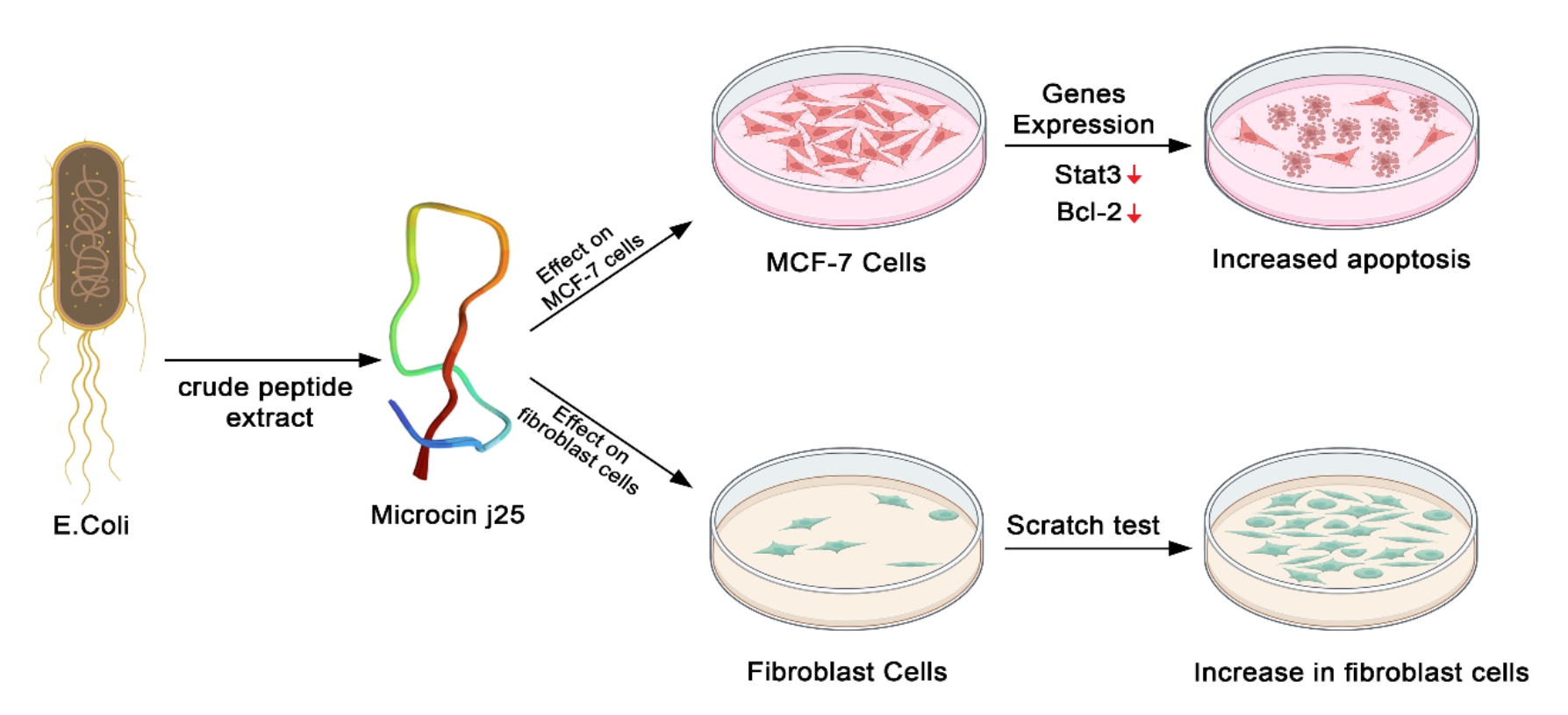
Due to their unique structural characteristics, bacteriocins have strong biological properties, such as the differentiation of cancer cells from non-cancer ones as well as antitumor activity [32]. The cytotoxic effects of E. coli. The efficacy of MccJ25 obtained from E. coli on MCF-7 breast cancer cells was assessed throughout 24, 48, and 72 h. The results showed the highest cytotoxicity (lowest IC50) of 1.081 µg/ml for MccJ25 concentration in 72h on MCF-7 cell lines, showing the highest significance among other concentrations at different times. As a result, microcins increased cancer cell mortality in a concentration-dependent manner. Consistent with our results, Chen et al indicated that the effect of H1-GW antimicrobial peptides inhibited the viability of liver cancer cell lines, such as (J5 HCC, Huh7, and Hep3B), in a dosage-dependent fashion. In contrast, normal fibroblast cells 3T3 exhibited markedly lower susceptibility to these antimicrobial peptides (AMPs) [66]. Additionally, the effect of 1CEa-Temporin as an antimicrobial peptides (AMPs), elicited cytotoxicity in a dose-dependant manner in human breast cancer cell lines, namely 231-MB-MDA and MCF-7 [33].
In a related investigation, MccJ25's cytotoxic effects on HT-29 human colorectal adenocarcinoma cell line were assessed via MTT assay. Results revealed that after 24 h of peptide exposure, HT-29 cell viability was at 83%, suggesting a minimal impact of the microcin on cancer cells [15]. Furthermore, an investigation regarding the treatment of RAW 264.7 and Caco-2 cells at varying levels of MccJ25 exhibited no statistically significant variations in the survival of cells and LDH-stimulating activity when compared to the control groups [56]. Soudy et al showed that MccJ25 caused no significant cytotoxicity in MCF-7 and MDA-MB-435 cells, while the MccJ25-18-4 (the breast cancer targeting peptide) conjugated to inhibit breast cancer cell growth [34].
Flow cytometry was used to determine the amount of cell apoptosis in MCF-7 breast cancer cells treated with isolate 83, in comparison with the population of control cells (without treatment). The current study's findings denote the confirmed initiation of apoptosis in MCF-7 cells upon treatment with compound 83. Within 24 hours of exposure to concentrations of 0.609 and 1.218 µg/ml, a marked induction of apoptosis occurred by 45.4% and 86%, respectively. Gaspard et al. posit that the anticancer properties of antimicrobial peptides (AMPs) can be largely attributed to their capacity for electrostatic interactions with the anionic membrane of cancer cells. This capability affords selective elimination of cancer cells[35]. Different studies have shown the effect of nisin on the induction of apoptosis into different cancer cells [35]. As an anti-apoptotic gene, the BCL2 gene regulates the apoptotic pathway. In addition, STAT3 proteins are contextually activated to respond to growth factors, cytokines or other polypeptide ligands. Moreover, they play a significant role in fundamental processes, including proliferation, development, differentiation, inflammation, and apoptosis. The findings indicated a decline in the manifestation of BCL2 and STAT3 genes relative to the control cohort, thereby validating the outcomes of the apoptotic process that instigates heightened cellular mortality[36].
After examining the effect of nisin on the stimulation of apoptosis in colon cancer cell lines, Ahmadi and colleagues discovered that the presence of nisin led to an induction of cell survival, as well as an increase in the expression of both BCL2 and BAX genes and proteins. Consequently, it was deduced that nisin has the potential to elicit apoptosis via intrinsic pathways, thereby leading to the death of cancerous cells [37].
Cell migration or the ability to repair physical damage was investigated by the scratch test in fibroblast cells. Therefore, in case of damage, normal cells of the animal's body were able to repair the physical damage, along with the capacity of cell migration. Medications capable of stimulating the motor effects of normal cells and making them perform the migration more quickly could be effective in repairing physical damage. This effect could be influenced by modifying the factors contributing to a faster division, thus filling the empty spaces at a faster pace. The results of our study demonstrate a significant upsurge in both cellular migration and repair capabilities following administration of purified proteins derived from isolate 83 in 24 and 48 h after exposure, as compared to the control sample in normal fibroblast cells. The present findings demonstrate the efficacy of the aforementioned peptide in repairing injured tissues as well as its favorable impact on viable cells. The analysis conducted by Soltani et al on hemolysis revealed that rat red blood cells were subject to lysis at concentrations exceeding 52 µg/ml by pediocin 1-PA, bactofencin A, and nisin. It was observed that MccJ25 did not exhibit any detrimental effects on these cells, which suggests that it does not pose a threat to the well-being of healthy cells. In addition, another study reported that MccJ25 had no adverse effects on normal human epidermal keratinocyte (NHEK) primary cells, with confirmed safety [38]. Pourahmadi et al, measured the impact of new BMAP27-Melittin conjugated peptide-nanoparticle against clinical isolates of Streptococcus mutans. The Biofilm Inhibitory Concentration (BIC) and Biofilm Eradication Concentration (BEC) of BMAP27-Melittin-NP against S. mutans were 2.1 and 3.8µg/mL. BMAP27-Melittin-nanoparticles demonstrated significant antibacterial and anti-biofilm effects against S. mutans [39].