Animals and experimental design
Male healthy and spontaneously hypertensive (SHR) Wistar Kyoto rats (obtained from the Military Medical Academy (Belgrade, Serbia), 8 weeks old, weight 250 ± 20 g, 6 animals in each group) were housed in polypropylene cages, filled with a layer of white pine shavings under controlled regular environmental conditions (temperature (22 ± 2°C), humidity (55 ± 10%), 12/12 h light/dark cycle) at the Center for Preclinical and Functional Research, Faculty of Medical Sciences, University of Kragujevac, Serbia. All animals had free access to food and water - ad libitum.
Experimental Protocol – Evaluation of ex vivo cardiac function
The hearts were excised and perfused using a Langendorff apparatus (Experimetria Ltd., Budapest, Hungary). After short-term narcosis by intraperitoneal injection of ketamine/xylazine (10 mg/kg and 5 mg/kg, respectively), rats were sacrificed by cervical dislocation, and the hearts was isolated and perfused with Krebs–Henseleit solution (KHS) (NaCl 118 mM, KCl 4.7 mM, CaCl2 × 2H2O 2.5 mM, MgSO4 × 7 H2O 1.7 mM, NaHCO3 25 mM, KH2PO4 1.2 mM, glucose 5.5 mM, equilibrated with 95% O2/ 5% CO2 and warmed to 37 °C, pH 7.4) through an aortic cannula on Langendorf apparatus. After establishing a normal heart rhythm, and placing the sensor (transducer BS473-0184, Experimetria Ltd., Budapest, Hungary) in the left ventricle, through an opening in the left atrium made by removing the left auricle and rupturing the mitral valves. After the establishment of the cardiac perfusion, the hearts underwent 20-min perfusion at CPP of 70 cmH2O in order to accomplish a stable rhythm and coronary flow (three measurements of the same value).
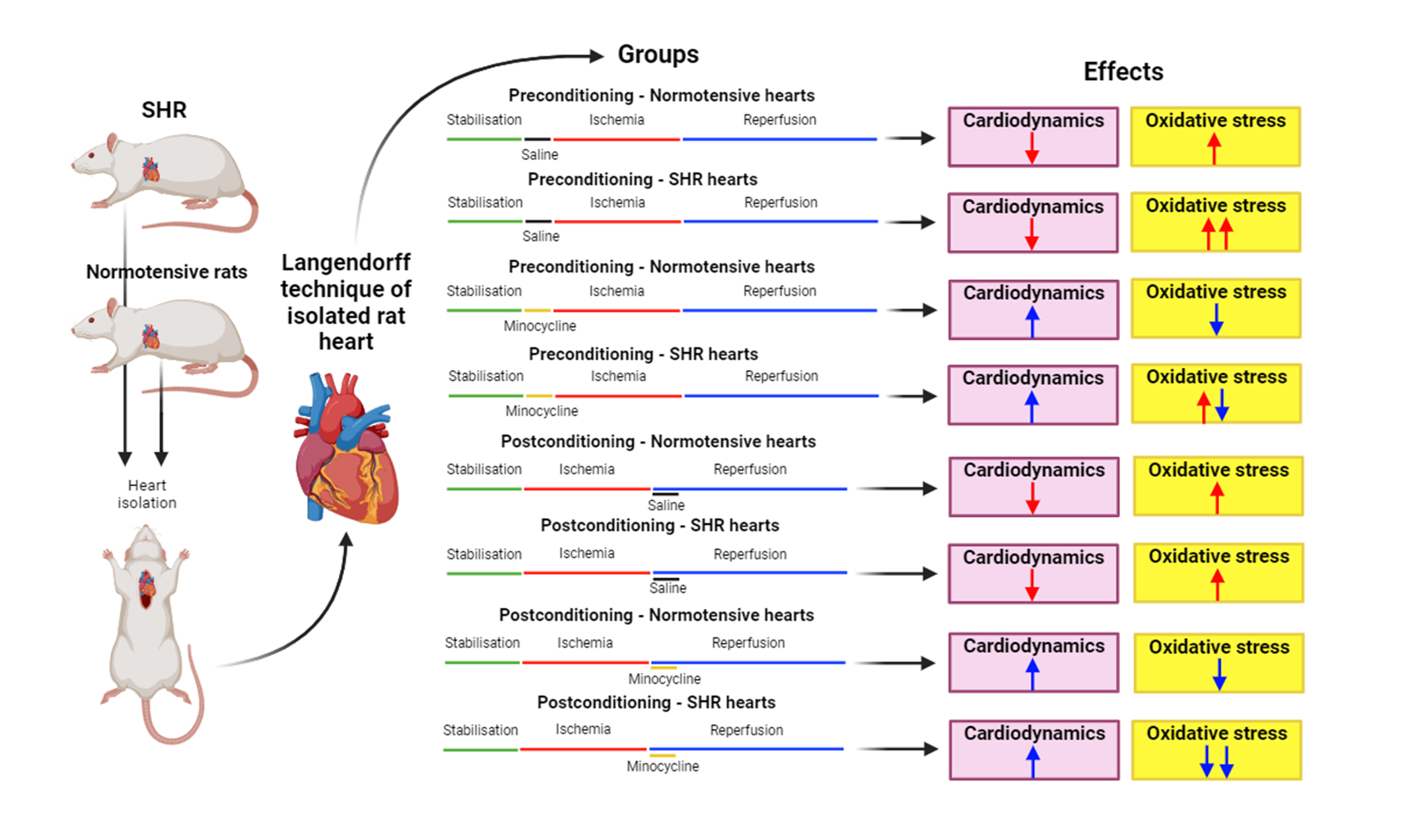
After the stabilization period, the hearts were exposed to global ischemia, perfusion was totally stopped for 20 min, after which it followed by 30 min of reperfusion. All parameters of cardiac function and coronary flow were measured in intervals of 5 min during the period of reperfusion (30 min). In the preconditioning (PreC) groups, after the stabilization period, the hearts were perfused with the saline or minocycline hydrochloride (dissolved in saline) at a dose of 1 μM for 5 min before global ischemia of 20 min which was followed by 30-min reperfusion. In the postconditioning (PostC) group, the hearts were perfused with a saline or minocycline hydrochloride (dissolved in saline) in the same dose, during the first 5 min of reperfusion. The applied dose of minocycline is based on previous studies of similar design [16,18,19].
According to the experimental protocol, healthy and SHR rats were divided into eight experimental groups:
- Healthy (normotensive) hearts preconditioned with saline and subjected to I/R injury (n = 8);
- SHR hearts preconditioned with saline and subjected to I/R injury (n = 8);
- Healthy (normotensive) hearts preconditioned with minocycline hydrochloride in a dose of 1 μM and subjected to I/R injury (n = 8);
- SHR hearts preconditioned with minocycline hydrochloride in a dose of 1 μM and subjected to I/R injury (n = 8);
- Healthy (normotensive) hearts postconditioned with saline and subjected to I/R injury (n = 8);
- SHR hearts postconditioned with saline and subjected to I/R injury (n = 8);
- Healthy (normotensive) hearts postconditioned minocycline hydrochloride in a dose of 1 μM and subjected to I/R injury (n = 8);
- SHR hearts postconditioned with minocycline hydrochloride in a dose of 1 μM and subjected to I/R injury x(n = 8).
The parameters of cardiac function and coronary flow were recorded in predefined points of interest. In PreC group: the last minute of stabilization period (S), the last minute of drug application (D – drug – saline or minocycline), the first and fifth minute of reperfusion, and further in intervals of 5 min until the end of the experiment (1, 5, 10, 15, 20, 25, and 30). In PostC group: the last minute of stabilization period (S), the first and fifth minute of reperfusion (PostC with minocycline takes place during the first 5 min of reperfusion), and further in intervals of 5 min until the end of the experiment (1, 5, 10, 15, 20, 25, and 30).
The following parameters of myocardial function were determined:
- The maximum rate of pressure development in the left ventricle (dp/dt max)
- The minimum rate of pressure development in the left ventricle (dp/dt mix)
- The systolic left ventricular pressure (SLVP)
- The diastolic left ventricular pressure (DLVP)
- The heart rate (HR)
- Coronary flow (CF) was measured flowmetrically.
Biochemical analysis
The samples of coronary venous effluent were collected in the same points of interest for determination of oxidative stress parameters. The following oxidative stress parameters were determined spectrophotometrically (UV-1800 UV–Vis Shimadzu Scientific Inc, Japan) using collected samples of the coronary venous:
- The index of lipid peroxidation, measured as thiobarbituric acid reactive substances (TBARS)
- The level of nitrites (NO2-).
- The level of the superoxide anion radical (O2-)
- The level of hydrogen peroxide (H2O2)
TBARS determination (Index of lipid peroxidation determination)
The degree of lipid peroxidation in coronary venous effluent was estimated by measuring TBARS using 1% thiobarbituric acid in 0.05 M sodium hydroxide incubated with a coronary venous effluent at 100oC for 15 min and then measured at a wavelength of 530 nm. KHS was used as a blank control (previously explained in Rankovic et al 2019) [20].
Nitrite level determination
Nitric oxide decomposes rapidly to form stable nitrite/nitrate products. Nitrite (NO2−) level was measured in order to indirectly assess nitric oxide level. Nitrites were quantified by the method according to Green using the Griess-reagent–1% sulfanilamide in 5% phosphoric acid/0.1% naphthalene ethylenediamine–dihydrochloride. In complete of 0.5 ml of coronary venous effluent was precipitated with 200 μl of 30% sulfosalicylic acid, then vortexed 30 min, and centrifuged at 3000 × g. Equal volumes of the supernatant and Griess’s reagent were added, incubated 10 min in the dark, and measured at a wavelength of 550 nm. KHS was used as a blank probe (previously explained in Rankovic et al 2019) [20].
Superoxide anion radical determination
The level of O2− were determined via nitro blue tetrazolium reaction in TRIS (tris(hydroxymethyl) aminomethane) buffer with the sample of coronary venous effluent, measured at 530 nm (previously explained in Rankovic et al 2019) [20].
Hydrogen peroxide determination
Determination of H2O2 was based on the oxidation of phenol red by hydrogen peroxide in a reaction catalyzed by horseradish peroxidase (HRPO) and measured at 610 nm. A total of 200 ml perfusate was precipitated with 800 mL of freshly prepared phenol red solution, along with 10 mL of 1:20 HRPO (made ex tempore) and subsequently added. For the blank probe, an adequate volume of KHS was used instead of coronary venous effluent (previously explained in Rankovic et al. 2019) [20].
Blood pressure
Blood pressure, systolic and diastolic, was measured immediately before animal sacrifice in order to confirm hypertension and normotension in the experimental groups. We used a tail-cuff noninvasive method blood pressure system (Rat Tail Cuff Method Blood Pressure Systems (MRBP-R),IITC Life Science Inc., Los Angeles, CA, USA) [21].
Pathohistological analysis of cardiac tissue
At the end of the ex vivo protocol on Langendorff apparatus, all hearts were fixed in 4% buffered paraformaldehyde solution for 24 h. Then, the tissues were dehydrated in increasing concentrations of alcohol (70%, 96%, and 100%), soaked in xylene on 60–70 °C for 60 min, and immersed in paraffin. Tissue sections (4 μm) were stained with hematoxylin–eosin, and examined under light microscopy (OlympusBX-51,OlympusEuropaGmbH,Germany) by two of the authors in a blinded fashion. Morphometric analysis included examination of longitudinal section diameter and cross-sectional area at × 200 magnification, using calibrated Axio Vision software (Zeiss, USA) [22,23].
Ethical approval
The protocol of this investigation was approved by the Ethics Committee for experimental animal well-being of the Faculty of Medical Sciences University of Kragujevac, Serbia (Approval No: 01–5301/7). All experimental processes were performed in accordance with the provisions prescribed acts (European Union Directive for the Protection of Vertebrate Animals used for Experimental and other Scientific Purposes 86/609/EEC and European Union Directive for the welfare of laboratory animals (2010/63/EU)) and the principles of Good Laboratory Practice.
Drugs
All reagents used in this experimental design were procured out of Sigma-Aldrich Chemie GmbH, Darmstadt, Germany.
Statistical analyses
All data were analyzed using SPSS Statistics 22 (SPSS, Chicago, IL). The results are expressed as the mean ± standard error (SE). In the PreC groups for statistical analysis, we used three specific points of interest: the last minute of stabilization (S), the last minute of drug application (saline or minocycline) (D), and the last minute of reperfusion (marked as 30). In the PostC groups for statistical analysis, we used three specific points of interest: the last minute of stabilization (S), the last minute of drug application (saline or minocycline) in the fifth minute of reperfusion (marked as 5), and the last minute of reperfusion (marked as 30). For analysis of differences within the same group, values of these three points were compared with each other. Differences between groups were examined by comparing the values of the same point of interest. Distribution of the data was checked by the Shapiro–Wilk test. Independent samples t-test (parametric) and Mann–Whitney U-test (nonparametric) as well as one-way ANOVA followed by Tukey’s multiple comparison post hoc test, and Kruskal–Wallis were used to assess the difference in estimated variables between the groups. Values of p lower than 0.05 were considered to be significant.