Permeability and cellular transport
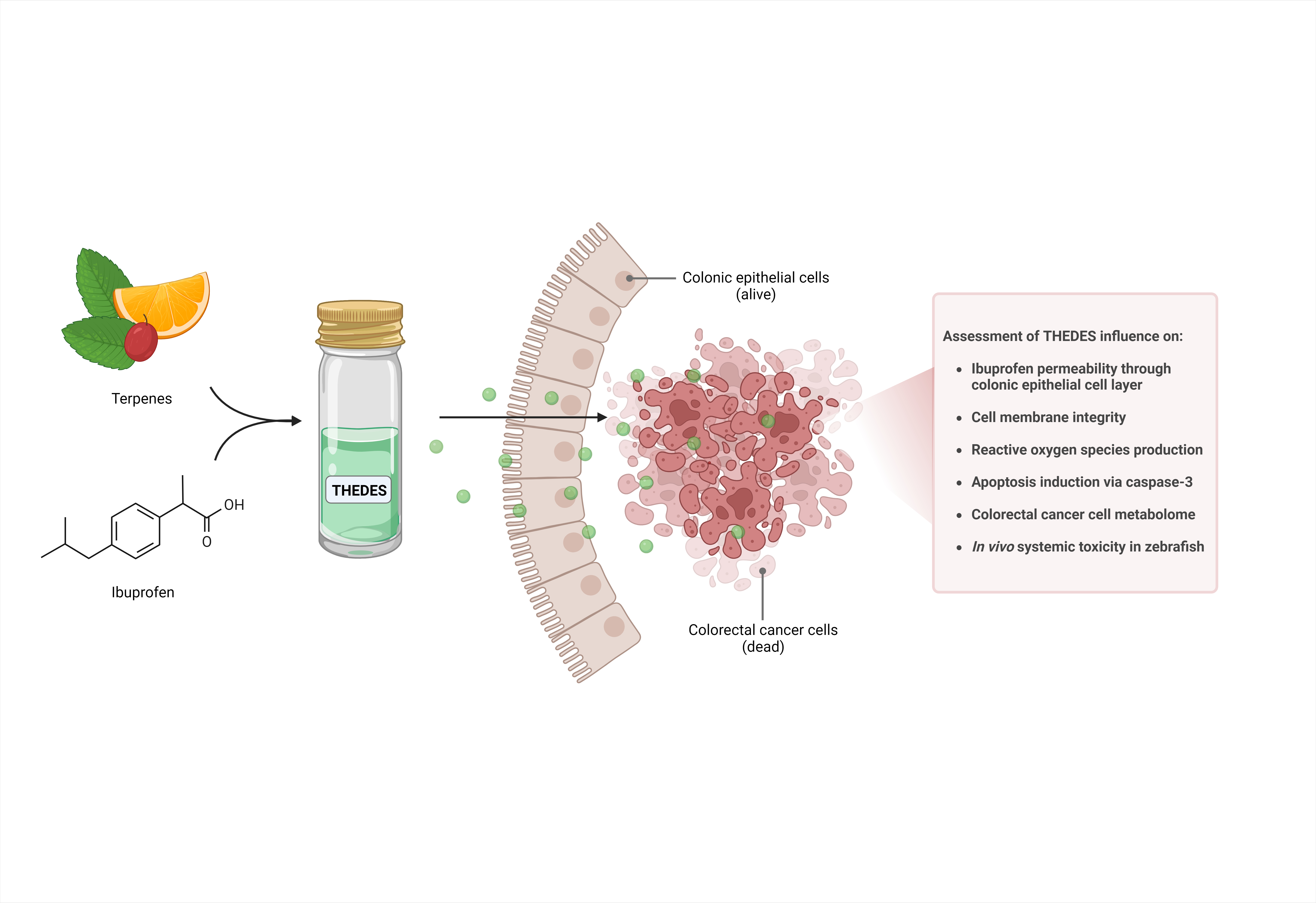
CRC carcinogenesis highly relies on inflammation, hence controlling such activity encompass a key aspect on the desired anti-CRC drug therapeutic profile 18,19. Non-steroidal anti-inflammatory drugs (NSAID) such as Ibu, not only have a well described anti-inflammatory activity, but have also been described as having preventive and anticancer activity by, for example, inhibiting the activation of nuclear β-catenin in CRC 28,29. Nevertheless, NSAIDs activity is often limited by its low bioavailability in physiological media, which is a consequence of its poor solubility although having high permeability (class II on biopharmaceutics classification system) 30. Thus, there is potential to optimize such APIs properties and eutecticity has been widely described for such performance enhancement 31–33. To push forward the understanding of THEDES based on the combination of a terpene with Ibu potential anticancer therapeutical role, its effects on Ibu permeability and cellular transport was herein investigated. Firstly, using a glass diffusion apparatus with a synthetic PES-U porous membrane (Table 1), it is possible to recognize that Thy:Ibu (3:1) promotes a significant increase of Ibu permeability (*p ≤ 0.05), however when combined with POH this API do not experience permeability increases (Figure S1 – A).
Table 1
Permeability results and diffusion coefficients obtained for Ibu in THEDES and in powder form using a synthetic PES-U membrane. Data indicated as mean ± SD.
| | Permeability (105 cm s− 1) | Diffusion coefficient (106 cm2 s− 1) |
| Thy:Ibu (3:1) | 5.76 ± 1.31 (*) | 2.52 ± 0.87 |
| POH:Ibu (3:1) | 4.45 ± 0.03 | 4.24 ± 0.93 (*) |
| POH:Ibu (8:1) | 1.92 ± 0.58 | 3.13 ± 1.15 |
| Me:Ibu (3:1) | 14.00 ± 1.53a | 4.32 ± 0.34a |
| Ibu | 3.52 ± 0.26 b | 1.30 ± 0.22 b |
a retrieved from 32.b retrieved from 25.
Furthermore, POH:Ibu (3:1) promotes a significant increase (*p ≤ 0.05) in Ibu’s diffusion coefficient, while Thy:Ibu (3:1) and POH:Ibu (8:1) does not (Figure S1 – B). Since the diffusion coefficient is a parameter correlating API amount diffused within time – with higher coefficients representing faster compound diffusion through the membrane 32,34 – from the obtained results it is possible to infer that with the combination of POH with Ibu as an eutectic system in a 3 to 1 molar ratio it is possible to obtain a faster Ibu diffusion through the membrane. This result is highlighted in Fig. 1 where the profile of Ibu permeability can be observed over time.
These permeability and diffusion results highlight that although these are all THEDES combining Ibu with a terpene, we see a difference not only in varying the terpene but also in varying its molar ratio within the eutectic formulation. This was also emphasised in a previous work, where combining Me with Ibu promotes a much higher permeability increase, while when using Safranal does not 25. In a second step, Ibu permeability and cellular transport were investigated using a Caco-2 cell-based transwell apparatus, which allows a more representative in vitro microenvironment reconstitution 31. The obtained results are presented in Table 2 where it is possible to observe that the eutectic formulations do not seem to promote beneficial effects on Ibu permeability, rather the opposite with a significant decrease observed for Thy:Ibu (3:1) (p ≤ 0.0001), POH:Ibu (3:1) (p ≤ 0.001) and Me:Ibu (3:1) (p ≤ 0.0001) (Figure S2).
Table 2
Estimate apparent permeability (Papp) and Ibu concentration on apical and basolateral side of the transwell apparatus, and cellular uptake obtained for Ibu in THEDES and in powder. Data indicated as mean ± SD.
| | Papp (105 cm s− 1) | Final concentration on apical side (mg mL− 1) | Final concentration on basolateral side (mg mL− 1) | Estimated cellular uptake (mg mL− 1) |
| Thy:Ibu (3:1) | 3.58 ± 1.1 (****) | 0.091 ± 0.00 | 0.003 ± 0.00 | 0.093 |
| POH:Ibu (3:1) | 0.93 ± 0.1 (****) | 0.008 ± 0.00 | 0.000 ± 0.00 | 0.031 |
| POH:Ibu (8:1) | 5.39 ± 0.2 | 0.134 ± 0.00 | 0.004 ± 0.00 | 0.056 |
| Me:Ibu (3:1) | 4.80 ± 0.1 (***) | 0.059 ± 0.00 | 0.002 ± 0.00 | 0.034 |
| Ibu | 5.72 ± 2.7 | 0.364 ± 0.02 | 0.011 ± 0.00 | 0.125 |
Interestingly, the permeability studies using normal colonic cells (Caco-2 model) revealed contrasting results in comparison with what was obtained using a synthetic membrane. This result is highlighted in Figure S2 – A, where the profile of Ibu permeability can be observed over time. In Fig. 2 it is possible to recognize that this result come as the effect of Ibu transcellular transport significantly increased when it is presented to the cell’s monolayer as a eutectic formulation. Moreover, it also provides the indication of an intracellular accumulation and metabolization, since the cellular uptake is significantly increased in all tested eutectic formulations, with highlight for the Thy-based one. Furthermore, the maintenance of TEER values (data not shown) reinforces the non-cytotoxicity of the tested concentrations towards normal colonic cells.
It has been described that Ibu is easily absorbed at the intestinal epithelium by passive transport 35. On the other hand, although terpenes such as Thy, POH and Me, transport is mainly passive, they may stimulate paracellular transport of large molecules by interacting with tight junctions 36,37. The observed results contrast with such reports since, in this case, combining a terpene with Ibu as a eutectic, do not stimulate paracellular but instead transcellular transport which ultimately, in this context, seem to contribute for less transport from the apical side to the basolateral side of the transwell plate. Pereira and coworkers also described this cellular uptake tendency for THEDES combining Lim with Ibu in 4 to 1 and 8 to 1 molar ratios, respectively 26. This increase in cellular uptake may be a hint for these THEDES effectiveness towards CRC cells.
THEDES bioactivity
For a better understanding on the reported selective cytotoxicity of THEDES based on the combination of Me, Thy, Lim, and POH with Ibu, towards CRC cells, several bioassays were performed and the results are herein described.
Intracellular ROS
Tumour-promoting inflammation is a significant contributor to carcinogenesis hence being considered as a cancer hallmark 38. ROS play an important role in modulating inflammatory pathways and when enhanced in tumour microenvironment, and specifically in human colonic mucosa, they contribute to the epithelial-mesenchymal transition which leads to increased proliferation and invasion 39,40. Thus, controlling ROS presence in CRC cells can represent a complementary strategy to cope with this type of cancer development and progression. In this work, HT29 cells were the model of choice since they have high levels of basal stress, hence presenting high levels of ROS production 40. When considering the obtained results for the POH-based system (Fig. 3 - B), although from previous reports its terpene precursor Lim in combination with Ibu revealed protective action towards oxidative stress 26, POH:Ibu (3:1) and POH:IBU (8:1) do not seem to influence ROS production at the tested concentrations. Neither their corresponding individual components. In contrast, in Thy:Ibu (3:1) (Fig. 3 - A) seems to promote a significant decrease (p ≤ 0.001), less than half, of oxidative species and so protecting HT29 cells from oxidative stress which may ultimately demonstrate an anti-inflammatory effect by inhibiting ROS production in CRC. When looking to its corresponding individual components, it is possible to observe the inverse tendency since they seem to increase ROS, which highlights the difference between the individual compounds action compared to them as a THEDES. This result may come as outcome of the cellular uptake tendency previously observed for the Thy-based system in the cellular transport studies. In a previous report, terpenes such as Me and safranal combined with Ibu were studied and revealed Me:Ibu (3:1) as a protective agent for oxidative stress in HT29, while Saf:Ibu (3:1) and Saf:Ibu (4:1) do not 25. It has been reported that low levels of ROS lead to downregulation on the inflammatory cascade, but it is also known that excessive high levels of ROS promote cancer cell death 39. Considering the obtained results, we hypothesise that the herein discussed THEDES do not induce cancer cell death via ROS increase, so its antiproliferative effect must have a different cause, but these THEDES may play an interesting role as protective agents towards oxidative stress and ultimately inflammation.
LDH release
There are several reports hypothesizing that DES-induced cancer cell death occurs via cell membrane disruption, following a necrotic form of cell death 24,33. In Fig. 4, it is possible to observe that all tested THEDES induce cancer cell death via its membrane disruption only at concentrations three times higher than their corresponding reported EC50. These results suggest that membrane disruption has not been the primary cause for HT29 cell death exposed to Thy:Ibu (3:1), POH:Ibu (3:1), POH:Ibu (8:1), Lim:Ibu (4:1) and Lim:Ibu (8:1).
In a previous report, Pereira and coworkers reported that Me:Ibu (3:1) promotes cell membrane disruption at EC50, so within the therapeutic window of the selectivity index, while Saf:Ibu (3:1) and Saf:Ibu (4:1) only induce disruption at higher concentrations than its EC50 25. The hydrophobic and/or amphipathic nature of the herein approach terpenes allows them to interact with the phospholipid cell membrane bilayer, but its hydrophobicity could prevent its full cellular uptake. Thus, the observed cancer cell death upon THEDES exposure could be related to alterations in cell membrane mechanical fluidity, compromising its stability, which is essential specially in cancer cells that have increased abnormal cell division rates. This alteration can be of most importance since this mechanical disturbance may cause membrane disruption and eventual cell death 41,42. These results suggest that the tested THEDES induce HT29 death via a different path and highlight that although all these THEDES are based on the combination of a terpene with Ibu, different combinations and even different molar ratios promote a different impact on this cancer cells.
Apoptosis via Caspase-3
For a better understanding on how these THEDES could be exerting the reported antiproliferative action towards HT29, it was evaluated if they could be inducing programmed cell death via the caspase-3 pathway. In Fig. 5 it is possible to observe that all tested THEDES appear to promote cell death by inducing apoptosis, when compared with the control of cell non exposed to THEDES. Interestingly, although there have been reports on terpenes and Ibu as inducers of apoptosis 43–45, in Fig. 5 it is observable that the compounds alone have minor or none effect and so this induction is enhanced by being as a eutectic formulation. This allows to take advantage of this compound’s bioactivity using a lower concentration.
These results follow the previously reported ones for Me:Ibu (3:1), Saf:Ibu (3:1) and Saf:Ibu (4:1) 25, suggesting that the main action of these THEDES towards cancer cells proliferation is, in fact, through the induction of apoptosis via caspase-3. On the other hand, it is interesting to mention that in another report Pereira and coworkers reported Lim:Ibu (4:1) and Lim:Ibu (8:1) as not promoting apoptosis via this pathway. So, although they are still a combination of a terpene and Ibu, they have different ways to exert its antiproliferative action towards HT29 cells. This once more emphasizes the heterogenic bioactive profile of these THEDES towards CRC cells.
Metabolomic landscape of CRC cells upon THEDES exposure
THEDES influence on HT 29 cells endometabolome
The influence of THEDES, combining a terpene with Ibu in different molar ratios, on CRC cells endometabolome was herein investigated by a GC-MS-based metabolomics approach. The GC-MS metabolomic analysis identified 16 metabolites in the intracellular environment, in which some were varying as a consequence of THEDES exposure (Table S1). To investigate this, first the analytical reproducibility of the GC-MS experimental data was confirmed by unsupervised statistical analysis (PCA). For this, all samples and QCs were included and the statistical analysis revealed well-defined QCs cluster (Figure S3 - A). Further, unsupervised (PCA) and supervised multivariate (PLS-DA) analyses were performed considering the comparison between the extracellular media collected from non-exposed (control) and exposed cells to the different THEDES. As expected from the previously obtained results in the different bioassays, different outcomes for the different combination of compounds and molar ratios composing THEDES were observed. The results for Me:Ibu (3:1) and Lim:Ibu (4:1) revealed a good separation between the control and the exposed cells, and POH:Ibu (8:1) also revealed a separation in the PLS-DA model although less robust (Q2 = 0.35). The cells exposed to Thy:Ibu (3:1), Lim:Ibu (8:1), POH:Ibu (8:1), did not reveal a separation between the control and exposed cells. Further, VIP score projection was performed (Fig. 6) to assess the metabolites responsible for group separation. Several metabolites such as lactate, oxalate, myo-inositol, 5-oxoproline, glycine, 1-octadecanol, glucose and palmitic acid revealed high importance (VIP > 1) to discriminate the THEDES exposed cells from control cells.
Among the 16 metabolites detected in the GC-MS analysis of the endometabolome, statistical significance, when comparing control cells with THEDES exposed cells, was only found for i) lactate, in cells exposed to Me:Ibu (3:1) and Lim:Ibu (4 :1) (Fig. 7), ii) myo-inositol, in cells exposed to Me:Ibu (3:1), POH:Ibu (8:1) and Lim:Ibu (4:1) (Fig. 7), iii) 1-octadecanol, in cells exposed to Lim:Ibu (4:1) (Fig. 7), iv) and palmitic acid, in cells exposed to POH:Ibu (8:1) (Fig. 7).
The effect size of each significantly altered metabolite is summarized in Table 3 with the potentially affected metabolic pathways.
Table 3
Altered metabolites and potentially affected metabolic pathways resultant from the endometabolome analysis of THEDES exposed cells compared with control cells.
| Metabolite | Alterations caused by each THEDES vs control | Metabolic pathways |
| Me:Ibu (3:1) | POH:Ibu (8:1) | Lim:Ibu (4:1) |
| Lactate | -2.35 ± 1.60 (*) | - | -3.69 ± 1.98 (*) | Glycolysis / Gluconeogenesis; Pyruvate metabolism |
| Myo-inositol | -3.76 ± 1.91 (*) | -2.98 ± 1.58 (*) | -2.08 ± 1.46 (*) | Galactose metabolism; Inositol phosphate metabolism; Biosynthesis of nucleotide sugars; Phosphatidylinositol signalling system |
| 1-Octadecanol | - | - | 3.89 ± 2.05 (*) | Plasmalogen Synthesis |
| Palmitic acid | - | 2.21 ± 1.37 (*) | - | Fatty acid metabolism |
From these results it is possible to observe that HT29 exposure to THEDES promote a heterogeneous outcome on this cancer cells endometabolome. HT29 exposure to Me:Ibu (3:1), POH:Ibu (8:1) and Lim:Ibu (4:1), promoted a deleterious effect in the levels of myo-inositol, with highlight for the Me-based system. This metabolite is involved in several key metabolic pathways for cells homeostasis and proliferation, mostly related to cell signalling and nucleotide synthesis 46. It works as a building block of a cellular language via: insulin signalling, PI3K/Akt signalling, endocytosis, vesicle trafficking, cell migration, proliferation and apoptosis 47. But also plays a pivotal role in nucleotide synthesis in the incorporation of adenine and guanosine 48. Thus, deregulation of this pathway may disrupt homeostasis and promote carcinogenesis 49. Hence, THEDES-induced myo-inositol depletion seem to compromise such metabolic pathway, downregulating it, which ultimately contributes to the induced cell death by apoptosis previously described. Furthermore, HT29 exposure to POH:Ibu (8:1) and Lim:Ibu (4:1) seem to interfere with lipid metabolism, upregulating it, resulting in a significant increase (p ≤ 0.05) of palmitic acid and 1-octadecanol, respectively, when compared to the control. Both catabolic and anabolic pathways of lipid metabolism play an important role in cell activity related to i) membrane structure and function, ii) intracellular signalling pathways, iii) transcription factor activity and consequent gene expression, iv) and production of bioactive lipid mediators 50. Considering cancer biology, fatty acids such as palmitic acid, are mostly recognized as substrates for energy production in these highly proliferating cells 51. Thus, the observed increase in palmitic acid could be associated with the previously reported Warburg effect, where cancer cells prefer anaerobic glycolysis pathway for energy production. To cope with the energy demand of fast proliferating cells, cancer cells may rely on fatty acids for energy production via β-oxidation 51. Nevertheless, it has also been reported their crucial role in cancer cells sensing since saturated and unsaturated fatty acids differentially regulate the transcriptional activity of the retinoic acid receptor RAR and PPARβ/δ. Meaning that saturated fatty acids, such as palmitic acid, by activating RAR and inhibiting PPARβ/δ, are capable to suppress cancer cell proliferation 52. Since in the previously obtained results it was observed that POH:Ibu (8:1), in this concentration, promotes cell death, we hypothesize that this could be the case of fatty acid synthesis working as suppressors of cancer cell proliferation instead of contributing for energy production. Further, the upregulation detected for the fatty alcohol 1-octadecanol suggests an altered fatty alcohol metabolism in the studied cells 53. This accumulation can be associated with a deficient activity of fatty aldehyde dehydrogenase (FALDH), which is an enzymatic component of fatty alcohol:NAD oxidoreductase (FAO) necessary for fatty alcohol metabolism 54. Furthermore, 1-octadecanol participates in the plasmalogen synthesis 55. Specifically for CRC, alterations in plasmalogen synthesis and the different plasmalogens subclasses have been associated with phospholipases C and D activity reduction and increased activity of phosphocholine cytidyltransferase, which results in generally elevated phospholipid and the phosphatidylethanolamine plasmalogen specie content 56,57. Since plasmalogens highly contribute to cell membrane fluidity and ultimately its stability, we hypothesize that the observed upregulation by POH:Ibu (8:1) of 1-octadecanol could contribute to an excessive upregulation of the plasmalogen synthesis that in the end result in membrane instability and resulting cell death induction previously observed. In contrast, from the depletion observed in lactate in HT29 cells exposed to Me:Ibu (3:1) and Lim:Ibu (4:1), it seems to be happening a deleterious effect on the anaerobic glycolysis pathway as lactate is its ultimate resulting metabolite since these cells can be considered to be in anaerobic-like conditions 58. Therefore, these THEDES seem to compromise cancer cell main choice for energy production and in this way compromising their viability.
THEDES influence in HT 29 cells exometabolome
Following the description on THEDES influence on HT29 cells endometabolome, it was investigated by GC-MS-based metabolomics which changes could be happening on this cell’s exometabolome. The GC-MS metabolomic analysis identified 17 metabolites (aldehydes and ketones) in the extracellular environment, in which some were varying as a consequence of THEDES exposure (Table S2). As previously described, first the analytical reproducibility of the experimental data was confirmed by PCA considering all samples and QCs. This analysis revealed a well-defined QCs cluster (Figure S3 - B). Thereafter, PCA and PLS-DA statistical analysis were performed considering the comparison between the intracellular metabolome of cells non-exposed (control) and exposed to the different THEDES. Once more, different results for the different combinations of compounds and molar ratios composing THEDES were obtained. The results for Thy:Ibu (3:1) revealed a good separation between the control and the exposed cells; POH:Ibu (3:1) and POH:Ibu (8:1) also revealed a separation in the PLS-DA models. In contrast, HT29 cells exposed to Me:Ibu (3:1), Lim:Ibu (4:1) and Lim:Ibu (8:1), did not revealed separation between the control and exposed cells in the PCA and PLS-DA models (Fig. 8). To better understand which could be the metabolites promoting such separation, a VIP score projection was performed, and in Fig. 8 it is possible to observe from VIP score 1 upwards several metabolites such as methylglyoxal, 4-methyl-2-pentanone, cyclohexanone, acetaldehyde and acetone.
Among the 17 metabolites detected in the GC-MS analysis of the extracellular environment, it was only found statistical significance for methylglyoxal, when comparing cells exposed to Thy:Ibu (3:1), POH:Ibu (3:1), and POH:Ibu (8:1) with the control (Fig. 9); and for acetaldehyde for the POH:Ibu (8:1) exposed cells compared with the control (Fig. 9).
The effect size of each significantly altered metabolite is summarized in Table 4 with the potentially affected metabolic pathways.
Table 4
Altered metabolites and potentially affected metabolic pathways resultant from the exometabolome analysis of THEDES exposed cells compared with control cells.
| Metabolite | Alterations caused by each THEDES vs control | Metabolic pathways |
| Thy:Ibu (3:1) | POH:Ibu (3:1) | POH:Ibu (8:1) |
| Methylglyoxal | 1.55 ± 1.07 (*) | 2.38 ± 1.24 (*) | 1.33 ± 1.03 (*) | Glycine, serine and threonine metabolism; Pyruvate metabolism; Propanoate metabolism | |
| Acetaldehyde | - | - | 1.58 ± 1.08 (*) | Glycolysis/Gluconeogenesis; D-Amino acid metabolism; Glycerophospholipid metabolism; Pyruvate metabolism | |
From these results it is possible to observe that HT29 exposure to these THEDES outcome is an augmentation of methylglyoxal presence in the extracellular environment and a lower acetaldehyde consumption from the external media. It has been reported that one of the key metabolic alterations on cancer cells is its preference for the anaerobic glycolysis energy production pathway, instead of the more efficient oxidative phosphorylation, regardless of oxygen availability 58. One of such consequences is the increased production of glycolysis intermediates, where among them there is methylglyoxal as a glucose-derived highly reactive dicarbonyl 59. Although this aldehyde is well-known to participate in glycine, serine and threonine metabolism 60, when talking about cancer cells methylglyoxal it has been hypothesised that its major role can be hormetic, thus working both as pro-tumorigenic and as an anti-cancer 61. At low concentration, methylglyoxal results in beneficial effects allowing cancer cell to avoid apoptosis and enhance cell proliferation, but when the threshold of dicarbonyl stress is exceeded it becomes the opposite and cell death is inevitable, due to DNA and protein synthesis inhibition, and cellular respiration 62. In Fig. 9 it is possible to observe that the control, consisting of cancer cells not exposed to THEDES, consumed methylglyoxal reducing its levels in comparison with the blank (cell culture media only). In contrast, the presence of high levels of methylglyoxal in extracellular medium of cells exposed to POH:Ibu (8:1), in comparison with the control, suggests that this THEDES system induce the uncontrolled production of such metabolite that ultimately lead to cell death, as observed in the previous sections were these THEDES induced apoptosis. Interestingly, although Thy:Ibu (3:1) and POH:Ibu (3:1) also induce apoptosis (Fig. 5), herein it was observed that these THEDES did not induce methylglyoxal production neither allowed its significant consumption by the cells. This could be related to the lower terpene presence in the eutectic system formulation. Furthermore, it is known that acetaldehyde participates in different key metabolic pathways such as glycolysis and pyruvate metabolism 60. However, it has been reported that increased levels of acetaldehyde promotes several deleterious effects in cells by forming protein, DNA, and phospholipid adducts, that result in DNA lesions 63. Hence, when looking to the results obtained for cells exposed to POH:Ibu (8:1) for the presence of acetaldehyde in the extracellular environment, in comparison to the control and blank it is possible to realize that there was a consumption from the external media by the control cells, which can be related to the acetaldehyde uptake for pyruvate metabolism for energy production 64. Nonetheless, cell exposure to this eutectic system seems to experience an impairment in acetaldehyde metabolism since its presence remains similar to the blank which could mean an intracellular accumulation of it and an impairment of aldehyde dehydrogenases that may result in cell death by acetaldehyde forming adducts.
THEDES effect in zebrafish
To push forward the establishment of the herein described THEDES as anticancer therapeutic agents, a perception on their effect on more complex models as a whole organism is mandatory. For that, THEDES influence on a battery of biomarkers (CAT, GST, SOD, Gpx, AChE, caspases, LPO and TAC), commonly used in organisms were analysed to provide a preliminary systemic toxicity assessment. Since a multibiomarker approach was used, the IBR index was used to better understand the consequence of THEDES and 5-FU (positive control) exposure on such biomarkers and differences among themselves 65. Lim:Ibu (4:1) was the only tested condition promoting a different IBR response with a 37.79 index value, whereas the rest of the tested conditions presented a similar IBR index of 27.10 (Table 5).
Table 5
IBR index values obtained for each biochemical biomarker after THEDES and 5-FU exposure, and negative control.
| Sample | Concentration (EC50, mM) | IBR index value |
| Negative control | - | 27.10 |
| 5-FU | 25.98 a | 27.10 |
| Me:Ibu (3:1) | 4.30 | 27.10 |
| Thy:Ibu (3:1) | 0.30 | 27.10 |
| POH:Ibu (3:1) | 1.32 | 27.10 |
| POH:Ibu (8:1) | 1.37 | 27.10 |
| Lim:Ibu (4:1) | 2.39 | 32.79 |
| Lim:Ibu (8:1) | 1.14 | 27.10 |
a retrieved from 66.
This isolated position of Lim:Ibu (4:1) seems to be related to a higher CAT activity, in comparison with the other compounds and biomarkers tested (Fig. 10).
Besides IBR, the multiple comparison statistical testing was also performed considering each individual biomarker. A significant increase (p ≤ 0.001) of CAT and GST activities was observed after 5-FU exposure, in comparison with the control (Figure S4 – A and B). CAT is an oxidoreductase found on peroxisomes, which are highly present in liver cells, and are mostly associated with high levels of hydrogen peroxide (H2O2) neutralization 67; furthermore, GST is found in cytosol and is mainly associated with detoxification by catalysing the conjugation of glutathione with xenobiotics, making them more water-soluble and easier to eliminate, but are also important mediators in oxidative stress responses 68. Thus, from the results obtained for these two biomarkers it is possible to hypothesize that 5-FU is inducing oxidative stress in the exposed animals. This was somewhat expected since although 5-FU is a standard drug for CRC treatment it presents a wide range of negative side effects such as oxidative stress induction 69. Contrasting results were observed in LPO since malondialdehyde (MDA) showed a significant decrease (p ≤ 0.05) after 5-FU exposure (Figure S3 – C), suggesting a decrease of oxidative stress regarding this biomarker. It was also observed a GST increase in animals exposed to Lim:Ibu (4:1) and POH:Ibu (3:1), although less significative (p ≤ 0.05) in comparison with 5-FU (Figure S3 – B). Similarly to CAT, GPx is also an oxidoreductase, but ubiquitous in cytoplasm and mitochondria, which catalyses the reduction of H2O2 to water and oxygen, and also the reduction of peroxide radicals to alcohols and oxygen 70. In animals exposed to Lim:Ibu (8:1), Thy:Ibu (3:1) and POH:Ibu (3:1) it was observed a GPx significative increase (p ≤ 0.05 and p ≤ 0.01) (Figure S3 – F), suggesting a response to fight the increased oxidative stress. Further, AChE is the main cholinesterase enzyme and is responsible for breaking down acetylcholine (Ach) neurotransmitter in synapses, which terminates signal transmission. Its incredibly fast turnover makes it one of the most efficient enzymes known and ultimately allows the precise control of nerve impulses in the nervous system 68. Hence, it is a biomarker for toxicity on the nervous system. From the obtained results it is possible to recognize a significant increase (p ≤ 0.05) for Thy:Ibu (3:1) and (p ≤ 0.001) POH:Ibu (3:1) on the enzymatic activity (Figure S3 – G). An AChE imbalance has severe effects depending on whether there is an excess or deficiency of this enzyme. AChE inhibition can lead to an Ach accumulation which can cause overstimulation of the nervous system, while its excess can lead to the rapid degradation of Ach and reduced signalling at cholinergic synapses. Both situations with severe impact related to the nervous system such as muscle spasms, paralysis, cognitive impairment, among others 68. AChE activity is often reduced by exposure to xenobiotics such as pesticides, but also in certain medical conditions such as dementia 71,72. Nevertheless, it has been reported that in Alzheimer's disease, prevention of AChE inhibition could happen via oxidative stress mediated compounds 73. So further studies on how these THEDES could beneficially influence such conditions could be of great interest. Finally, although the individual multiple comparison statistical testing revealed significant differences between the control and animals exposed to 5-FU and THEDES for certain biomarkers, it is important to highlight that in such experiments external elements, such as gender, age and genetic predispositions, may also contribute to the obtained results, therefore it is reinforced the need for a statistical data analysis that better encompasses such diversity. In this way, the Spearman's rank correlation coefficient was determined. However, it did not reveal any significant correlation between the tested biomarkers upon exposure to the different systems (Figure S3 – I), which could be corroborated by the previously reported IBR indexes.