The study shows that chronic interval training leads to significant changes in gene expression in connection with muscle growth and muscle atrophy. In the MIIT group, the expression of the myostatin gene was reduced by 41% and that of the atrogin gene by 87%. In the HIIT group, these decreases were even greater at 66% for the myostatin gene and 93% for the atrogin gene. In addition, there was a significant increase in the expression of the follistatin gene, with the MIIT group showing an increase of 114% and the HIIT group an increase of 267%. These results suggest that the intensity of chronic interval training may have differential effects on gene expression related to muscle growth and atrophy, potentially promoting hypertrophy and reducing atrophy. The study reveals that chronic interval training led to significant changes in gene expression associated with muscle growth and muscle atrophy. In the MIIT group, the expression of the myostatin gene decreased by 41%, while the expression of the atrogin gene decreased by 87%. In the HIIT group, these reductions were even more pronounced, with a 66% decrease in the myostatin gene expression and a 93% decrease in the atrogin gene expression. Additionally, there was a substantial increase in the expression of the follistatin gene, with the MIIT group showing a 114% increase and the HIIT group showing a 267% increase. These findings suggest that the intensity of chronic interval training may have varying effects on gene expression related to muscle growth and atrophy, potentially promoting hypertrophy and reducing atrophy. Moreover, the research indicates significant differences in certain physiological factors between high-intensity interval training (HIIT) and moderate-intensity interval training (MIIT). Although there were no significant differences in hypertrophic and atrophic factors between the two, there is a notable distinction in the follistatin to myostatin ratio, implying that overall exercise intensity has a greater impact on the balance of these factors. Additionally, higher exercise intensity appears to have a greater impact on insulin sensitivity. In healthy male rats, the HIIT group showed a substantial increase of 27.5%, while the MIIT group only showed a 2.5% increase. There are also significant differences in insulin sensitivity between chronic exercise groups. Acute interval training, however, does not affect myostatin and follistatin gene expression, but it does influence the expression of atrogin, a gene associated with muscle atrophy. Moreover, our study reported a significant decrease in atrogin gene expression in both the acute MIIT group (73% reduction) and the acute HIIT group (70% reduction). Furthermore, the study highlights that acute training has an impact on the ratio of myostatin and follistatin gene expression, even though there is no significant difference in their individual expression between the control and acute exercise groups. Additionally, the study indicates that while there may not be a significant difference in the gene expression of myostatin and follistatin between the control group and the acute exercise groups, there is an impact on their ratio. It was observed that the ratio increased by 2.5 times in the acute moderate-intensity interval training (MIIT) group and by 2 times in the acute high-intensity interval training (HIIT) group. This suggests that acute exercise may not have a significant effect on follistatin and myostatin alone, but it can influence their ratio, potentially affecting muscle hypertrophy and atrophy processes in rats.
The results of the study suggest that the expression of the myostatin gene decreased significantly in the chronic exercise group undergoing High-Intensity Interval Training (HIIT) compared to the Moderate-Intensity Interval Training (MIIT) group and the control group. The findings indicate that higher intensity exercise, such as HIIT, may be necessary to effectively reduce myostatin levels, which is considered a negative factor in muscle growth. This information suggests that High-Intensity Interval Training (HIIT) can impact muscle myostatin levels. Consistent with our findings, the study by Biglari et al. (2018) demonstrated a 68% reduction in muscle myostatin in male rats after 8 weeks of HIIT training[1], while Bradley et al. found a significant decrease in myostatin levels after 6 weeks of HIIT training in both men and women[2]. However, the study by Rashid Lamir et al. (2016) indicated that 8 weeks of resistance training did not lead to significant changes in the expression of the myostatin gene in cardiac muscle in healthy male Wistar rats[3]. This illustrates the differing effects of different types of training on myostatin levels, highlighting the importance of considering the specific mode of exercise when interpreting research findings. In their study rats participated in resistance training for 8 weeks, with three sessions per week[3]. Their study indicates that 8 weeks of resistance training did not lead to significant changes in the expression of the myostatin gene in cardiac muscle[3]. Additionally, it suggests that acute exercise did not result in significant changes in myostatin gene expression among the exercise groups, except for a partial decrease observed in the MIIT and HIIT groups[3]. Our research findings indicate that chronic exercise affects myostatin gene expression, while single-session exercise does not have a significant impact. However, a study by Allen et al. (2011) demonstrated a decrease in myostatin mRNA levels after an endurance exercise session, which contradicts our research [4]. The difference in findings between our research and Allen's study could be attributed to variations in exercise protocols and the timing of sample collection from the participants. Additionally, it is noted that physical exercises may impact myostatin regulation by deactivating signaling pathways such as myostatin and SMAD2/3, while activating GSK-3B/B-Catenin signaling pathways [5]. The results suggests that exercise intensity plays a crucial role in increasing muscle protein synthesis [20]. According to the information shared, myostatin expression may have a threshold, and until the mechanical tension force in skeletal muscle reaches this threshold, the susceptibility to exercise-induced effects on myostatin may not occur significantly [20]. Additionally, it is suggested that low-intensity interval training may not have a significant effect on this process. Myostatin and follistatin are two proteins that play a significant role in regulating the growth and development of skeletal muscles. Myostatin is a negative regulator of muscle growth, meaning that it inhibits muscle cell proliferation and differentiation, while follistatin is a myostatin antagonist, promoting muscle growth[6, 7].
Research suggests that imbalances in these proteins can contribute to the development of conditions such as diabetes and obesity[4]. Specifically, decreased myostatin levels have been linked to insulin resistance, and there is evidence indicating that myostatin plasma and muscle protein levels decrease in people with insulin resistance as a result of aerobic activity, showing a strong connection with insulin sensitivity [4, 8]. Furthermore, the hypertrophy of muscle fibers, which can be influenced by myostatin and follistatin levels, has the potential to improve glucose tolerance and insulin resistance by increasing energy consumption and enhancing fatty acid oxidation [9]. The results indicate that deleting the myostatin gene in mice leads to specific physiological effects such as skeletal muscle hypertrophy, reduced fat accumulation, and prevention of insulin resistance[10]. Additionally, evidence indicates that exercise can regulate myostatin, potentially leading to improved glucose regulation and increased insulin sensitivity[4]. Moreover, it has been suggested that myostatin may influence insulin sensitivity in muscles by signaling through Akt phosphorylation[11]. One proposed mechanism by which myostatin stimulation could induce hypoglycemia is by increasing the activity of various glucose-regulating proteins, including GLUT1, GLUT4, IL-6, hexokinase, and phosphorylated adenosine monophosphate-activated protein kinase (AMPK), ultimately enhancing cellular glucose uptake [12]. The findings also highlight an interesting relationship between exercise intensity, insulin sensitivity, and myostatin expression. The data suggests that high-intensity interval training (HIIT) significantly increases insulin sensitivity, potentially offering better prospects for preventing diabetes and insulin resistance compared to moderate-intensity interval training (MIIT). Additionally, the research indicates that increased myostatin expression is inversely related to insulin sensitivity, irrespective of obesity status. However, the study also revealed that a single-session exercise did not have a significant impact on insulin sensitivity [4]. These findings underscore the potential benefits of high-intensity interval training for improving insulin sensitivity and potentially preventing insulin resistance and diabetes.
The research article summarized above examines the effects of different types of exercise on the expression of follistatin. It discovered that there was no significant difference between high-intensity interval training (HIIT) and moderate-intensity interval training in terms of follistatin levels. However, there was a notable increase in follistatin levels in the exercise groups compared to the control group. Previous studies have produced conflicting results regarding the impact of exercise on follistatin expression. Some studies found no significant changes in follistatin mRNA expression in response to certain exercises, while others reported an increase in follistatin secretion during activities such as cycling, knee flexion, and swimming [13][16]. The current study suggests that there are inconsistent findings in research regarding the effects of exercise on protein or gene expression. These inconsistencies may be due to differences in exercise type, intensity, sampling time, and measurement methods. The research specifically highlights a limited exploration of the effects of exercise methods such as High-Intensity Interval Training (HIIT) and a scarcity of studies comparing the acute and chronic effects of interval training on myostatin and follistatin. Additionally, the text mentions conflicting results in previous investigations of the impact of exercise on mRNA expression of follistatin in skeletal muscle [14]. Similarly, it indicates that resistance exercise in men did not show regulation of follistatin, but older men had higher baseline levels of follistatin mRNA compared to young men [15]. Acute exercise resulted in increased gene expression of follistatin in the exercise groups compared to the control group, although the increase was not statistically significant. However, chronic high-intensity interval training (HIIT) and moderate-intensity interval training were shown to significantly increase gene expression of follistatin, which could potentially have a positive effect on skeletal muscle hypertrophy. The results indicate that follistatin, a protein that may play a role in regulating energy homeostasis and skeletal muscle changes during exercise and recovery, is influenced by various factors such as the glucagon-to-insulin ratio and the type of exercise performed [16]. Specifically, the study highlights that follistatin levels increase during post-exercise recovery [13] and that high-intensity interval training (HIIT) may be particularly effective in increasing follistatin levels, potentially more so than resistance exercises [17]. This implies that physical activity, especially interval training, could be a beneficial approach for enhancing follistatin gene expression [18].
In the present study, we measured the expression ratio of the follistatin gene to myostatin, which is significant because myostatin has a dual function within muscle cells. Myostatin increases the levels of FOXO1, which contributes to protein breakdown and apoptosis, while also decreasing the levels of mTOR, the main regulator of protein synthesis [19]. On the other hand, follistatin inhibits myostatin expression through complex molecular and cellular mechanisms and is recognized as a competitive inhibitor of myostatin [19–21]. The study suggests that blocking myostatin activity could be a potential therapeutic approach to enhance muscle strength, especially in conditions like diabetes [22, 23]. The focus of the study is on the ratio between hypertrophic factors such as follistatin and atrophic factors like myostatin, as this is believed to better indicate health and performance [24]. The findings indicate that both high-intensity interval training (HIIT) and moderate-intensity interval training (MIIT) can increase the follistatin-to-myostatin ratio, with higher intensity exercise showing a greater impact on this ratio. This implies that more intense physical activity may have a more positive effect on muscle hypertrophy and atrophy factors. Importantly, the study also reports that acute HIIT and MIIT did not significantly impact the follistatin-to-myostatin ratio when compared to the control group.
Moreover, this research article delves into the role of exercise intensity in controlling muscle atrophy. It emphasizes the importance of autophagy, a cellular process responsible for breaking down proteins in skeletal muscles, and how myostatin signaling can activate this pathway, resulting in the formation of autophagosomes. The article demonstrates the impact of exercise on muscle protein synthesis and the ongoing debate about the optimal exercise intensity for achieving maximum results. It highlights the effectiveness of high-intensity interval training (HIIT) protocols in reducing the expression of myostatin and atrogin, which are linked to muscle atrophy. The article suggests that myostatin regulates muscle fiber size through its interaction with protein synthesis and degradation pathways [13]. Furthermore, it indicates that consistent exercise can suppress factors like atrogin and prevent muscle atrophy, thus highlighting the potential benefits of regular exercise [14]. However, further research is needed to compare the effects of moderate and high-intensity exercise on skeletal muscle adaptation in diseases.
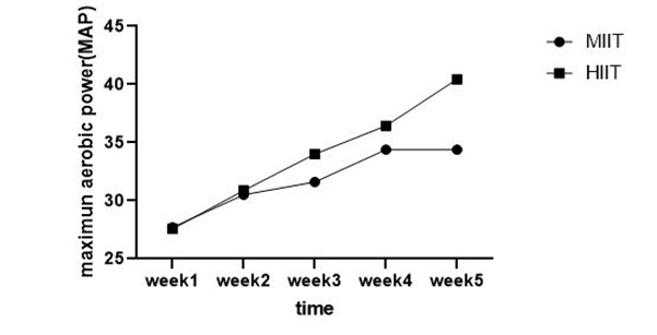
The research article summarized in this text explores the relationship between exercise intensity and myostatin levels. Previous studies have reported a decrease in myostatin levels and an increase in muscle mass following resistance exercise. However, limited research has focused on the association between interval training and myostatin [2, 15]. This study examines the gene expression of myostatin in the skeletal muscle of healthy rats, as previous research has shown higher myostatin expression in skeletal muscle compared to fatty tissue [15]. Additionally, other research has indicated that the expression of myostatin in fast-twitch skeletal muscles of rats is higher than in slow-twitch muscles. Therefore, this study uses the Gastrocnemius muscles of rats as a model for fast-twitch contraction tissue [16, 17]. The results of previous studies on the effects of exercise on myostatin are contradictory, with some showing no impact on myostatin levels in older adults after six weeks of high-intensity interval training (HIIT) [1, 18]. However, this current study demonstrates that HIIT leads to a reduction in myostatin expression in rats. It suggests that age is a significant factor affecting responsiveness to exercise programs, as older adults have shown less responsiveness to HIIT compared to younger individuals. Therefore, future researchers should consider the age of participants, as it may influence their response to exercise [2].
The reason for the discrepancies between these studies and the explanations provided above is not clear. However, these differences could possibly be attributed to variations in the exercise regimens used. Factors such as rest periods, repetitions, contraction intensity, and exercise status may have varied between the two studies. Additionally, the genetic background of the rats used in the studies could have also contributed to these differences. Furthermore, variations in the timing of sample collection after single-session or chronic exercise, as well as potential confounding effects of differences in myokine circulation or systemic blood clearance between individuals or different exercise samples, may also account for the disparities [7]. Other studies have observed differences in the expression of myostatin and follistatin genes, indicating that the concentration of these proteins is regulated not only at the transcriptional level but also by post-transcriptional mechanisms [13]. Therefore, one limitation mentioned is the lack of measurement of myostatin, follistatin, and atrogene protein levels, which could provide valuable insights as significant changes can occur from gene expression to protein levels. It is also worth noting that other studies have shown that significant changes can occur from gene expression to protein levels [9]. Furthermore, this study has various limitations that should be acknowledged. Specifically, we did not measure muscle cell dimensions morphologically, which prevents us from definitively stating whether hypertrophy has occurred. However, it is important to note that despite these limitations, our findings provide valuable insights into the regulatory mechanisms of myostatin and follistatin expression.