Between the 1st of March and the 25th of April 2020, 56 patients with respiratory symptoms and positivity to SARS-COV2 confirmed with RT-PCR underwent CT at our Institute. Fourteen patients underwent CTPA in the suspicion of pulmonary embolism and the other 42 patients underwent non enhanced chest CT. Fourteen patients were excluded due to severe motion artifacts at CT, with the other 42 Patients formed group COV-P (12 Females, 30 Males, mean age 64.5±13.5 years). Most of them had moderate to critical COVID–19 pneumonia, as demonstrated also by their average PaO2/FiO2 ratio 241±117 (available in 35 patients at admission). Only 12 over 42 patients had blood oxygen saturation >94% while breathing ambient air.
Group NL (17 Females, 24 Males, mean±SD age 65.3±14.5 years) included 42 patients without respiratory symptoms who underwent MDCT at our Institute for follow-up for pathologies not involving the lungs; 30 patients underwent the exam before December 2019, and 12 patients between the 4th and the 7th of May.
Group DTE (18 females, 23 males, mean age 66.0±17.1 years) included 42 patients who underwent CTPA in the suspicion of pulmonary embolism with evidence of endoluminal defects not involving the main pulmonary arteries but only their distal branches.
Group Bact-P (14 females, 28 males, mean age 59.8±13.7 years) included 42 Patients who underwent MDCT of the chest for pulmonary infection, and in whom a bacterium was isolated at blood cultures or BAL.
Group Fung-P (9 females, 33 males, mean age 57.0±14.7 years) included 42 patients who underwent MDCT of the chest in the suspicion of pulmonary infection and in whom a fungus was isolated at blood cultures or BAL or beta-glucan positivity was demonstrated.
Age but not sex was slightly different between the groups (p<0.05 for Fung-P vs. DTE and NT, for Bat-P vs DTE and NT, for COV-P vs Fung-P).
The V/B RATIO of the segmental branches of healthy lung parenchyma in group COV-P was significantly higher than the one of all the other groups (p<0.0001; table 1, graph1, Fig.1). The V/B RATIO of the subsegmental branches of healthy lung parenchyma in group COV-P was the highest between the groups and significantly superior to that of groups NL, DTE, and Fung-P (p≤0.0001; table 1, graph 2).
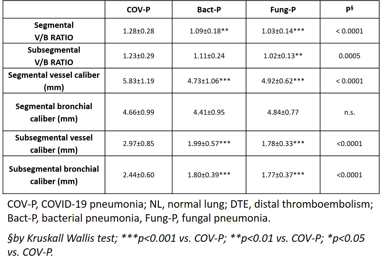
When analyzing the measure of the segmental V/B RATIO in patients of groups COV-P, Bact-P and Fung-P in the area of lung opacification, the values in group COV-P were always the highest than the corresponding ones in the two other groups (table 2, graphs 3, Fig.1).
V/B RATIO values of the subsegmental branches in the area of lung opacification in group COV-P were the highest between the groups and significantly superior to that of Fung-P (p = 0.0003; table 2, graph 4).
Among the groups of patients with pulmonary infections (COV-P, Bact-P and Fung-P), considering the caliber of the segmental and subsegmental branches of pulmonary arteries in the area of lung opacification, the calibers in group COV-P were significantly higher than those in groups Bact-P and Fung-P. No significantly difference was found in segmental bronchi calibers, while the caliber of subsegmental bronchi was significatively higher in group COV-P than in groups Bact-P, and Fung-P (p<0.0001).