Study patients with PC and pancreatic tissue samples
Chronic pancreatitis tissue samples (15) and 40 pancreatic cancer and accompanying non-tumour tissue samples were obtained from patients at the First Affiliated Yijishan Hospital of Wannan Medical College (Wuhu, Anhui province, China) between October 2020 and September 2022. Simultaneously, 40 PC, 15 CP and 40 NP samples were gathered. The clinicopathological data for the patients with PC in this study are presented in Table 1. PC and corresponding paracancerous tissues were collected from surgical resections or biopsies of patients undergoing palliative surgery using a perioperative biopsy gun (MC1816, BARD Magnum, NJ, USA). CP tissues were obtained from patients who had undergone surgery for pancreatic duct stones.
Table 1
Clinicopathological correlations of PC patients according to LncRNA-MANCR expression levels.
| Feature | LncRNA-MANCR expression | p-value |
| Low | High | Total |
| Total cases | 20 | 20 | 40 | |
| Age(y) | | | | |
| ≤ 60 | 11 | 13 | 24 | 0.206 |
| > 60 | 9 | 7 | 16 | |
| Gender | | | | |
| Female | 8 | 11 | 19 | 0.752 |
| Male | 12 | 9 | 21 | |
| Tumor size( cm ) | | | | |
| ≤ 2 | 12 | 6 | 18 | 0.527 |
| > 2 | 8 | 14 | 22 | |
| Histological Grade | | | | |
| High/Moderate | 18 | 5 | 23 | 0.343 |
| Low | 2 | 15 | 17 | |
| TNM stage | | | | |
| I ~ II | 10 | 5 | 15 | 0.114 |
| III ~ IV | 10 | 15 | 25 | |
| Lymphatic metastasis | | | | |
| Positive | 3 | 10 | 14 | 0.027* |
| Negative | 17 | 10 | 26 | |
| Vascular invasion | | | | |
| Positive | 6 | 8 | 14 | 0.058 |
| Negative | 14 | 12 | 26 | |
| Distant metastasis | | | | |
| Positive | 2 | 10 | 12 | 0.011* |
| Negative | 18 | 10 | 28 | |
Cell Culture
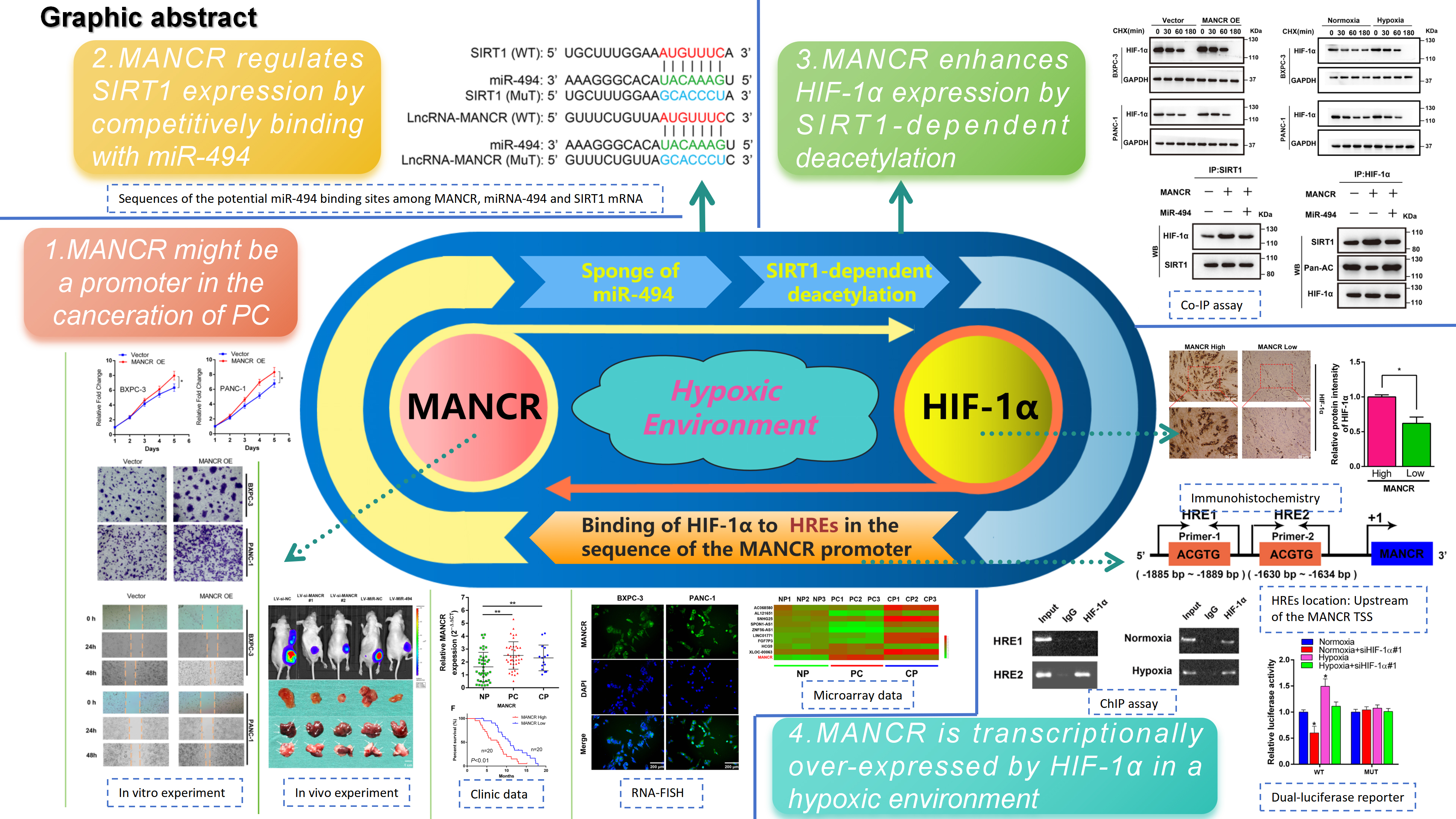
The PC cell lines (BXPC-3, PANC-1, ASPC-1, and SW1990) were obtained from the American Type Culture Collection located in Rockville, MD, USA. Human pancreatic duct epithelial (HPDE) cells were acquired from the Beijing Be-Na Culture Collection (Beijing, China). The cell lines were cultured in RPMI-1640 (Gibco, Grand Island, NY, USA) supplemented with 10% characterised foetal bovine serum (Gibco, USA) and a penicillin/streptomycin solution (Gibco, USA) under standard conditions in a 37℃ incubator (5% CO2). Hypoxic conditions were constructed as described in our previous research16,17,23 in two ways: hypoxia modular incubator chamber with 1% O2 or stationary concentration (400 mmol/L) of CoCl2 solution-mediated co-culture. The protocols are described in detail to guarantee the accuracy of the models under hypoxic conditions.
Plasmids and small interfering RNA transfection
All the small interfering RNAs (siRNAs) targeting MANCR, miR-494, HIF-1α, and SIRT1 were devised by Ribo Biological Company (Guangzhou, China). The plasmids pcDNA-MANCR, pcDNA-HIF-1α, pcDNA-SIRT1, and the miRNA-494 mimic were designed and acquired from the Gene-Chem Company located in Shanghai, China, and the empty plasmid was utilised as a negative control. Lentivirus vectors containing the MANCR-siRNA sequence (LV-siMANCR) and miRNA-494 sequence (LV-miR-494) were acquired from GeneChem (Shanghai, China). Proteins and total RNA were extracted for further analysis 48 h after transfection. The siRNA sequences used in this study are listed in Supplementary Table 1.
Reverse transcription quantitative polymerase chain reaction
RNA was extracted from tissues or cells using RNAiso Plus (TAKARA, Dalian, China) according to the manufacturer’s instructions. SYBR Premix Ex Taq II (TAKARA) was used for reverse transcription quantitative polymerase chain reaction (RT-qPCR) analysis according to the manufacturer’s protocol. All RT-qPCR data were validated in at least three independent experiments. Table S2 shows the primer sequences used in this study.
Western blot and co-immunoprecipitation
Western blot (WB) experiments were conducted in repeated trials as previously outlined23–25. The antibodies were rabbit anti-SIRT1 (1:1,000, catalog#ab189494, Abcam, Cambridge, MA, USA), HIF-1α (1:1,000, catalog#ab51608, Abcam), histone deacetylase (HDAC)1 (1:1,000, catalog# ab281585, Abcam), E-cadherin (1:1,000, catalog#ab212059, Abcam), vimentin (1:1,000, catalog#ab137321, Abcam), PAN-AC (1:1,000, catalog#A2391, ABclonal, Woburn, MA, USA), glyceraldehyde 3-phosphate dehydrogenase (1:1,000, catalog#10494-1-AP, Proteintech, Chicago, IL, USA). During the co-immunoprecipitation experiment, the lysates were incubated with control mouse/rabbit immunoglobulin (Ig)G or primary antibodies at a constant temperature of 4°C overnight. Following this, Protein A/G PLUS-Agarose (Santa Cruz, catalogue #sc2003, Helena, MT, USA) was added and incubated at 4°C for 2 h. The agarose was collected and washed with lysis buffer. Technical abbreviations have been defined for the first time. Equal sample volumes were evaluated using western blot analysis. All WB data were replicated in at least three independent experiments.
MTT assay for cell proliferation and viability
A 2,5-diphenyl-2H-tetrazolium bromide (MTT) assay was conducted to evaluate the proliferation and viability of PC cells. PC cells were seeded in a 96-well plate at a density of 2000 cells/well, viability was assessed after a 2-day incubation period, and proliferation was monitored for seven days. The cells were treated with 20 µl of MTT solution (5 mg/ml) per well for 4 h. Subsequently, the MTT solution was discarded, and 150 µl dimethylsulfoxide (Sigma-Aldrich, Santa clara, CA, USA) was added to each well. Finally, after the crystals dissolved, the absorbance was recorded at 570 nm using an ELISA reader (ThermoFisher Scientific, Waltham, MA, USA).
Wound healing and transwell invasion assays
A wound-healing assay was conducted by seeding PC cells into 12-well plates to form a confluent monolayer. The monolayer was scratched using a micropipette. Cells were grown in a serum-free solution and cell migration was detected by measuring the width of wound scratches at different time points. For the invasion assay, 5×10⁴ cells were applied to the upper side of a chamber (Corning, Corning, NY, USA) pre-coated with 200 mg/mL Matrigel (Corning). The number of cells in nine fields were then evaluated using a microscope.
Chromatin immunoprecipitation
The chromatin immunoprecipitation (ChIP) analysis was conducted using Abcam's ChIP Kit (catalogue# ab500) and anti-HIF-1α (1:1,000, catalogue#ab51608) antibody as per the manufacturer's instructions16,17. Rabbit IgG (catalogue # ab172730; Abcam) was used as the control. The bound DNA was amplified using PCR, and the resulting PCR products were separated by gel electrophoresis on a 2% agarose gel. The primer sequences are listed in Table S2.
RNA-fluorescence in situ hybridization
The fluorescence in situ hybridization (FISH) Tag™ RNA Multicolour Kit (Invitrogen, Waltham, MA, USA) and MAXIscript® Kit (ThermoFisher Scientific) were purchased and manipulated to perceive MANCR in PC cells. All RNA-FISH procedures were performed following the given instructions16,17. A green, fluorescent probe was used to identify MANCR, and 4,6-diamidino-2-phenylindole was used to stain cell nuclei.
Luciferase reporter assays
A pGL3-based luciferase reporter plasmid was utilised in this experiment to ensure binding between the 3’ untranslated region (UTR) of SIRT1 mRNA and miR-494. It contained the potential binding sequence of the 3′UTR of SIRT1 mRNA, in both the wild type (WT) and mutant type (MUT) variations. Co-transfection of these plasmids was performed with miR-494 mimic, inh-miR-494, MANCR-siRNA, pcDNA-MANCR, and their respective NC, into BXPC-3 cells in a 96-well plate. To assess MANCR promoter activity, BXPC-3 cells were transfected with a pGL3-based plasmid containing the promoter sequence that included the hypoxia response element (HRE) sequence (WT) or the mutant HRE sequence (MUT). Additionally, HIF1α-siRNA and the corresponding NC were co-transfected. All experiments were independently conducted in triplicate.
Immunohistochemistry
Sections (4-µm thick) of pancreatic tissues embedded in paraffin blocks were obtained from patients diagnosed with pancreatic cancer. A previously described method17 shows that experience summarisation includes tissue dehydration, dewaxing, and rehydration. Anti-SIRT1 antibody (1:1,000, catalogue #ab189494, Abcam) or anti-HIF-1α antibody (1:1,000, catalogue #ab51608, Abcam) and horse radish peroxidase-conjugated secondary antibody (1:200, rabbit anti-goat, catalogue #PR30011, Proteintech) were incubated with tissues. Immunohistochemical staining was performed under a biological microscope (Olympus, Tokyo, Japan). Each sample's intensity score was recorded as previously described17 on a scale of 0–3: 0, negative (no colour); 1, weak (pale yellow); 2, moderate (yellow); and 3, strong (brown). A total score > 3 in a section was considered as a positive expression.
Xenograft Assay
Four-week-old male BALB/c hairless mice aged 4 weeks (n = 5 per group) were obtained from the Beijing HFK Bio-Technology Laboratory. To demonstrate the in vivo effects of MANCR and miR-494, we subcutaneously implanted BXPC-3 cells (2 × 10⁶ cells/mouse) that had been stably transfected with lentivirus containing negative control (LV-control), si-LV-MANCR#1, si-LV-MANCR#2, LV-miR494, or sequences into the right flank of nude BALB/c mice. Each group comprised five mice kept for 27 days. Tumour weight and volume (0.5 × L × W2; where L is length and W is width) was measured and recorded every three days. Tumour tissues, lungs, and liver samples were excised from the mice and subsequently stained with haematoxylin and eosin (H&E). Rigorous procedures were employed throughout the experiments to ensure accuracy, and the expression of MANCR and SIRT1 was measured in five sections of metastatic tissue from each mouse.
Statistical analysis
SPSS v21.0 software (IBM, Chicago, IL, USA) was used for statistical analyses and the results were expressed as the means ± standard deviation. The Student's t-test was conducted to evaluate the disparities between the two cohorts. The correlation between MANCR and clinical characteristics of PC tissue were examined by conducting χ2 tests. In addition, Spearman’s correlation analysis was used to determine the correlation between MANCR and SIRT1, HIF-1α, and miR-494. Furthermore, the survival of patients with PC was recorded using Kaplan–Meier analysis with log-rank tests. All graphs were generated using GraphPad Prism 6 software (GraphPad Software Inc., San Diego, CA, USA).