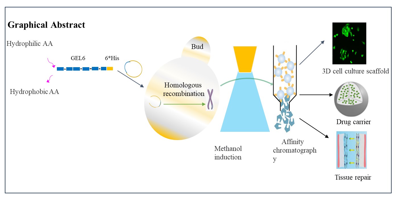
Construction of recombinant strains
The expression vector pPICza-B-gel6 was extracted, and subsequent double digestion with Xho I and Not I yielded two DNA bands that matched the expected sizes (1294bp and 3519bp), confirming the correct construction of the expression vector containing the gel6 gene (Figure 2A). Afterwards, this properly constructed vector was linearized and introduced into GS115 for transformation. The results of pPICZα-gel6 linearization are presented in Figure 2B, demonstrating successful linearization. Subsequently, the linearized vector was electro-transformed into GS115 and plated on YPDZ medium supplemented with 200μg/mL Zeocin, followed by incubation at 28℃ for 2~3 days. After monoclonal growth, a total of 20 clones were randomly selected for PCR amplification. Agarose gel electrophoresis analysis (Figure 2C) confirmed the presence of an amplified fragment consistent with the theoretically predicted size of 540bp.
From these identified clones, ten single colonies were randomly chosen and inoculated into a culture medium (2mL) for induction and cultivation. Following a culture period of 48 hours, protein secretion and expression were assessed using SDS-PAGE and Western blot techniques. Consistent with these analyses, all ten clones expressed the target protein at a molecular weight slightly larger than the expected value of 35.67kDa (Figure 3). Among them, clone (4#) exhibited higher expression levels and was subsequently utilized for shaking flask fermentation.
Expression and purification of rhGEL6
The clones exhibiting the highest level of expression were selected for shaker culture (culture system: 200 mL) and induced for 72 h. SDS-PAGE and Western blot were used to assess rhGEL6 purification and protein secretion, respectively (Fig.3). According to the results of SDS-PAGE, only one protein band with a molecular weight of 55kDa was observed in the elution sample, indicating high protein purity (Fig.4). Additionally, based on the Western blot analysis of both the cultured supernatant and induced intracellular products, it was found that most of the target proteins were secreted into the fermentation supernatant, while a small fraction remained within the cells (Fig.4). The purified protein concentration measured by BCA assay was determined to be 0.95 mg/mL. These findings demonstrate that nearly all rhGEL6 designed in this study was successfully secreted extracellularly by P. Pastoris. Furthermore, it is noteworthy that the purified protein concentration achieved in our 200 mL culture system reached an impressive value of 0.95 mg mL-1, surpassing previously reported maximum expression levels documented in literature.
Characterization
Amino acid analysis of rhGEL6
The analysis of amino acid composition can be utilized to comprehend the chemical structural characteristics of polypeptide proteins and provide scientific insights for investigating protein structure and function. The results show that the observed number and molar percentage of amino acids in rhGEL6 are in accordance with theoretical expectations, as presented in Table 3 (Additional file 2: Fig. S2, Tables S2).
Table3 Amino acid analysis of rhGEL6
|
Amino
acid
|
AA number
of theories
|
Measured sample
|
|
AA
mole
|
AA
number
|
Error
(%)
|
Mole
(%)
|
|
Asp
|
24
|
921.92 a
|
23.64 a
|
-1.49a
|
5.21 a
|
|
Glu
|
48
|
1800.43 b
|
46.17b
|
-3.81b
|
10.17b
|
|
Ser
|
48
|
1769.81
|
45.39
|
-5.44
|
10.00
|
|
Gly
|
210
|
9147.96
|
234.61
|
11.72
|
51.67
|
|
His
|
6
|
269.15
|
6.90
|
15.04
|
1.52
|
|
Arg
|
30
|
1234.96
|
31.67
|
5.57
|
6.98
|
|
Thr
|
0
|
-
|
-
|
-
|
-
|
|
Ala
|
12
|
506.95
|
13.00
|
8.34
|
2.86
|
|
Pro
|
36
|
1538.87
|
39.47
|
9.63
|
8.69
|
|
Tyr
|
0
|
-
|
-
|
-
|
-
|
|
Val
|
0
|
-
|
-
|
-
|
-
|
|
Met
|
0
|
-
|
-
|
-
|
-
|
|
Cys
|
0
|
-
|
-
|
-
|
-
|
|
Ile
|
0
|
-
|
-
|
-
|
-
|
|
Leu
|
0
|
-
|
-
|
-
|
-
|
|
Phe
|
0
|
-
|
-
|
-
|
-
|
|
Trp
|
0
|
-
|
-
|
-
|
-
|
|
Lys
|
12
|
513.99
|
13.18
|
9.85
|
2.90
|
a, b Asn and Gln will generate Asp and Glu, respectively, after acid hydrolysis
treatment, so the values in the table are Asn + Asp and Gln + Glu
Molecular weight determination of rhGEL6
Molecular mass is a crucial characteristic parameter for protein samples, serving as a prerequisite for the identification of new proteins and subsequent research. In this study, the theoretical molecular weight of rhGEL6 was calculated to be 36.49 kDa; however, SDS-PAGE results (Fig.3 and Fig.4) indicated that the target protein's molecular weight ranged from 43-55 kDa. Therefore, MALDI-TOF/TOF molecular weight determination was performed on the sample to identify its actual molecular weight, which turned out to be consistent with the theoretical value (36462 Da) shown in Figure 5. The apparent discrepancy between electrophoresis and actual molecular weights may be attributed to gelatin's Gly-X-Y amino acid composition.
The observed apparent molecular weight of the target band is larger than the theoretical value (36.49 kDa), which is commonly observed in other studies related to recombinant protein expression (Butkowski, Noelken, & Hudson, 1982; Zhi Xiang Xiang et al., 2022). A reasonable explanation for this discrepancy is that gelatin contains a substantial number of hydrophilic residues, leading to reduced binding affinity between the protein and SDS and a decreased negative charge on the target protein. Consequently, this results in slower migration rate and shorter migration distance (Ponnuswamy, Prabhakaran, & Manavalan, 1980; Toshihiko, HAYASHI, Yutaka, & NAGAI, 1980). Moreover, yeast possesses a comprehensive post-modification system where glycosylation, phosphorylation, and hydroxylation of specific residues contribute to an increase in apparent molecular weight. To confirm whether this band indeed represents the target protein, western blotting analysis was conducted. In Figure 3A, no expression was detected in the control group while a distinct band appeared at 55 kDa in the experimental group, providing evidence that this band corresponds to our intended target protein.
SEM analysis of the rhGEL6 hydrogel
The Eppendorf tube containing 200 µL of rhGEL6 solution was incubated for over 30 mins, resulting in the formation of hydrogel at the bottom of the tubes (Fig. 6A). Figure 6 presents two SEM spectra with magnifications of 500 or 2000 times, revealing a distinct porous structure in the rhGEL6 hydrogel. The pore size ranges up to 100μm, predominantly falling within the range of 30-70 μm. Moreover, the average diameter of the interconnected hydrogel measures between 2- 3μm.
The 3D cell culture of the rhGEL6 hydrogel
The findings from Calcein-AM/PI fluorescence staining demonstrated that HepG2 cells cultivated in two-dimensional conditions displayed a spindle-shaped morphology, while those grown in three-dimensional settings primarily exhibited a rounded shape. Inside the hydrogel scaffold, the cells underwent proliferation and gradually formed clusters over time (see Fig. 7). The three-dimensional culture environment facilitated enhanced cell viability, indicated by an augmented count of viable cells with green fluorescence and an absence of deceased ones with red fluorescence. These observations underscored the exceptional biocompatibility of hydrogel as a scaffold for three-dimensional cell culture.
Compared to natural type III collagen, the rhGEL6 exhibits enhanced water solubility due to its higher glycine, serine, arginine, and lysine content. Additionally, it possesses a greater positive charge as its theoretical isoelectric point (pI) closely aligns with that of the cell adhesion coating agent poly-lysine. This property promotes cell adhesion in culture and improves the survival rate of primary cells. Furthermore, rhGEL6 has self-crosslinking properties that enable it to form a hydrogel with a distinct three-dimensional porous network structure. These characteristics indicate its potential as a biomedical material and lay the foundation for its application in tissue engineering.