1. Screening of nanoemulsions (NEs) and nanocapsules (NCs)
Considering that our overall objective was the production of a library of NEs and polymeric NCs for the efficient delivery of mRNA, as a first step to the development of nasal formulations, a wide range of components that could potentially enhance immune responses was selected. For the selection of adequate candidates, we proposed several requirements, including (A) particle size below 200 nm, preferably close to 100 nm; (B) uniform particle size distribution; (C) high mRNA association capacity; (D) stability during storage or lyophilization potential; and (E) alignment with regulatory requirements (e.g. low ethanol content, or the use of compounds already approved).
In the case of NEs, different combinations and molar ratios between the different components were explored, as depicted in Table 1. These nanocarriers were composed of a combination of cationic or ionizable lipids (DOTAP and C12-200, respectively), a helper lipid (DOPE), an oil (Vitamin E or Captex800 NF), and a surfactant (Tween® 80). These different lipidic components were combined in different proportions and the resulting prototypes were analyzed in terms of mRNA encapsulation, particle size, colloidal stability, or transfection efficiency.
Table 1. Summary of the lipid compositions and molar ratios betwe e n the different components of the nanoemulsions investigated
|
Code
|
Lipid composition
|
Molar composition (%)
|
|
NE-1
|
DOTAP: DOPE: Vit E
|
16.8: 15.7: 67.5
|
|
NE-2
|
40.7: 11.2: 48.1
|
|
NE-3
|
DOTAP: DOPE: Vit E: T80
|
38.1: 10.5: 45.1: 6.4
|
|
NE-4
|
17.3: 8.1: 69.7: 4.9
|
|
NE-5
|
46.2: 9: 35.6: 9.3
|
|
NE-6
|
DOTAP: Captex8000 NF: T80
|
29: 43: 28
|
|
NE-7
|
DOTAP: DOPE: Captex8000 NF
|
22.5: 10.6: 66.9
|
|
NE-8
|
DOTAP: DOPE: Captex8000 NF: T80
|
25.5: 12: 37.8: 24.7
|
|
NE-9
|
21.1: 9.9: 59.7: 9.4
|
|
NE-10
|
29.9: 8.8: 53: 8.3
|
|
NE-11
|
DOTAP: C12-200: DOPE: Captex8000 NF: T80
|
20.4: 9.6: 3.1: 57.8: 9.1
|
|
NE-12
|
19.8: 6.1: 9.3: 56: 8.8
|
|
NE-13
|
4: 16: 10: 60: 10
|
Abbreviations: C12-200: 1,1’-((2-(4-(2-((2-(bis(2-hydroxydodecyl)amino)ethyl) (2-hydroxydodecyl)amino) ethyl)piperazin-1-yl)azanediyl)bis(dodecan-2-ol) DOPE: 1,2-dioleoyl-sn-glycero-3-phosphoethanolamine. DOTAP: 1,2-dioleoyl-3-trimethylammonium propane. NE: nanoemulsion. T80: Tween® 80. Vit E: D, L-α-tocopherol.
Following mRNA entrapment, these NE-mRNA carriers were used for the preparation of NC-mRNA nanosystems, by adding a polymeric layer over the pre-formed NE-mRNAs. The nomenclature for the resulting NC-mRNAs was derived from the initial NE name code, for easy identification of the core composition. As detailed Table 2, different polymers were explored, including protamine (PR), dextran sulphate (DX), chitosan (CS) and PEG-PGA (PP), aiming to modify the surface of the nanocarriers and enhance their uptake by dendritic cells and, hence, their immunogenic properties. These polymers have previously been used as adjuvant and antigen delivery systems, capable of activating different T and B cell responses [28–32].
Table 2. Summary of the lipid and polymeric compositons, and the molar and weight-to-weight ratio between the different components of the nanoocapsules investigated
|
Code
|
Initial NE
|
Molar composition (%)
|
Polymer coating
|
mRNA:polymer (w/w)
|
|
NC-1-PR
|
DOTAP: DOPE: Vit E
|
16.8: 15.7: 67.5
|
Protamine
|
1:1
|
|
NC-3-PR
|
DOTAP: DOPE: Vit E: T80
|
38.1: 10.5: 45.1: 6.4
|
1:1
|
|
NC-4-PR
|
17.3: 8.1: 69.7: 4.9
|
1:1
|
|
NC-7-PR
|
DOTAP: DOPE: Captex8000 NF
|
22.5: 10.6: 66.9
|
1:1
|
|
NC-8-PR
|
DOTAP: DOPE: Captex8000 NF: T80
|
25.5: 12: 37.8: 24.7
|
1:1
|
|
NC-9-PR
|
21.1: 9.9: 59.7: 9.4
|
1:1
|
|
NC-3-DX
|
DOTAP: DOPE: Vit E: T80
|
38.1: 10.5: 45.1: 6.4
|
Dextran Sulphate
|
1:1
|
|
NC-4-DX
|
17.3: 8.1: 69.7: 4.9
|
1:1 or 1:2
|
|
NC-5-DX
|
46.2: 9: 35.6: 9.3
|
1:2
|
|
NC-3-CS
|
DOTAP: DOPE: Vit E: T80
|
38.1: 10.5: 45.1: 6.4
|
Chitosan
|
1:1
|
|
NC-4-CS
|
17.3: 8.1: 69.7: 4.9
|
1:1
|
|
NC-4-PP
|
DOTAP: DOPE: Vit E: T80
|
17.3: 8.1: 69.7: 4.9
|
PEG-PGA
|
1:12
|
|
NC-5-PP
|
46.2: 9: 35.6: 9.3
|
1:12
|
Abbreviations: CS: chitosan. DOPE: 1,2-dioleoyl-sn-glycero-3-phosphoethanolamine. DOTAP: 1,2-dioleoyl-3-trimethylammonium propane. DX: dextran sulphate. NC: nanocapsule. NE: nanoemulsion. PEG-PGA or PP: PEG (5 kDa)-b-PGA (10) Na. PR: protamine. T80: Tween® 80. Vit E: D, L-α-tocopherol. w/w ratio: weight-to-weight ratio between polymer and mRNA content.
Table 3. Physicochemical properties of blank NEs.
|
Code
|
Size (nm)
|
PDI
|
Z-Pot (mV)
|
|
NE-3
|
102 ± 13
|
0.40 ± 0.05
|
+56 ± 5
|
|
NE-4
|
104 ± 15
|
0.30 ± 0.04
|
+55 ± 3
|
|
NE-5
|
Formulated in a single step by microfluidics
|
|
NE-9
|
108 ± 9
|
0.15 ± 0.03
|
+56 ± 8
|
|
NE-10
|
Formulated in a single step by microfluidics
|
|
NE-11
|
113 ± 6
|
0.20 ± 0.01
|
+58 ± 2
|
|
NE-12
|
131 ± 15
|
0.16 ± 0.01
|
+56 ± 2
|
|
NE-13
|
Formulated in a single step by microfluidics
|
Abbreviations: NE: nanoemulsion. PDI: polydispersity index. Values represent the mean ± standard deviation (n ≥ 3, unless indicated otherwise).
1.1. Preparation of mRNA-loaded nanoemulsions (NEs)
NE-mRNAs were prepared using two distinct strategies. In the first approach, blank NEs were prepared by solvent-displacement technique, resulting in nanodroplets with a size in the range of 100 to 130 nm and a highly positive surface charge (Table 3). In a second step, mRNA was complexed onto the NE by electrostatic interactions between the positive charge of the cationic lipid and the negative charge of the nucleic acid. On the other hand, certain prototypes were prepared in a single-step microfluidic mixing of both aqueous and organic phases, leading to the simultaneous formation of the NEs and the complexation of mRNA. These two formulation strategies were performed with different mRNAs, including mGFP, mLuc, and mOVA, leading to different physicochemical characteristics (Table 4). Overall, the use of microfluidic mixing led to the formation of nanostructures with a smaller particle size (60-100 nm) than the bulk mixing of components (100-150 nm). These variations suggested potential changes in the internal structure of the NEs and the organization of the mRNA molecules, potentially due to the rapid mixing enabled by the microfluidic device.
Furthermore, as expected, the incorporation of negatively charged mRNA onto the surface of the previously formed blank NEs led to a significant reduction of their positive charge. The resulting surface charges of NE-mRNAs were slightly negative at low N/P ratios, and highly positive at high N/P ratios. The N/P ratio also influenced the EE% values, with higher values obtained at a higher N/P ratio. For example, NE-1, -6, -7, and -8 were prepared at an N/P ratio of 0.6:1, leading to particle sizes between 115-150 nm, negative surface charges (-5 to -10 mV), and low EE% values (below 50% in all cases). Additionally, different types of mRNA exhibited different particle diameters and PDI due to their distinct interaction with the cationic DOTAP lipid.
Selected NE-mRNAs were subjected to a lyophilization process, intended to preserve the physicochemical properties of the nanosystems and the integrity of the mRNA cargo upon long-time storage (Supplementary Table 1). Different cryoprotectants, added at different percentages, were tested. In general, the incorporation of sucrose (10-20% w/v) allowed the maintenance of the physicochemical properties of the NE-mRNAs upon resuspension, suggesting the possibility of long-term storage for these nanocarriers.
Table 4. Physicochemical properties of NE-mRNA nanocarriers, using different types of mRNAs and different N/P ratios.
|
Code
|
Type of mRNA
|
Formulation method
|
N/P ratio
|
Size (nm)
|
PDI
|
ζ-Potential (mV)
|
EE (%)
|
|
NE-3
|
mGFP
|
Bulk mixing
|
2:1
|
108 ± 13
|
0.14 ± 0.03
|
+39 ± 2
|
100
|
|
NE-4
|
mGFP
|
Bulk mixing
|
2:1
|
127 ± 13
|
0.09 ± 0.03
|
+43 ± 4
|
100
|
|
mLuc
|
Bulk mixing
|
2:1
|
138 ± 18
|
0.14 ± 0.06
|
+42 ± 2
|
100
|
|
mLuc
|
Bulk mixing
|
4:1
|
89 ± 2
|
0.21 ± 0.02
|
+48 ± 6
|
100
|
|
mLuc
|
Microfluidics
|
4:1
|
84 ± 11
|
0.21 ± 0.02
|
+54 ± 3
|
100
|
|
mOVA
|
Bulk mixing
|
2:1
|
121 ± 9
|
0.10 ± 0.01
|
+40 ± 3
|
100
|
|
NE-5
|
mGFP
|
Microfluidics
|
4:1
|
62 ± 13
|
0.24 ± 0.07
|
+53 ± 7
|
100
|
|
NE-9
|
mGFP
|
Bulk mixing
|
4:1
|
97 ± 1
|
0.19 ± 0.02
|
+51 ± 3
|
100
|
|
mLuc
|
Bulk mixing
|
4:1
|
104 ± 1
|
0.17 ± 0.01
|
+52 ± 2
|
100
|
|
NE-10
|
mGFP
|
Bulk mixing
|
4:1
|
65 ± 1
|
0.2 ± 0.01
|
+44 ± 1
|
100
|
|
NE-11
|
mGFP
|
Microfluidics
|
4:1
|
107 ± 3
|
0.18 ± 0.02
|
+53 ± 2
|
100
|
|
NE-12
|
mGFP
|
Bulk mixing
|
4:1
|
123 ± 2
|
0.17 ± 0.01
|
+49 ± 1
|
100
|
|
NE-13
|
mOVA
|
Microfluidics
|
2:1
|
116 ± 1
|
0.17 ± 0.01
|
+49 ± 1
|
75
|
Abbreviations: EE: encapsulation efficiency. mGFP: mRNA encoding for GFP. mLuc: mRNA encoding for luciferase. mOVA: mRNA encoding ovalbumin protein. NE: nanoemulsion. N/P ratio: nitrogen to phosphate ratio. PDI: polydispersity index. Values represent the mean ± standard deviation (n ≥ 3, unless indicated otherwise).
1.2. Preparation of mRNA-loaded nanocapsules (NCs)
The resulting NE-mRNA nanocarriers were subsequently coated with various polymers to form a polymer shell, intended to modulate the surface properties of the resulting NC-mRNAs (Table 5). Different w/w ratios between the polymer and the mRNA were evaluated for each NC-mRNA nanosystem, based on the amount of polymer required to effectively modify the surface properties while preserving particle stability. The formation of the polymer shell around the oily nanodroplets led to significant changes in the physicochemical properties. In order to facilitate the interaction of polymers with NE-mRNAs formulations, the net charge of NE-mRNAs was adjusted by the N/P ratio. Namely, NC-PR-mRNAs and NC-CS-mRNAs were prepared at N/P ratios between 0.6:1 to 0.9:1, resulting in nanoparticles of 130-190 nm, low PDI and positive surface charges (+25 to +30 mV), ensuring anionic surface charge on the NE-mRNAs and allowing the adequate absorption of the positively charged polymers.
Overall, the particle diameters observed in the NC-mRNA nanosystems ranged from 70 to 240 nm, primarily dependent on the method used for the initial preparation of NE-mRNAs. Significant differences were observed for different mRNA molecules (e.g. overall, particle size observed when mLuc is encapsulated is greater than when other mRNAs are used). This suggests a different interaction between the NE, the mRNA and the polymer, mainly dependent on the type of mRNA used.
Selected NC-mRNAs were lyophilized as an assessment of their long-term preservation upon storage in these conditions (Supplementary Table 1). After screening of different cryoprotectants, a preliminary preference by certain polymers was observed. For example, NC-PR-mRNAs were capable of maintaining their physicochemical properties when using trehalose 10%, while this behavior was observed in NC-DX-mRNAs when lyophilized with sucrose 10%.
Table 5. Physicochemical properties of NC-mRNA nanocarriers, using different types of mRNA, polymers and RNA to polymer ratios.
|
Code
|
Type of mRNA
|
RNA:polymer ratio (w/w)
|
Size (nm)
|
PDI
|
Z-Pot (mV)
|
EE (%)
|
|
NC-3-PR
|
mGFP
|
1:1
|
152 ± 9
|
0.12 ± 0.01
|
+24 ± 1
|
100
|
|
NC-4-PR
|
mGFP
|
1:1
|
153 ± 16
|
0.11 ± 0.03
|
+27 ± 2
|
75
|
|
NC-3-DX
|
mGFP
|
1:1
|
144
|
0.09
|
-26
|
100
|
|
NC-4-DX
|
mLuc
|
1:1
|
154 ± 8
|
0.14 ± 0.04
|
-14 ± 1
|
100
|
|
1:2
|
126 ± 3
|
0.11 ± 0.02
|
-21 ± 2
|
100
|
|
mOVA
|
1:2
|
132 ± 3
|
0.07 ± 0.03
|
-16 ± 2
|
100
|
|
NC-5-DXm
|
mGFP
|
1:2
|
93 ± 9
|
0.2 ± 0.01
|
-6 ± 7
|
100
|
|
NC-5-PPm
|
mGFP
|
1:12
|
70 ± 1
|
0.17 ± 0.01
|
+13 ± 5
|
-
|
Abbreviations: CS: chitosan. DX: dextran sulphate. EE: encapsulation efficiency. m: base NE-mRNA prepared by microfluidics. mGFP: mRNA encoding for GFP. mLuc: mRNA encoding for luciferase. mOVA: mRNA encoding ovalbumin protein. NC: nanocapsule. PDI: polydispersity index. PP: PEG-PGA or PEG (5kDa)-b-PGA (10) (Na). PR: protamine sulphate EP. w/w ratio: weight-to-weight ratio between polymer and mRNA coating. Values represent the mean ± standard deviation (n ≥ 3, unless indicated otherwise).
2. In vitro assessment of cytotoxicity and transfection efficiency of NE-mGFP and NC-mGFP
HeLa cells were selected as a model for easy transfection and evaluation of cellular toxicity. These cells have also been used in different preclinical vaccine studies [33,34]. Different NE-mGFP and NC-mGFP candidates were incubated with HeLa cells for 4 hours, and toxicity and efficient translation into the florescence reporter protein were assessed 24 hours after transfection.
The results depicted in Figure 1 show a concentration-dependent toxicity in the range evaluated, with a maximum reduction in viability of approximately 25% observed at the highest doses investigated (200 and 100 ng of mRNA) for most of the tested nanosystems. No significant differences in cell viability were observed among the different NE-mGPF and NC-mGFP formulations tested. Notably, NC-mGFP exhibited slightly lower toxicity compared to NE-mGFP (for instance, NC-4-PR, NC-4-DX, and NC-4-CS resulted in better cell viability profiles than NE-4; and NE-9 showed lower viability than NC-9-PR). These findings suggest that the polymeric shell surrounding the NE-mGFP to form the NC-mRNA prototypes contributes to reducing the overall high surface charge of the NE-mGFP. Additionally, NC-mGFP with more neutral surface charges has the potential to yield better cell viability profiles. This reduction of cellular viability, primarily driven by the positive charge, aligns with previously reported findings [35,36].
To evaluate transfection efficiency, two metrics were utilized: the percentage of GFP-positive cells, indicating the proportion of cells capable of expressing GFP; and mean fluorescence intensity (MFI), which quantifies the amount of fluorescence emitted by these cells. In general, NE-mGFP formulations (depicted in Figure 2, top) exhibited higher percentages of GFP-positive cells and MFI compared to NC-mGFP formulations, especially at the lower RNA concentrations (Figure 2, bottom). In all instances, a notable dose-dependent increase in transfection efficiency was observed. Among the various NE-mGFP formulations, NE-3-mGFP, NE-4-mGFP, and NE-9-mGFP showed the highest transfection efficiency. This might be related to the presence of Tween® 80 in their composition and, also, to the appropriate combination of DOTAP and DOPE [37,38].
Interestingly, NC-mGFP formulations exhibited different behavior depending on the polymer shell, with different significances depending on the concentration tested. For instance, both NC-3-PR-mGFP and NC-3-CS-mGFP exhibited superior percentages of GFP-positive cells compared to NC-3-DX-mGFP at the highest concentration tested. Furthermore, significant differences were observed among NC-PR formulations. NC-3-PR-mGFP and NC-4-PR-mGFP resulted in greater GFP-positive cell levels than NC-1-PR-mGFP, NC-7-PR-mGFP, or NC-9-PR-mGFP. These findings indicate that the NE-mGFP used to form the NC-mGFP formulations have the potential to determine their final cellular transfection potential, possible due to the lipidic composition of the oily nanodroplets, driving the internal cellular fate of the nanocarriers. On the other hand, NC-4-DX-mGFP markedly outperformed NC-3-DX-mGFP, despite both utilizing DX as the polymeric shell. These disparities between nanosystems were not evident were comparing NE-3-mGFP and NE-4-mGFP, thus suggesting that the entanglement of the polymeric shell may influence the transfection efficiency of the nanocarriers.
Selected formulations, all prepared via microfluidic mixing, were tested in HeLa cells using a larger number of cells in 24-well plates, including NE-5-mGFP, NE-9-mGFP, NE-10-mGFP, NE-11-mGFP, NE-12-mGFP, NC-5-DX-mGFP, and NC-5-PP-mGFP (refer to Supplementary Figure 2). No significant cellular toxicity was observed for none of the concentrations tested. Regarding transfection efficiency, notorious differences were found between NE-9-mGFP and NE-10-mGFP (containing DOTAP as complexing lipid) as compared with NE-11-mGFP and NE-12-mGFP (containing a combination of DOTAP and C12-200 as complexing agents). These results highlight that the combination of DOTAP with an ionizable lipid (such as C12-200) could have positive effects on the transfection efficiency of nanocarriers. C12-200 is a multi-tailed ionizable lipidoid known for its ability to adopt a cone-shaped structure, with the potential to enhance the disruption of endosomes upon cellular uptake [39,40].
3. Intramuscular administration of nanocarriers with mLuc
The preliminary assessment of the in vivo transfection efficiency of selected mRNA nanocarriers, was performed using mRNA encoding for luciferase (mLuc). Figure 3, shows the expression of the reporter fluorescence protein following intramuscular administration in mice. The results showed that, among the NE-mLuc formulations tested (NE-4-mLuc, NE-9-mLuc and NE-12-mLuc), NE-4-mLuc exhibited the highest capacity to produce luciferase. On the other hand, the response was similar for the NCs and NEs. However, when comparing formulations with the same polymer shell but different NE cores (e.g. NC-4-PP-mLuc and NC-5-PP-mLuc), some differences were observed. In line with the in vitro results (Figure 2), even when using the most potent NE-mLuc (such as NE-4-mLuc), the choice of polymer can modulate the transfection efficiency of the resulting NC-mLuc nanocarrier to some extent, although not exceeding the performance of NE-mLuc.
IVIS images corresponding to this study can be found in Supplementary Figure 3.
4. Immune response assessment of intranasal administration of NE-13 and NC-4-DX containing mOVA
Based on the synergy observed for the combination of DOTAP and C12-200 observed on in vitro studies, an optimized version of the previous NE-11 and NE-12 formulations was developed. In the earlier iterations, the quantity of DOTAP significantly outweighed that of C12-200. In the new optimized version, named NE-13, the proportion of C12-200 was substantially higher than that of DOTAP. This adjustment aimed to exploit the enhanced endosomal escape capabilities offered by ionizable lipids, such as C12-200. Upon cellular uptake, this lipid becomes protonated within acidic endosomes, thereby interacting with endosomal phospholipids. This interaction induces a phase transition from a highly stable bilayer structure to an inverted hexagonal HII phase, capable of rupturing the endosomal membrane and facilitating the release of mRNA cargo into the cytosol [41].
On the other hand, based on the aforementioned in vitro and in vivo studies, NC-4-DX emerged as a promising nanocarrier without need of subsequent optimization. Its favorable cellular safety and transfection profile, both in vitro and in vivo, coupled with its slightly negative surface charge (in contrast to the highly positive surface charges observed in NE-mRNAs), position it as a promising candidate for further exploration.
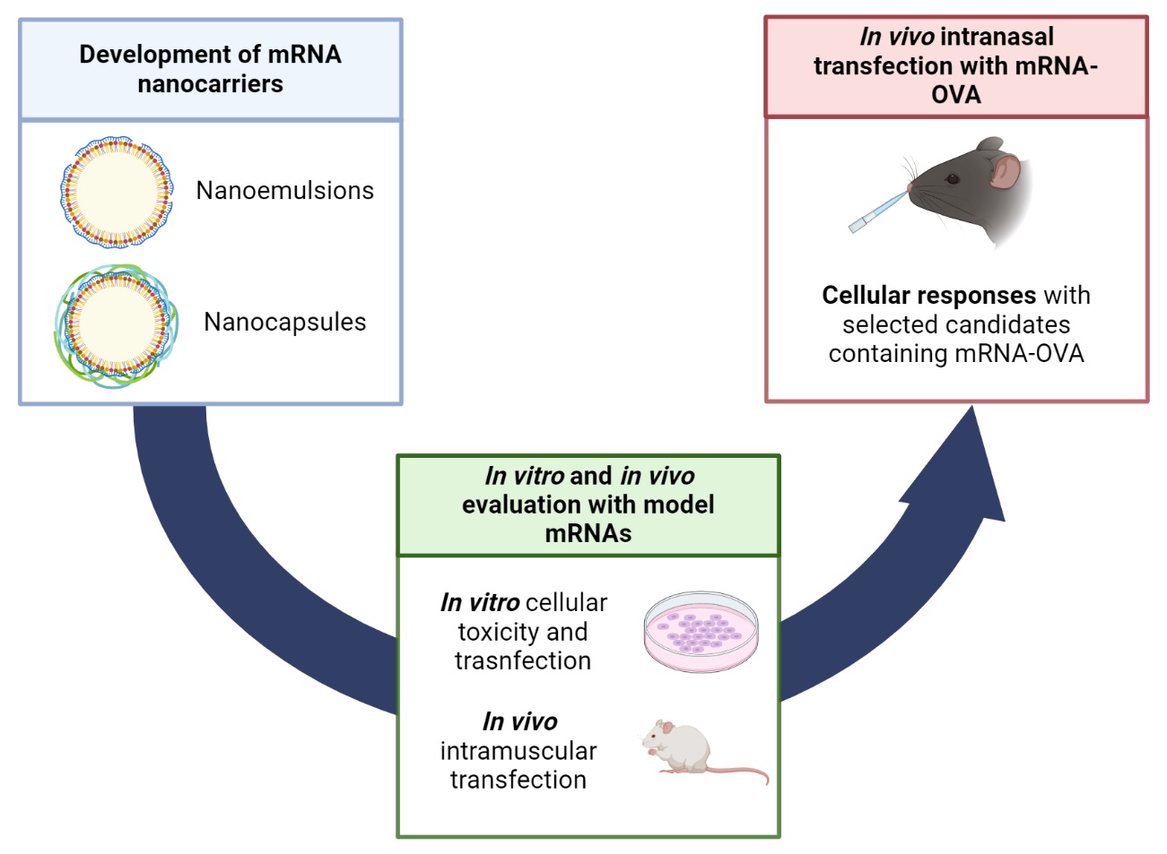
These two selected candidates were utilized to encapsulate mRNA encoding for ovalbumin (mOVA) [42]. To evaluate in vivo immune responses induced by NE-13 and NC-4-DX, animals were intranasally administered twice (day 0 and 7).
The adaptative immune system is designed to provide protection from recurring infections, and can be activated through humoral responses (mainly, antibodies) or cellular responses (T cells). Cellular response can induce specialized T cells, known as cytotoxic T cells (CTLs), or clusters of differentiation 8 (CD8+) T cells. Ideally, the nanosystems developed should be able to induce robust Major Histocompatibility Complex I (MHC-I)-mediated CD8+ T cell responses [43,44]. To evaluate this desirable immune response, detection of antigen-specific CD8+ T cells was performed by MHC I Dextramer-PE staining in PBMCs (collected on day 7, Figure 4, left) and splenocytes (collected on day 10, Figure 4, right).
In blood samples, both formulations, NE-13-mOVA and NC-4-DX-mOVA, resulted in an increase in antigen-specific CD8+ T cell numbers (gated as CD45+/CD8+/Dextramer+), as compared with the control group (Figure 4, left). However, although the percentage of antigen-specific CD8+ T cells is similar for both formulations, only NC-4-DX-mOVA has the capacity to induce a statistically significant increase. At day 10 post-administration, splenocytes were harvested and results showed a similar tendency as at day 7 (Figure 4, right). Compared with the control group, both formulations induced an in vivo immune response by increasing the percentage of OVA-specific CD8+ T cells. Further, NC-4-DX-mOVA resulted in the formulation with the highest immunological effect, as previously observed for day 7.
To measure CD8+ T cell responses, the quantity of interferon-gamma (IFN-γ) induced in splenocytes following intranasal administration of NE-13-mOVA and NC-4-DX-mOVA (Figure 5). In accordance with blood sample results, immune cells were stimulated by both NE-13-mOVA and NC-4-DX-mOVA formulations, with the latter having a higher evident stimulatory effect. Further, IFN-γ production was higher after OVA stimulation than in unstimulated immune cells for both formulations.
Overall, these results indicated that both NE-13-mOVA and NC-4-DX-mOVA are promising delivery systems for mRNA vaccination, inducing robust CD8+ T cell activation. Furthermore, this study shows consistent cellular immune responses for both OVA-specific CD8+ T cell activation and IFN-γ release, determined by Dextramer and IFN- γ ELISpot assay, two powerful technologies for accurately assessing cellular immune responses after vaccination [45].