Synthesis of SNF-Cy5.5
To maintain the original structure of peptide SNFYMPL and productive conjugation with Cy5.5-NHS, a GGGSK linker was added to the C-terminus of SNFYMPL. The peptide-linker SNFYMPLGGGSK were synthesized through using standard solid-phase fluorenylmethoxycarbonyl chemistry as previously reported (12). Cy5.5 monofunctional NHS ester was purchased from GE Healthcare (Piscataway, NJ, USA).
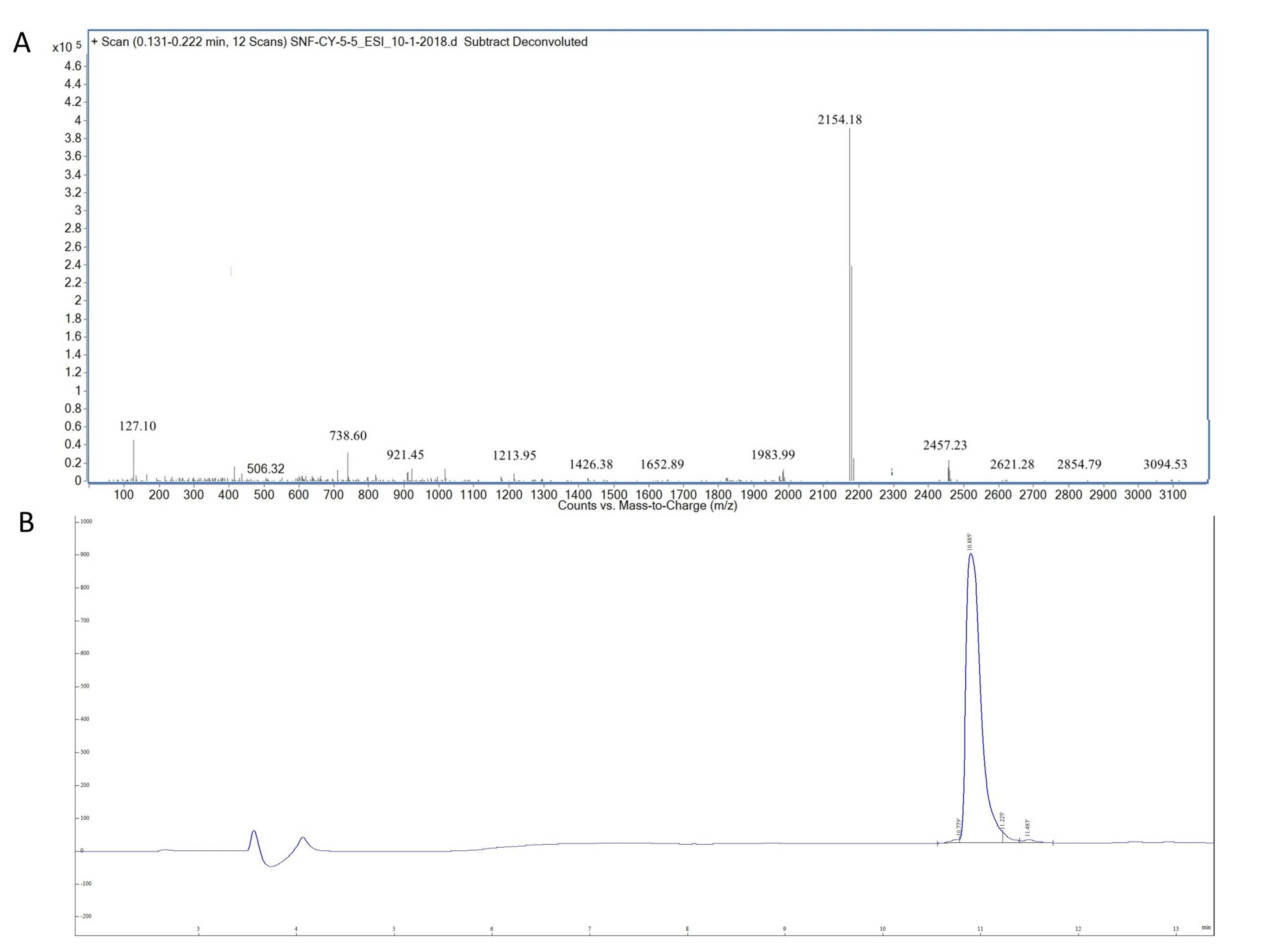
Cy5.5 monofunctional NHS ester (0.50mg, 0.443 μmol. 1 equiv.) was dissolved in 200μl of Dimethyl sulfoxide(DMSO)and mixed with SNF peptide (0.54mg, 0.443μmol, 1 equiv) in the dark. Then triethylamine (5µl) was added to the mixed solution. The reaction mixture was stirred overnight in dark at room temperature. After removal of the solvent, The crude product was dissolved in a solution of 1:1 acetonitrile and H2O and purified by HPLC with a C18 column (25 cm×10 mm) using a gradient mobile phase beginning from 5% solvent A (0.1% trifluoroacetic acid(TFA) in acetonitrile) and 95%solvent B (0.1% TFA) in water) to 70% solvent A and 30%solvent B at 30 min. Finally, the coloured peak was collected and lyophilized. The ultimate purity of the peptides was confirmed with an analytical C18-column. SNF-Cy5.5 was also analyzed by mass spectrometry.
Cell culture and condition
The human-derived esophageal adenocarcinoma cell line OE33 and immortalized epithelial cell line Het-1A were purchased from ATCC (Manassas, VA). OE33 cells were cultured in RPMI 1640 (Gibco) containing 10% FBS (Gibco), and 100 U/mL streptomycin/penicillin. Het-1A cells were cultured in Bronchial Epithelial Cell Growth Medium (BEBM), along with the additives obtained from Lonza/Clonetics Corporation as a kit (Catalog No.CC-3170). All cells were incubated at 37°C in a 5% CO2 humidified atmosphere.
Fluorescence imaging
OE33 and Het-1A cells were grown in 4-chamber slides for 24h. OE33 and Het-1A cells were grown in 4-chamber slides for 24 h, then the cells were washed 3 times in PBS and fixed with acetone on ice for 15 min. Blocking was performed by adding 10% normal rabbit serum. The cells were then incubated with 10μmol/L of the SNF-Cy5.5 at dark. In the block group, the cells were incubated with 10 μmol/l of SNF-Cy5.5 and 50 μmol/l of unlabeled SNF peptide. The cells were washed with PBS for 3 times after fluorescently-labeled peptide. 4',6-diamidino-2-phenylindole (DAPI) (Invitrogen, CA, USA) was dropped onto the cell chambers and incubated with cells for 15mins. After a drop of Prolong Gold anti-fade reagent was added onto the slides, the images were acquired using a FLUOVIEW FV1000 laser scanning confocal microscope (Olympus, Tokyo, Japan).
Flow cytometry analysis
We also performed flow cytometry to validate Preferential binding of the SNF-Cy5.5 to OE33 cells. Cell triplicates counting about 106 were prepared. OE33 cells were the cells were trypsinized, centrifuged at 800 rpm for 5 mins, then collected and washed with ice-cold PBS thrice. The cells were incubated with SNF-Cy5.5, SNF-Cy5.5 with block peptide and PBS at the concentration as previously stated for 30 minutes on ice. These cells were then washed by ice-cold PBS thrice to clean unspecific binding. Finally, fluorescence-activated cell sorting (FACS) was used to record and analyze the results.
Animal model
Athymic female nude male BALB/c mice (4–6 weeks, 20–25 g) were obtained from the Laboratory Animal Center of Fourth Military Medical University (FMMU), with the care and treatment of these animals performed in accordance with FMMU animal protocols. When female athymic nude mice grew until about 4–6 weeks old, weighing about 20–25 g, about 5 × 10⁶ OE33 cells suspended in 200 μL of serum-free RPMI 1640 were injected into the right shoulder of mice. The cells were allowed to grow 2 weeks until tumors’ size was was 5–10 mm. The animal studies were registered and approved by the Animal Welfare and Ethics Committee of Fourth Military Medical School.
Targeting ability of SNF-Cy5.5 in vivo
In vivo imaging system (IVIS) Imaging System (IVIS Kinetics, Caliper Life Sciences, Hopkinton, MA, USA), was used to assess the tumor targeting efficacy of the SNF-Cy5.5. A Cy5.5 fiter set and Identical illumination settings (eg. lamp voltage, fiters, f/stop, field of views, binning) were used to acquire all images. The mice in the experimental group (n = 3) received a 100μM of SNF-Cy5.5 tail-intravenously and then exposed to optical imaging of IVIS at various period points after injection. The mice from the block group (n = 3) received a 100μM of SNF-Cy5.5 mixed with unlabeled SNF peptide (1000μM) as previously described (5). After IRI fluorescent imaging of the tumor-bearing mice, the mice were euthanized using Carbon dioxide. Various organs, such as tumor, heart and liver, were excised and subjected to ex vivo fluorescence imaging. The bioluminescent intensities of the regions of interest (ROIs) were measured by Living Image version 4.2 Software. Fluorescent intensity was reported as photons per second per centimeter squared per steradian (p/s/cm2/sr).
Data analysis
All the data are described as means ± standard deviation (SD). Statistical analysis was performed using a Student’s t test. To determine tumor contrast, mean fluorescence intensities of the tumor (T) area at the right shoulder of the animal and of the normal tissue (N) at the surrounding tissue were calculated using the ROI function Dividing T by N yielded the contrast between tumor and normal tissue. Statistical analysis of the data was performed using SPSS 23.0 software (Chicago, IL, USA). If p-value <0.05, Statistical significance was defined.