Materials
Lecithin, Cholesterol (Aladdin), 1,2-Distearoyl-sn-glycero-3-phosphoethanolamine-N-[methoxy(polyethylene glycol)-2000] (DSPE-PEG), Atorvastatin (McLean), peptides (flame trite), N-hydroxysuccinimide (NHS), 1- Ethyl − 3- (3-dimethylaminopropyl) carbondiimide hydrochloride (EDC, Sigma). anhydrous dimethylsulfoxide (DMSO), fatty acid methyl ester sulfonate (MES), anhydrous dimethylformamide (DMF), chloroform (Tris-Hcl) Human high-oxidized low-density lipoprotein (ox-LDL), Cyanine5 NHS ester (Cy5), Recombinant murine interferon-γ (TNF-γ).
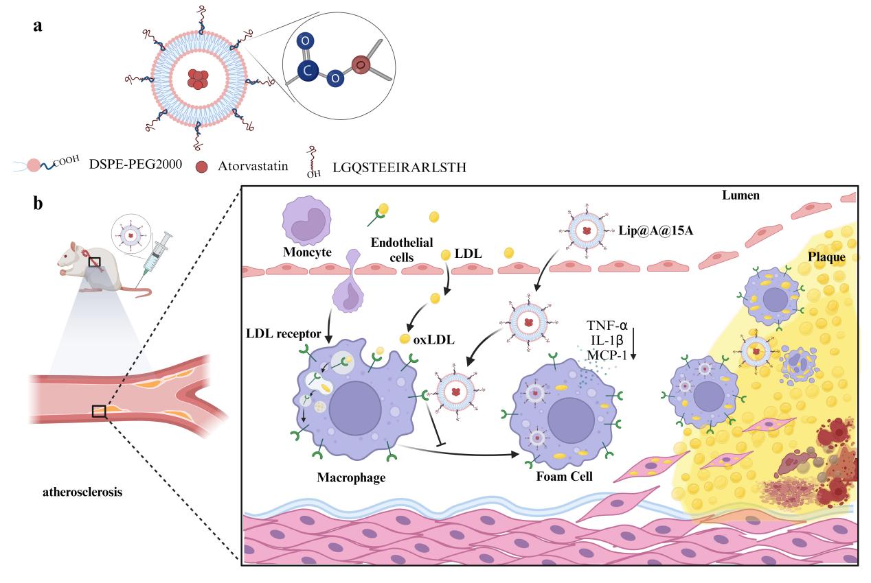
Synthesis of Atorvastatin-loaded liposome modified peptide (Lip@A@15A).
1. Synthesis of Atorvastatin loaded liposome (Lip@A). Lip@A was prepared using a thin-film dispersion method. Atorvastatin, lecithin, DSPE-mPEG-COOH, cholesterol, and chloroform were dissolved adequately by ultrasonication, followed by evaporation at 70 ℃ at 30 rpm. Then the solution was hydrated at 60 ℃ at 17 rpm for 40 min and then homogenized by ultrasonic probes for 9 min under ice bath, followed by washing. After passing through a 0.22µm microporous membrane, the Lip@A was prepared.
2. Synthesis of peptide (15A). The ApoE domain, which spans residues 136–150 in the N-terminal domain, is critical for the specific binding to LDLR. The amino acid sequence LGQSTEEIRARLSTH was queried using UniProtKB and synthesized by a company (Jiangsu Qiangyao).
3. Drug content(DC)and encapsulation efficiency(EE). The amount of Atorvastatin was determined by high-performance liquid chromatography (HPLC) using an Apollo C18 column at a UV absorption wavelength of 240 nm. The drug loading and encapsulation efficiencies of Atorvastatin in the liposomes were calculated.
4. Preparation of peptide-modified Lip@A. Lip@A was conjugated to MES and diluted with deionized water. The peptide and EDC were then added. The reaction was performed overnight at 37°C on a shaker at 150 rpm. After centrifugation and washing, the supernatant was used to measure the peptide coupling rate using bicinchoninic acid (BCA), and the remaining Lip@A@15A was collected.
Fluorescence spectra
The fluorescence spectra of Cy5 and Cy5 modified liposome (Lip@Cy5) were obtained using a fluorescence spectrophotometer.
Characterization of Nanoparticles
Size, size distribution profiles, polydispersity index (PDI), and zeta-potential of nanoparticles were quantified using a Malvern Zetasizer NanoZS instrument at 25°C. Transmission electron microscopy (TEM) was performed using a JEM-1400 PLUS microscope (JEOL, Tokyo, Japan).
Cytotoxicity Evaluation
Mouse macrophages (RAW264.7) were cultured in 96-well plates in Dulbecco’s modified Eagle’s medium (DMEM) with fetal bovine serum (FBS), penicillin, and streptomycin. After the cells were treated with various concentrations of the nanomaterials, cell viability was assayed using the Cell Counting Kit-8 assay (CCK-8).
The study of cellular uptake of nanomaterials
RAW264.7 cells were cultured in 12-well plates and incubated for 12h in medium different nanomaterials containing Cy5. After centrifugation and washing, the cells were harvested for flow cytometry. RAW264.7 cells were cultured in confocal dishes and incubated for 12h in different medium nanomaterials containing Cy5. Then RAW264.7 cells were stained with 4, 6-Diamidino-2-phenylindole (DAPI). Confocal laser scanning microscopy (CLSM) was performed to acquire fluorescent images.
Inhibition of generation of foam cells and Anti-Inflammatory functions of nanomaterials
RAW264.7 cells were cultured in 24-well plates and treated with medium, 100 ng/mL LPS and 10ng/mL IFN-γ and nanomaterials, separately. Supernatants were harvested and concentrations of Tumor Necrosis Factor-α (TNF-α), Interleukin-1β (IL-1β), and Monocyte chemotactic protein-1 (MCP-1) were detected using Enzyme-linked Immunosorbent Assay (ELISA). Finally, all groups were incubated with ox-LDL and stained with Oil Red O (ORO) and hematoxylin. The cells were observed under a light microscope.
Animals
All the procedures and protocols were approved by the Institutional Animal Ethics Committee of the National Center for Nanoscience and Technology. Male C57BL/6 and Apolipoprotein E-deficient (ApoE-/-) mice (approximately 6 weeks old) were fed a high-fat and high-cholesterol diet. Mice were purchased from Beijing Viton Lever.
Distribution and anti-atherosclerosis effect of nanomaterials in Vivo
C57BL/6 and ApoE-/-mice were fed a high-fat, high-cholesterol diet for 3 months. The nanomaterials were then administered via intravenous (i.v.) injection to mice. The mice were euthanized and the aortas and vital organs, including the heart, liver, spleen, lung, and kidney, were separated and imaged using an In Vivo Imaging System (IVIS).
ORO Staining and immunohistochemistry of plaque of entire aortas and aortic root
ApoE-/- mice were randomly assigned and different treatments were conducted for additional 2 months. The mice were intravenously injected with saline or various nanomaterials. The Mice were euthanized, and the entire aorta and aortic root were collected and stained with ORO. The aortic roots were immunohistochemically stained with Verhoeff’s Van Gieson (EVG), macrophage differentiation antigen-3 (Mas-3), and monocyte chemotactic protein-1 (MCP-1).
Statistical analysis
All data are presented as mean ± standard error (SE), The ANOVA test for experiments with multiple groups, while a two-tailed, unpaired t-test was used for data with two groups. * means P < 0.05, ** means P < 0.01, *** means P < 0.001.