Ethical statement for animal experiments
All animal experiments were approved by the University Animal Care and Use Committee of the University of Tsukuba (approval no. 21-273, 22-167, and 23-128) and conducted at the laboratory animal resource centers of the University of Tsukuba in accordance with the guidelines of the institutional ethics committee.
Mice
C57BL/6J and BALB/c mice were purchased from the CLEA Japan Inc. (Tokyo, Japan). Ifng–/– mice (B6.129S7-Ifngtm1Ts/J, strain 002287)45, and Rag2–/–Il2rg–/– mice (C;129S4-Rag2tm1.1FlvIl2rgtm1.1Flv/J, strain 014593)17 were purchased from Jackson Laboratory (Bar Harbor, Maine, USA). Rag2–/– mice (C;129S6-Rag2tm1FwaN12, strain 601)46 were purchased from Taconic Biosciences (Rensselear, New York, USA). Ahnak-deficient B6. Cg-AhnaktmJ1tak mice (strain RBRC01958)47 were obtained from RIKEN BRC Inc. (Tsukuba, Japan). All mice were housed and maintained under specific pathogen-free conditions.
Liver ischemia-reperfusion (IR)
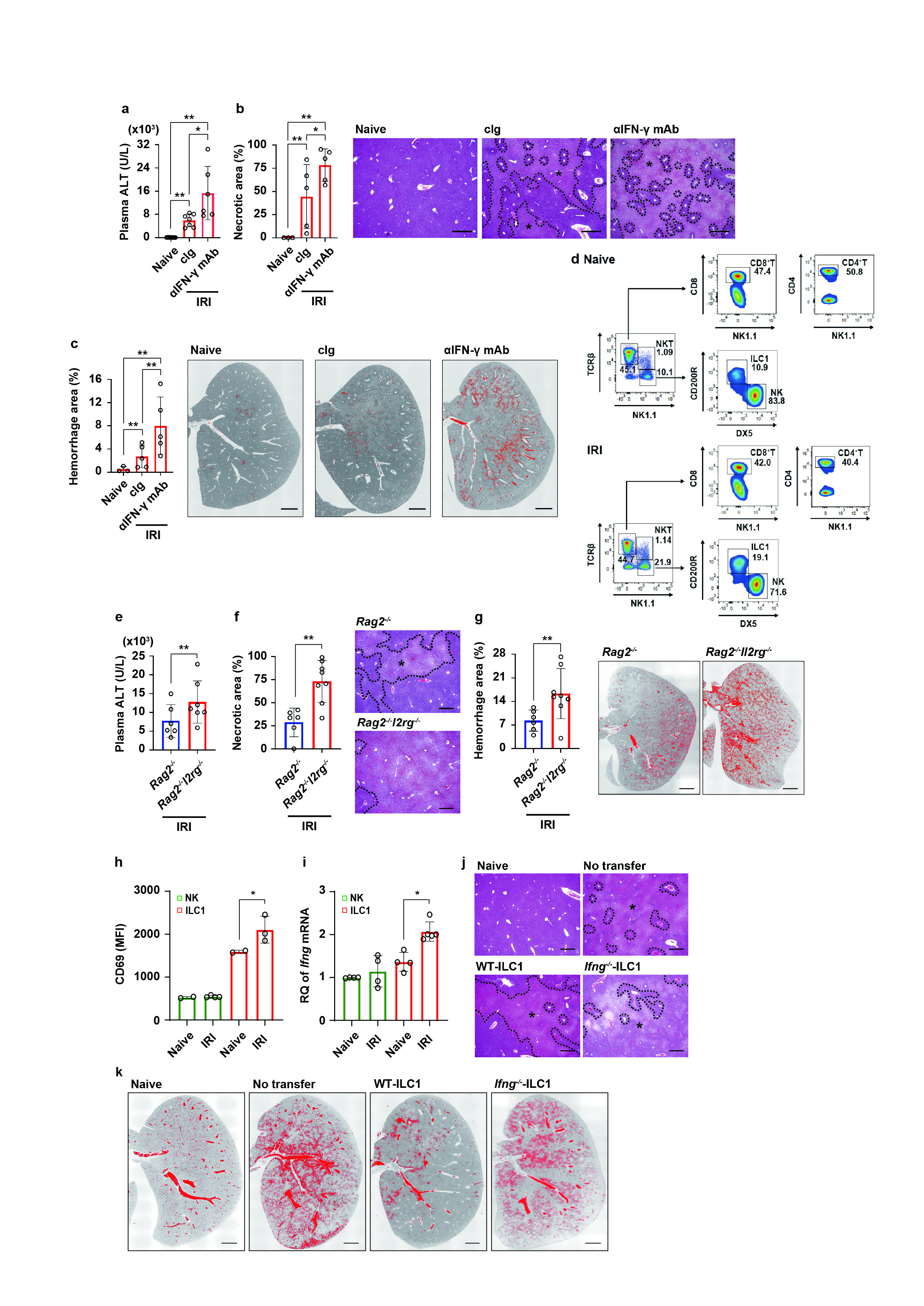
Mice were anesthetized by the intraperitoneal injection of the mixture of 0.3 mg/kg medetomidine, 4 mg/kg midazolam, and 5 mg/kg butorphanol in saline. After the midline supraumbilical incision, the liver can be exposed by the retractors. The connective tissue around the portal vein, hepatic artery, and common vile duct was carefully removed. The triad was then clamped, and mice were kept the body temperature at 37˚C on the heat controller. Sixty minutes after ischemia, circulation was restored into the left and median lobes for 1, 3, 5, 6, and 24 h according to each individual study48.
To block cytokines signaling, 100 µg of neutralizing mAbs against mouse IFN-g (clone R4-6A2, Bio X Cell, New Hampshire, USA) or mouse IL-10 (clone JES5-2A5, Bio X Cell, New Hampshire, USA) were injected into mice intraperitoneally before reperfusion.
Cecal ligation and puncture (CLP)
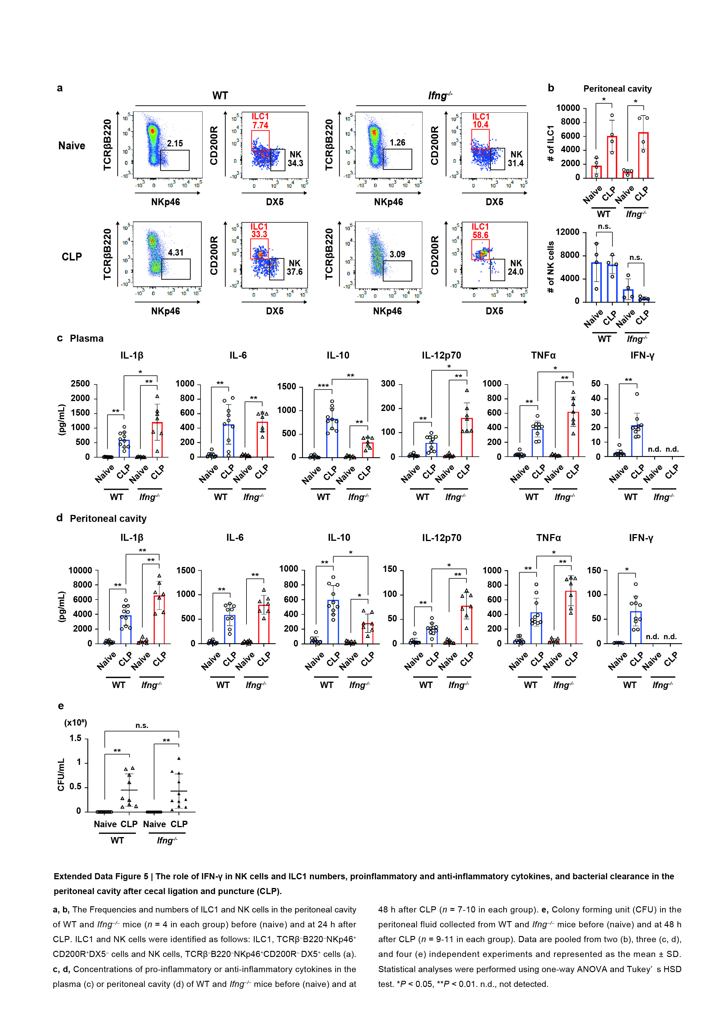
To induce the systemic inflammatory responses, polymicrobial sepsis was induced by CLP according to the general guidelines.24 Briefly, female C57BL/6J mice were anesthetized by the inhalation of isoflurane, and a 1 cm incision was made in the abdomen. The cecum was exposed, ligated at the bifurcation by a 4-0 suture, and punctured once with a 22 Gauge needle. The abdominal musculature and skin were closed with simple running sutures. Body weight loss and mortality were monitored for 6 days.
Preparation of lymphocytes in the liver and peritoneal cavity
Ice-cold phosphate phosphate-buffered saline (PBS) was injected into the portal vein for perfusion, and the liver tissue was isolated and mechanically ground into cell suspension by using a slide glass (Sanyo, Tokyo, Japan). The liver cells were washed with RPMI-1640 culture medium (Sigma-Aldrich, Missouri, U.S.A.), followed by PBS, then resuspended with 40% Percoll PLUS density gradient medium (GE Healthcare, Chicago, Illinois, USA), overlaid on 60% Percoll PLUS medium, and centrifuged at 800 x g for 35 min at 21˚C. Buffy coats were collected and washed with RPMI-1640 medium, followed by PBS supplemented with 5% fetal bovine serum (FBS, Thermo Fisher Scientific, Massachusetts, USA), and used as liver lymphocytes. Peritoneal lymphocytes were collected after gentle abdominal massage for 1 min with 2 ml PBS. Each lymphocyte was identified as follows; ILC1, NK1.1+TCRβ–B220–DX5–CD200R+ or NK1.1+TCRβ–B220–DX5–CD49a+ lymphocytes; NK cells, NK1.1+TCRβ–B220–DX5+CD200R– or NK1.1+TCRβ–B220–DX5+CD49a– lymphocytes; CD8+ T cells, NK1.1–TCRβ+CD8a+ lymphocytes; CD4+ T cells, NK1.1–TCRβ+CD4+ lymphocytes; NKT cells, NK1.1+TCRβ+ lymphocytes.
Adoptive transfer of ILC1
ILC1 (NK1.1+TCRβ–B220–DX5–CD200R+) and NK cells (NK1.1+TCRβ–B220–DX5+CD200R–) were sorted from the liver of WT and Ifng–/– mice by using BD FACS Aria (BD Biosciences, San Jose, California, USA). ILC1 (1 x 10⁶) were cultured in 96-well round-bottom plates for 24 h under 5% CO2 and 37˚C in RPMI-1640 culture medium supplemented with 10% heat-inactivated FCS, 2 mM glutamate, 100 U penicillin, 100 µg/ml streptomycin, 10 mM HEPES, 1 mM sodium pyruvate, 100 µM MEM non-essential amino acids, and 500 nM 2-mercaptoethanol in the presence of 25 ng/mL rmIL-7 for 24 h. Hundred or 50 thousand ILC1 together with 4 µg rmIL-15 were i.v. injected into male Rag2–/–Il2rg–/– mice 24 h before liver IR or CLP, respectively.
Preparation of myeloid cells in the liver, blood circulation, and peritoneal cavity
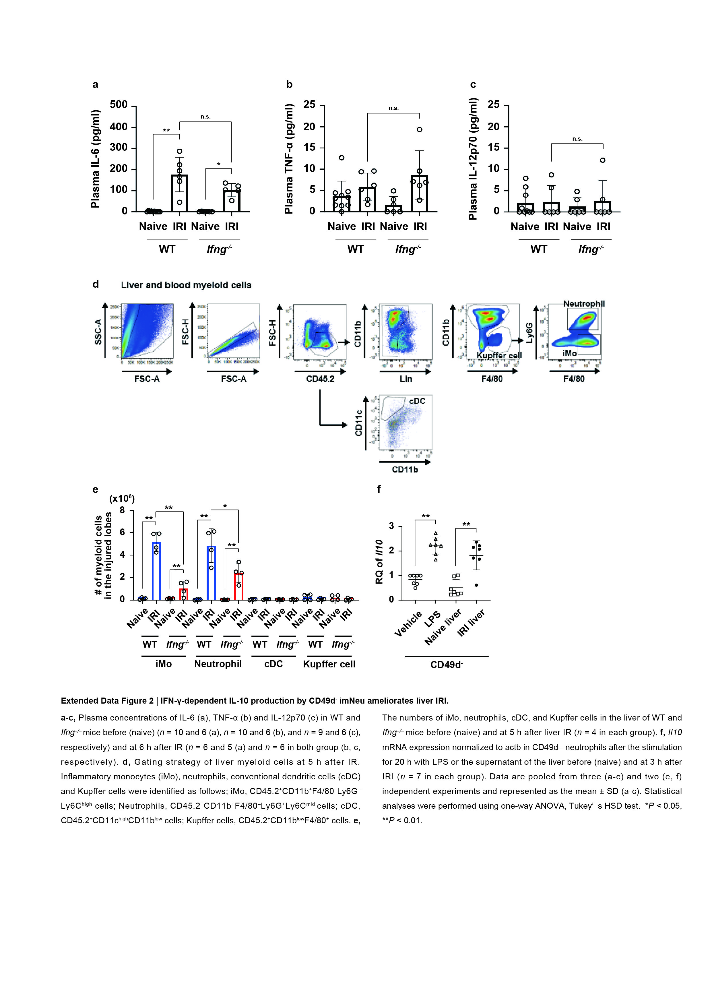
The liver was perfused with ice-cold PBS and incubated in the RPMI-1640 medium containing 500 CDU/mL Clostridium histolyticum-derived collagenase type IV (Sigma-Aldrich, Missouri, U.S.A.), 150 U/mL DNase I, 10 nmol CaCl2, and 4 nmol MgCl2 at 37˚C for 30 min with gentle shaking. One ml of blood was collected from the heart and diluted in 4 ml PBS (-) in the presence of heparin. The peritoneal cavity was injected with 2 ml PBS, and peritoneal cells were collected after the gentle abdominal massage for 1 min. Neutrophils and inflammatory monocytes in the liver, blood, and peritoneal cavity were identified as CD45.2+Lin–CD11bhighF4/80–Ly6CmidLy6G+ cells and CD45.2+Lin–CD11bhighF4/80–Ly6ChighLy6G– cells, respectively. Kupffer cells and conventional dendritic cells in the liver were identified as CD45.2+Lin–CD11b–/lowF4/80+ cells and CD45.2+CD11b–CD11chigh cells, respectively.
Effect of IFN-g on neutrophil influx from the BM in vivo
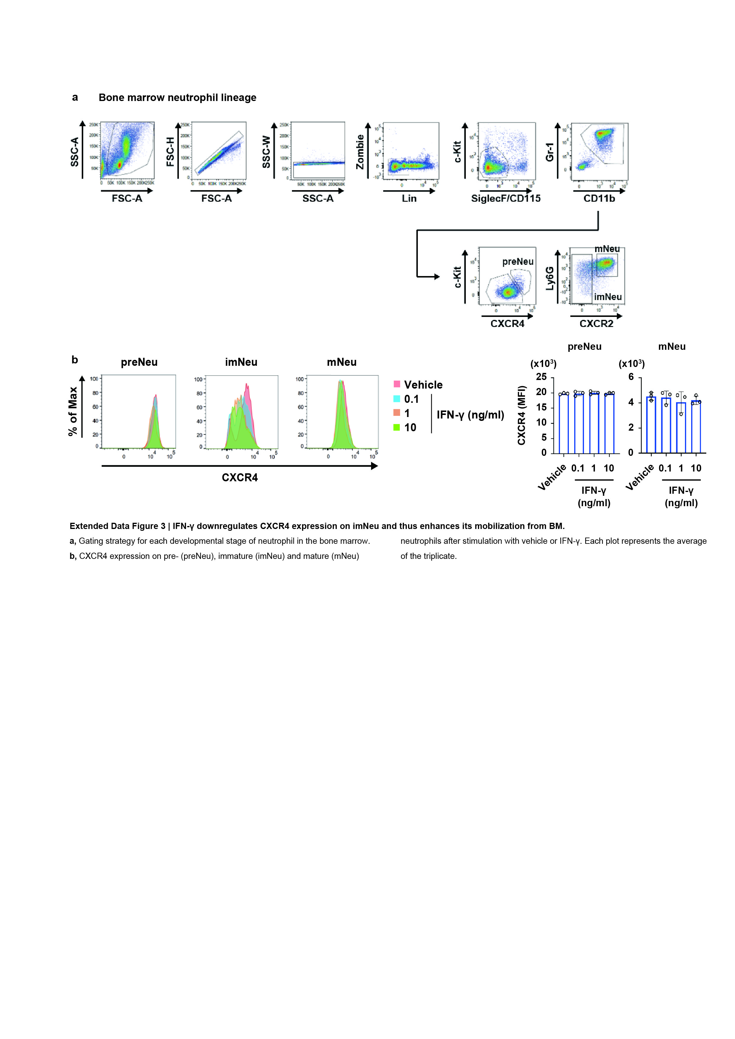
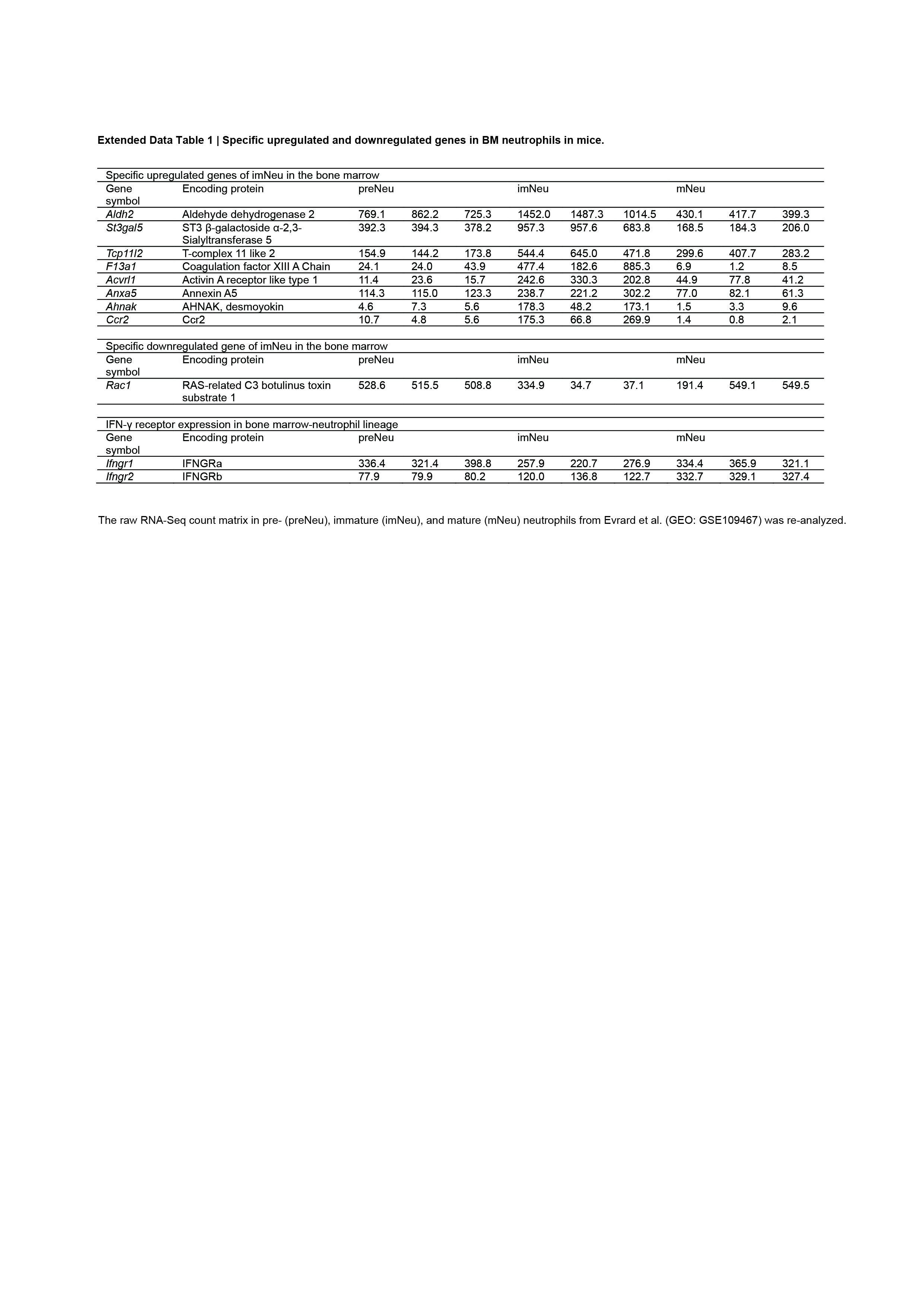
To investigate the effect of IFN-g on neutrophil influx from the BM, IFN-g or vehicle were i.v. injected into Ifng–/– mice. The dosage of IFN-g injected was calculated to reach the plasma level of IFN-g at 10 ng/ml using the following formula: total dosage of IFN-g = body weight x 72 ml/kg (whole blood volume) x 10 ng/ml. BM and whole blood were collected from the femora and heart of Ifng–/– mice, respectively, 24 h after the injection of vehicle or IFN-g. Red blood cells were lysed by an ammonium-chloride-potassium lysis buffer for 1 min, and imNeu and mNeu in the BM and blood were identified by flow cytometry as follows: imNeu, Lin–CD115–SiglecF–c-Kit–Gr-1lowCD11b+CXCR4–/lowCXCR2– cells and CD45.2+Lin-CD11bhighF4/80-Ly6CmidLy6Glow cells, respectively, and mNeu, Lin–CD115–SiglecF–c-Kit–Gr-1+CD11b+CXCR4–/lowLy6G+CXCR2+ cells and CD45.2+Lin–CD11bhighF4/80–Ly6CmidLy6Ghigh cells, respectively.
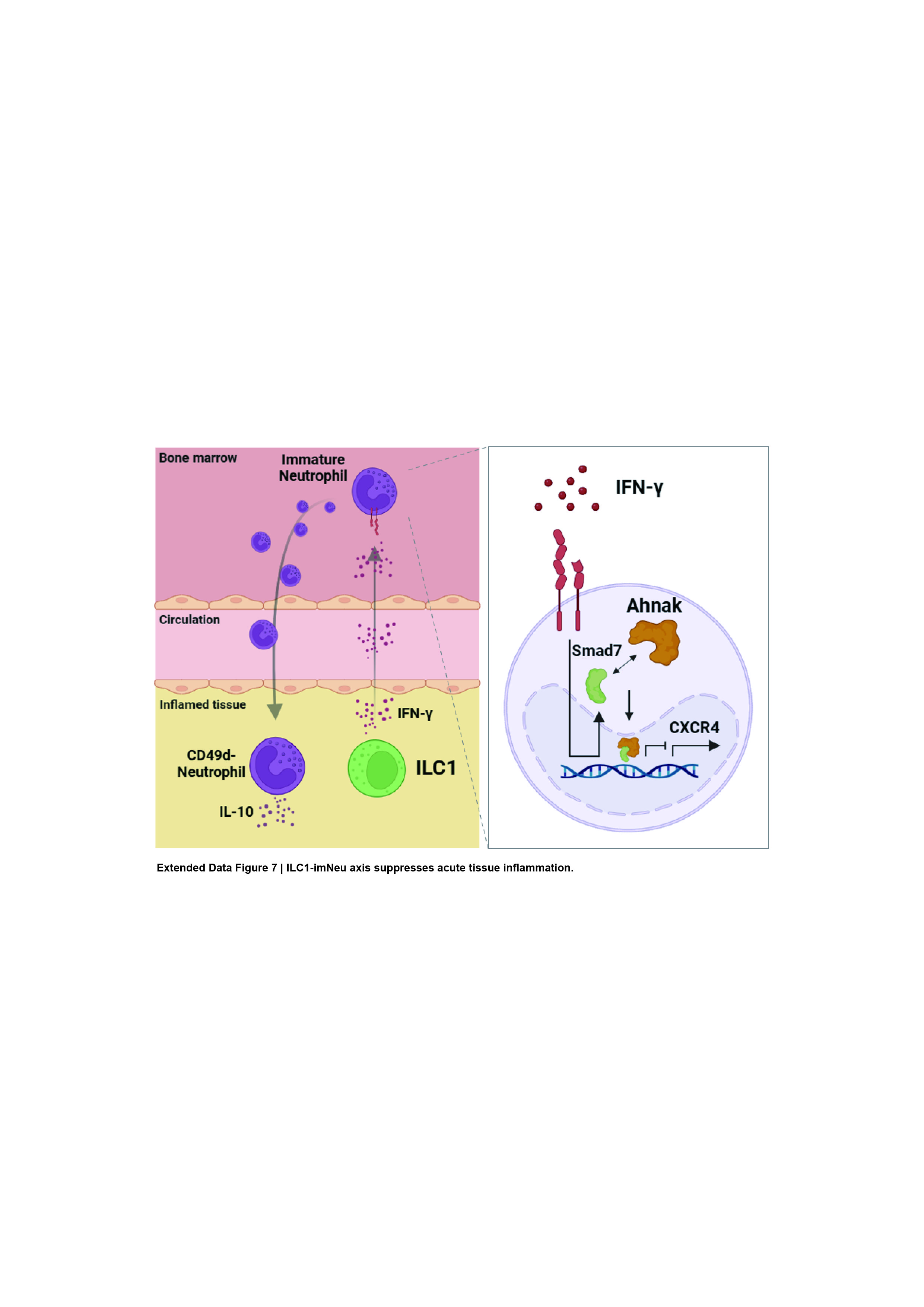
Effect of IFN-g on CXCR4 expression on BM neutrophils in vitro
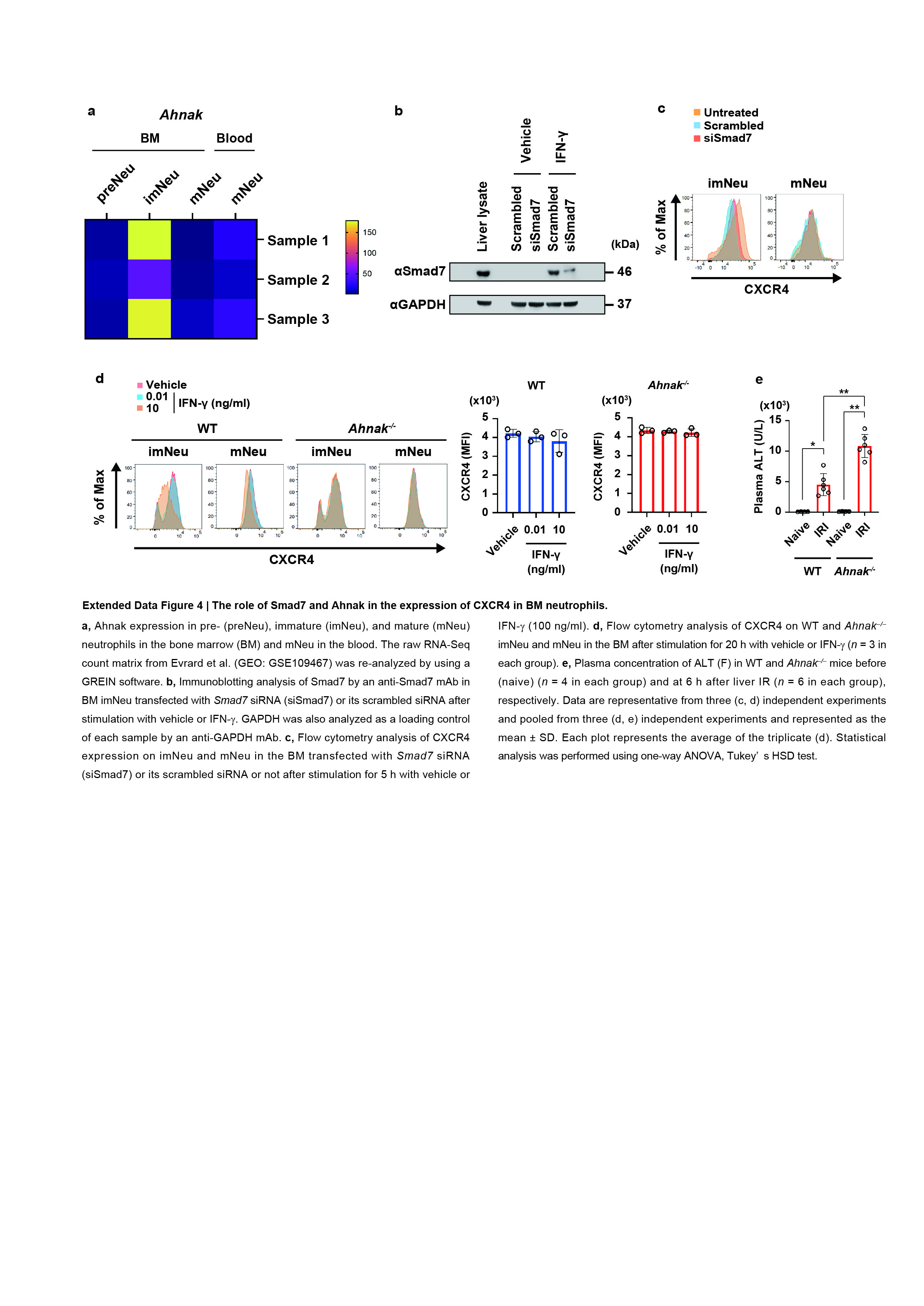
Pre-neutrophil (preNeu), imNeu and mNeu were sorted from the BM of WT, Ifng–/–, and Ahnak–/– mice by flow cytometry according to the following gating strategies: preNeu, Lin–CD115–SiglecF–c-KitmidGr-1+CD11b+CXCR4high cells; imNeu, Lin–CD115–SiglecF–c-Kit–Gr-1lowCD11b+CXCR4–/lowCXCR2– cells; mNeu, Lin–CD115–SiglecF–c-Kit–Gr-1+CD11b+CXCR4–/lowLy6G+CXCR2+ cells. Each neutrophil subsets were cultured in RPMI-1640 medium in the presence or absence of IFN-g for 5 or 20 h and analyzed for CXCR4 expression by flow cytometry. BM imNeu or mNeu (106 cells) were also transfected with 30 pmol Smad7 siRNA or its scrambled siRNA by using lipofectamine RNAiMAX transfection reagent (Thermo Fisher Scientific, Massachusetts, U.S.A.) for 20 h according to the manufacturer’s instruction with slight modification, stimulated with IFN-g for 5 h, and then analyzed for CXCR4 expression by flow cytometry.
Cytokine Assay
Blood was collected in the presence of heparin 6 and 48 h after IR and CLP, respectively. Plasma samples were prepared by centrifugation at 3,000 rpm for 15 min at 4˚C. Peritoneal fluid was collected from the peritoneal cavity after injection with 1 ml PBS and gentle massage 30 times and centrifuged at 3,000 rpm for 15 min at 4˚C. IL-1b, IL-6, IL-10, IL-12p70, TNF-a, and IFN-γ in the plasma and peritoneal fluid were quantified using BD Cytometric Bead Array Mouse Flex Sets (BD Bioscience) or Milliplex® assay kits (Sigma-Aldrich) according to the manufacturer’s instructions.
Immunoblotting
For immunoblotting of IL-10 in the liver, the liver from naive mice or 6 h after IR were lysed with 1% Nonidet P-40 supplemented with 20 U/ml aprotinin and protease inhibitor cocktail (Roche, Basel, Switzerland). After SDS-polyacrylamide gel electrophoresis (SDS-PAGE), separated proteins were transferred onto the polyvinylidene difluoride (PVDF) membranes by electroblotting, incubated with 5% bovine serum albumin (BSA), and immunoblotted with rat anti-mouse IL-10 (R&D Systems, Minneapolis, U.S.A.) or rabbit anti-mouse GAPDH (clone: 14C10, Cell Signaling Technology, Massachusetts, U.S.A.). Horseradish peroxidase (HRP)-conjugated goat anti-rat IgG was then incubated for 20 min with gentle shaking, and HRP was detected by ECL western blotting substrate (Thermo Fisher Scientific, Massachusetts, U.S.A.). For immunoblotting of Ahnak and Smad7, the cytoplasmic protein was extracted from BM neutrophil, separated by SDS-PAGE, and transferred onto PVDF membranes. The membrane was incubated with 5% rabbit non-immunized serum or 5% BSA, and the immunoblotted with 0.5 µg/ml rabbit anti-mouse Ahnak or 1 µg/ml anti-mouse Smad7 antibodies. The membrane was then incubated with HRP-conjugated anti-rabbit polymer IgG (Perkin Elmer, Massachusetts, U.S.A.) or HRP-conjugated sheep anti-mouse IgG (Cytiva, Amersham, U.K.) for 30 min at RT, and HRP was detected by ECL western blotting substrate. The PVDF membranes were stripped with RetoreÔ Western Blot Stripping Buffer (Thermo Fisher Scientific, Massachusetts, U.S.A.) for 20 min at RT, incubated with 3% BSA for 1 h, and reblotted with anti-GAPDH mAb (clone: 14C10, Cell Signaling Technology, Massachusetts, U.S.A.) overnight at 4˚C, followed by incubation with HRP-conjugated donkey anti-rabbit IgG. HRP was detected by the ECL western blotting substrate.
Far-western blotting
To assess the direct binding of Ahnak and Smad7 proteins, 5 x 10⁶ cells of immature and mNeu were sorted from the BM of naive mice and lysed in the cytoplasmic protein extraction reagent (Thermo Fisher Scientific, Massachusetts, U.S.A.) followed by IFN-γ stimulation for 20 h in vitro. Separated proteins by SDS-PAGE were transferred onto the PVDF membranes by electroblotting and refolded using 6 M guanidine hydrochloride buffer supplemented with 20 mM Tris, 1 mM dithiothreitol, 1 mM EDTA, 10% glycerol, 0.1% Tween 20, and 0.1 M sodium chloride for 30 min with gentle shaking. Endogenous Ahnak protein were then bound with refolded prey protein overnight at 4˚C, followed by the blocking of non-specific binding sites. One µg/ml anti-mouse Smad7 mAb was immunoblotted and HRP-conjugated donkey anti-rabbit IgG was incubated for 30 min at RT. HRP was detected by ECL western blotting substrate (Thermo Fisher Scientific, Massachusetts, U.S.A.). The PVDF membranes were stripped for 15 min at 57˚C and reblotted to detect Smad7 and GAPDH.
Histology
Injured left and median lobes were collected from naive and subsequent 6 h after hepatic IR and fixed overnight in 4% paraformaldehyde. Paraffin-embedded cross sections were stained with hematoxylin and eosin (H&E) or eosin Y alone. To examine necrotic area, congestion, vacuolization, and necrosis were graded as 0 (no-symptom), 1 (minimal), 2 (0-30%), 3 (31-60%), and 4 (60% over) for a sign and symptom of acute liver injury49. Erythrocytes in parenchyma were extracted as hemorrhage area, and quantified using Image J software.
cDNA synthesis and qPCR
cDNA was synthesized and total RNA was extracted by using Isogen reagent (Nippon Gene, Tokyo, Japan). cDNA was synthesized by using a high-capacity RNA-to-cDNA kit (Applied Biosystems, Carlsbad, CA). Expression of genes was measured through quantitative real-time PCR analysis by using SYBR Green master mix (Applied Biosystems) and appropriate primers. Primer sequences of the target genes are as follows: mouse Ifng, forward, 5’- ACA GCA AGG CGA AAA AGG ATG -3’, reverse, 5’- TGG TGG ACC ACT CGG ATG A -3’; mouse Il10, forward, 5’- GCT GGA CAA CAT ACT GCT AAC C -3’, reverse, 5’- ATT TCC GAT AAG GCT TGG CAA -3’; mouse actb, forward, 5’- ACT GTC GAG TCG CGT CCA -3’, reverse, 5’- GCA GCG ATA TCG TCA TCC AT -3’; human CXCR4, forward, 5’- GGG CAA TGG ATT GGT CAT CCT-3’, reverse, 5’- TGC AGC CTG TAC TTG TCC G -3’; human SMAD7, forward, 5’-TTC CTC CGC TGA AAC AGG G-3’, reverse, 5’-CCT CCC AGT ATG CCA CCA C-3’; human AHNAK, forward, 5’-TAC CCT TCC TAA GGC TGA CAT T-3’, reverse, 5’-TTG GAC CCT TGA GTT TTG CAT-3’; human GAPDH, forward, 5’- CTT CAC CAC CAT GGA GAA GGC -3’, reverse, 5’- GGC ATG GAC TGT GGT CAT GAG -3’. The thermal cycling conditions comprised an initial denaturation step at 95˚C for 10 min, followed by 50 cycles at 95˚C for 15 sec and 60˚C for 1 min.
Chemotaxis assay
Relative fluorescent units were quantified with
For chemotaxis assay, we used QCM chemotaxis 96-well cell migration assay kit (Merck, New Jersey, U.S.A.). Briefly, RPMI-1640 medium containing 10 pg/ml, 10 ng/ml or 1 µg/ml IFN-γ were poured into the 96-well feeder tray. Two hundred thousand imNeu sorted from the BM were resuspended in 100 µl of RPMI-1640 medium, placed onto the migration chamber with 3 µm of microporous membrane, and incubated for 20 h at 37˚under 5% CO2, and then removed from the plate assembly, followed by flipping out to discard cell suspension. A new feeder trey containing 150 µl of cell detachment solution were inserted into the trey, and cells were removed completely from the underside of the migration chamber by incubation for 30 min at 37˚C with gentle shaking and tilting several times, and then lysed with CyQuant GR Dye-contained lysis buffer. The lysates were analyzed for a fluorescence using 480/520 nm filter set according to the manufacturer’s instruction.
Bacterial colony formation unit
For determination of bacterial burden in the peritoneal cavity, mice were sacrificed at 48 h after CLP. Peritoneal fluid dilutions were performed, and the samples were seeded on tryptic soy agar plates. The colony numbers were counted 24 h after incubation at 37 °C.
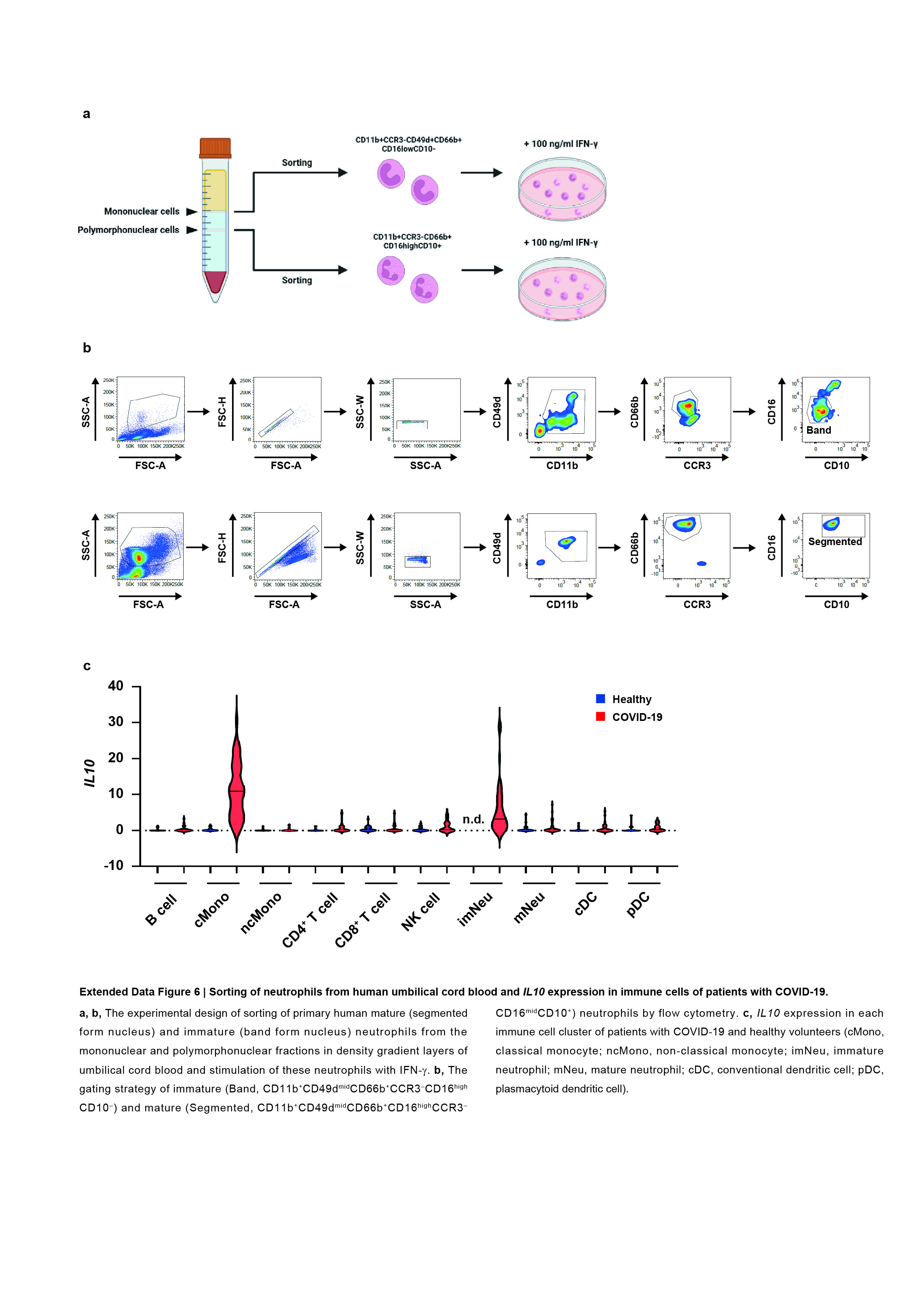
Reanalysis of single-cell RNA-Seq of PBMC in patients with COVID-19
Expression of AHNAK, SMAD7, CXCR4, IL10 and other neutrophil signature genes in the PBMC of patients with COVID-19 or healthy volunteers was investigated by re-analysis of publicly deposited data. Public single-cell RNA-seq databases of PBMC in patients with COVID-19 are available on NCBI Expression Omnibus (GEO) and European Genome-phenome Archive, including GSE150728, GEO155673, GSE149689, and EGAS00001004571 (or Schulte-Schrepping_2020_COVID19_10x_PBMC under FastGenomic)50. Original and processed data can be downloaded on Mondeley Data (http://cellxgene.cziscience.com/collections/b9fc3d70-5a72-4479-a046-c2cc1ab19efc).
Statistics
Statistical analyses were performed by one-way ANOVA with Tukey’s HSD test or unpaired student t test. P values less than 0.05 were considered statistically significant. All results are presented as mean ± SD of at least two independent experiments.
Data availability statement
Original single-cell RNA-Seq datasets of PBMC from healthy volunteers and patients with COVID-19 are available on NCBI Expression Omnibus (GEO) and European Genome-phenome Archive, including GSE150728, GEO155673, GSE149689, and EGAS00001004571 (or Schulte-Schrepping_2020_COVID19_10x_PBMC under FastGenomic).
Methods References
45. Dalton, D. K. et al. Multiple defects of immune cell function in mice with disrupted interferon-gamma genes. Science 259, 1739–1742 (1993).
46. Hao, Z. & Rajewsky, K. Homeostasis of peripheral B cells in the absence of B cell influx from the bone marrow. J Exp Med 194, 1151–1164 (2001).
47. Kouno, M. et al. Ahnak/Desmoyokin is dispensable for proliferation, differentiation, and maintenance of integrity in mouse epidermis. J Invest Dermatol 123, 700–707 (2004).
48. Abe, Y. et al. Mouse model of liver ischemia and reperfusion injury: method for studying reactive oxygen and nitrogen metabolites in vivo. Free Radic Biol and Med 46, 1–7 (2009).
49. Suzuki, S., Toledo-Pereyra, L. H., Rodriguez, F. J. & Cejalvo, D. Neutrophil infiltration as an important factor in liver ischemia and reperfusion injury. Modulating effects of FK506 and cyclosporine. Transplantation 55, 1265–1272 (1993).
50. Jin, K. et al. An interactive single cell web portal identifies gene and cell networks in COVID-19 host responses. iScience 24, 103115 (2021).