Clinical specimens
Twelve paired of intrahepatic cholangiocarcinoma and para-carcinoma tissues were collected from January 2016 to December 2019 in hepatological surgery department, affiliated hospital of Qiangdao Unversity via surgery resection. The patients had not received chemotherapy or radiotherapy treatment. The specimens were identified as cholangiocarcionma by pathological examination. Informed consent was obtained from each patient. The specimen collection and experimental procedure were line with Declaration of Helsinki, and approved by Ethics Committee of Affiliated Hospital of Qingdao University.
The expression levels of MT1JP and miR-18a-5p in the cholangiocarcinoma specimens were detected with real-time PCR.
The expression level of MT1JP in another thirty intrahepatic cholangiocarcinoma samples was analyzed with the patients’ clinicalpathological characteristics by Chi-square test.
Real-time PCR
Total RNA was extracted with TRIpure reagent (BioTeke, Beijing, China), and reversely transcribed into cDNA with M-MLV reverse transcriptase (BioTeke), in presence of Olig(dT) and random, or specific miRNA primers (GenScript, Nanjing, China). The cDNA was used for real-time PCR to detect the expression levels of MT1JP and miR-18a-5p, and mRNA level of FBP1 with 2×Power Taq PCR MasterMix (BioTeke) and SYBR Green (Sigma, St. Louis, USA). The data were analyzed using 2-ΔΔCt method. β-actin served as the internal control of MT1JP, and 5S served as the internal control of miR-18a-5p. The sequence information of primers was shown in Table 1.
Western blot
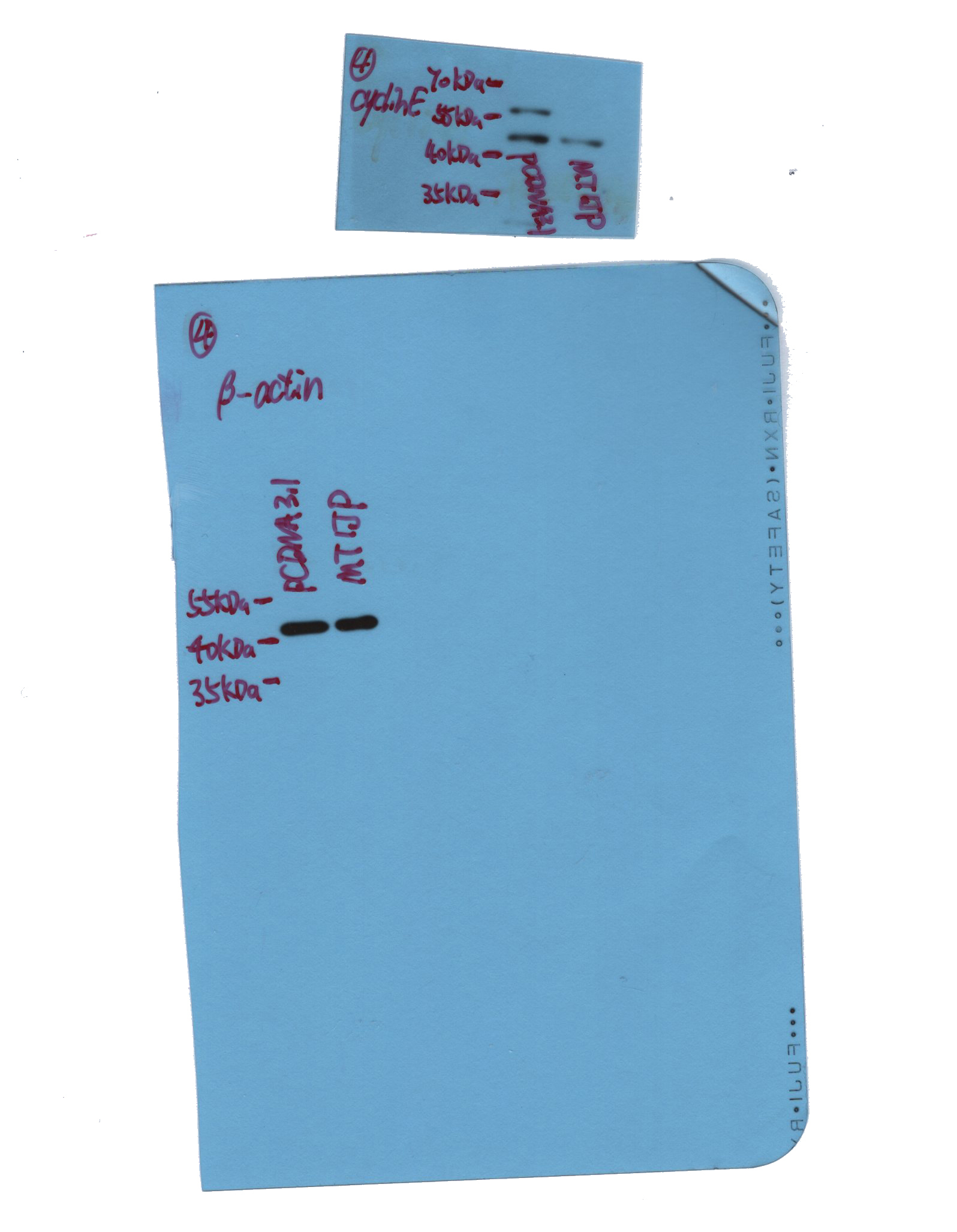
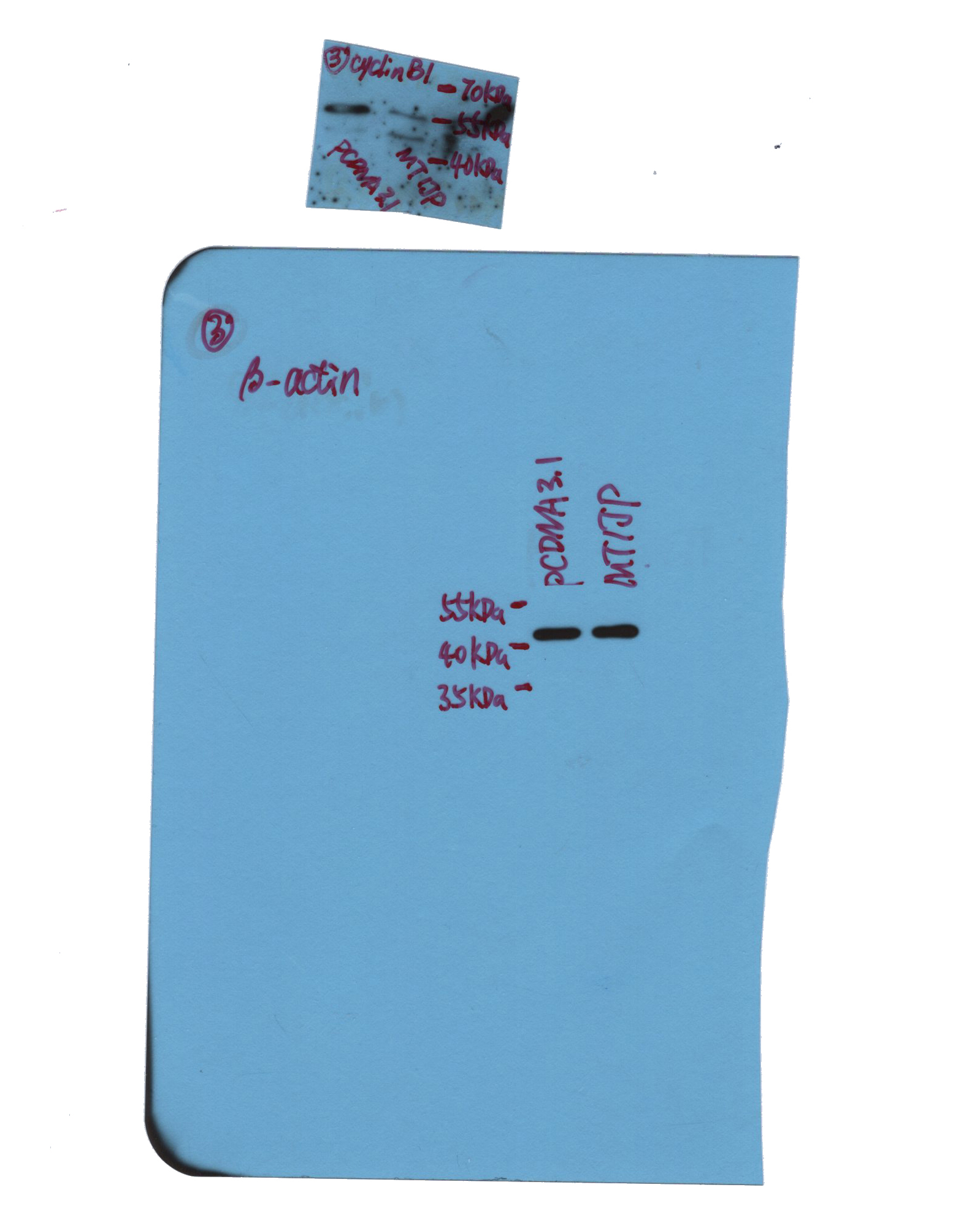
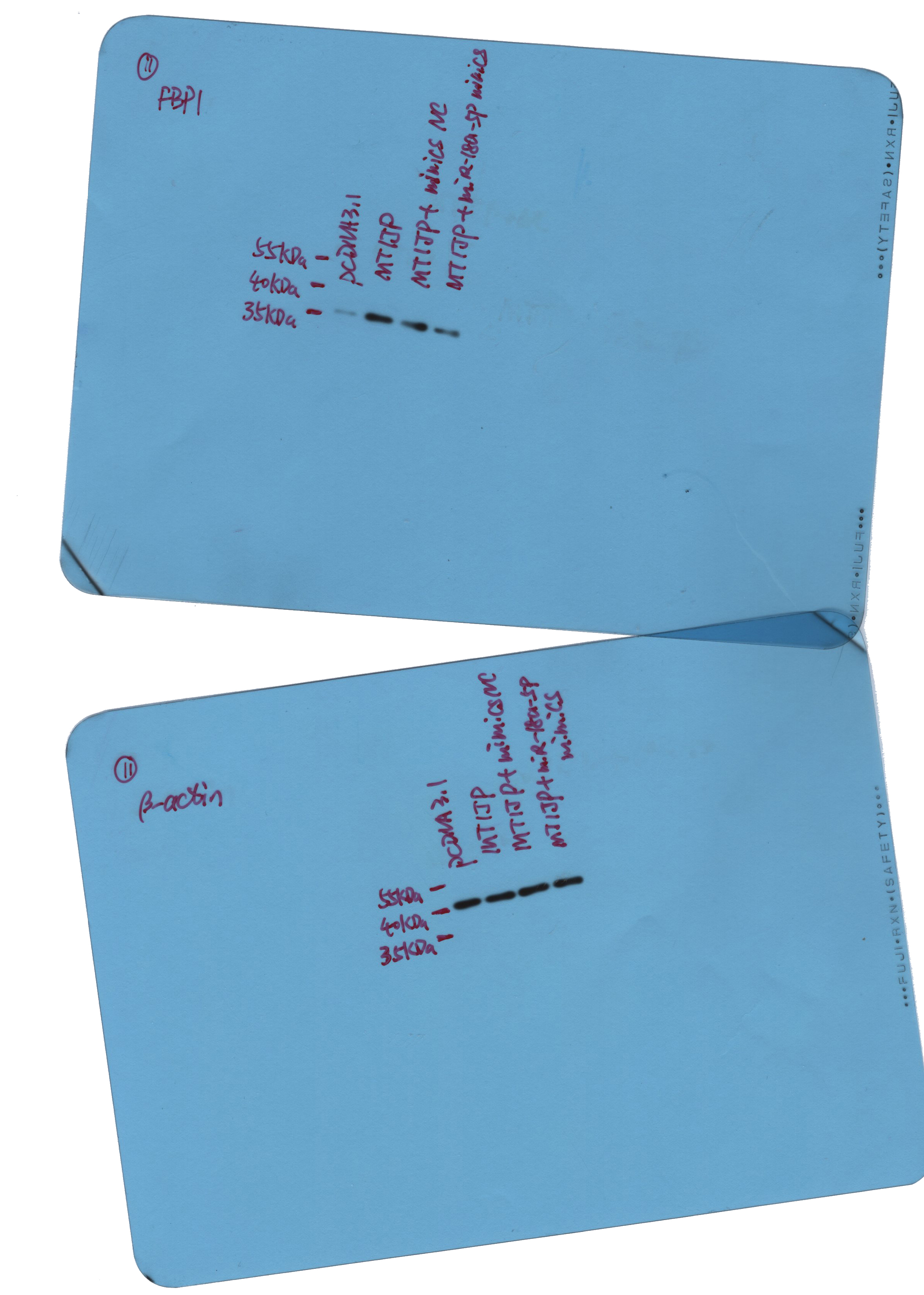
The protein was extracted using lysis buffer supplemented with 1 mM PMSF (Beyotime, Haimen, China), and the concentration was determined with BCA protein assay kit (Beyotime). Then the protein was separated with SDS-PAGE, and transferred onto polyvinylidene difluoride membrane (Millipore, Boston, USA). The membrane was blocked with skim milk (YILI, Hohhot, Inner Mongolia, China) at room temperature for 1 h, and incubated with following antibodies at 4 ℃ overnight: rabbit anti-proliferating cell nuclear antigen (PCNA) (1:1000; cat. no. A0264, Abclonal, Wuhan, Hubei, China), rabbit anti-caspase-3 (1:1000; cat. no. A19654, Abclonal), rabbit anti-poly ADP-ribose polymerase (PARP) (1:1000; cat. no. A0942, Abclonal), rabbit anti-cyclin E (1:1000; cat. no. AF0144, Affinity, Changzhou, Jiangsu, China), rabbit anti-cyclin B1 (1:1000; cat. no. A2056, Abclonal), rabbit anti-fructose-1,6-bisphosphatase 1 (FBP1) (1:5000; cat. no. 12842-1-AP, Proteintech, Wuhan, Hubei, China) or mouse anti-β-actin (1:1000; cat. no. sc-47778, Santa Cruz, USA). After rinsing with TBST, the membrane was incubated with goat anti-rabbit or mouse secondary antibody (1:5000; cat. no. A0208, A0216, Beyotime) at 37 ℃ for 45 min. The protein in the membrane was reacted with ECL reagent (Beyotime), followed with a signal exposure in the dark. The optical density of the blotting bands was analyzed with Gel-Pro-Analyzer software.
Cell culture
Primary intrahepatic cholangetic epithelial cells were purchased from iCell (Shanghai, China), and cultured with primary epithelial cell medium (iCell) at 37 ℃ with 5% CO2. The primary intrahepatic cholangeitc epithelial cells were identified by immunofluorescent staining of cytokeratin 19 (CK19), and the results were shown in figure S12.
Cholangiocarcinoma cell lines HCCC-9810 and RBE were purchased from Procell (Wuhan, China), and HUCCT1 from Zhongqiaoxinzhou (Shanghai, China). The cells were cultured in RPMI-1640 medium (Gibco BRL, Gaithersburg, USA) supplemented with 10% fetal bovine serum (FBS) (Hyclone, Logan, USA) at 37 ℃ with 5% CO2.
Cell transfection was performed with Lipofectamine 2000 reagent (Invitrogen, Carlsbad, USA) in serum-free medium according to the manufacturer’s protocol.
To obtain the stably transfected cell line, HCCC-9810 cells were transfected with MT1JP overexpression plasmid, and treated with 400 μg/ml G418 for 2 weeks. The single cells were selected out, and cultured without G418. After verification of MT1JP at RNA levels, the MT1JP-stably expressed cell lines were obtained.
Cell counting kit-8 (CCK-8) assay
CCK-8 assay was performed to measure the cell viability. The cells were cultured in 96-well plates at 37 ℃ with 5% CO2. After culture for 0 h, 24 h, 48 h or 72 h, the cells were incubated with CCK-8 reagent (10 μl per well) (KeyGEN, Nanjing, China) for 2 h. The optical density of medium was detected with a microplate reader (BioTek, Winooski, VT, USA) at 450 nm.
Flow cytometry
Flow cytometry was used for detection of apoptosis and cell cycle.
The cells were collected, and incubated with Annextin V-FITC reagent and propidium iodide at room temperature for 20 min in the dark. Then the cells were detected by flow cytometer (BD, Franklin Lakers, NJ, USA).
For cell cycle detection, the cells were collected and immobilized with 70% ethanol at 4 ℃ overnight. Then the cells were incubated with PI/RNaseA buffer at room temperature for 60 min in the dark, and used for detection by flow cytometer.
Transwell assay
Transwell assay was used for detection of migration and invasion.
The cells were collected and counted. About 3×10³ cells were seeded into the upper chamber with serum-free medium, and the lower chamber was added with medium with 30% FBS. After culture for 24 h, the cells on the reverse surface of transwell membrane was fixed with 4% paraformaldehyde (Aladdin, Shanghai, China) and stained with 0.4% crystal violet (Amresco, Solon, OH, USA). The cells were photographed and counted under a microscope at 100× magnification.
For invasion analysis, the polycarbonate membrane of transwell chambers (Corning, NY, USA) was pre-coated with Matrigel at 37 ℃. Approximately 1.5×10⁴ cells were seeded into upper chamber with serum-free medium, and medium with 30% FBS were added into the lower chamber. After culture for 24 h, the cells on the reverse surface was fixed and stained, and the cell number was counted under a microscope.
Dual-luciferase assay
The binding between MT1JP and miR-18a-5p was analyzed by bioinformatic website RNAhybrid (https://bibiserv.cebitec.uni-bielefeld.de/rnahybrid/) [13]. The MT1JP sequence containing miR-18a-5p-bound region or mutant sequence was cloned into pmirGLO vector with NheI and XhoI sites, and cotransfected into 293T cells with miR-18a-5p mimics. Twenty-four hours later, the cells were lysed, and the luciferase activity of pmirGLO vector was detected.
The candidate targets of miR-18a-5p were predicted by bioinformatic website targetscan (http://www.targetscan.org/vert_71/). The 3’UTR sequence of FBP1 containing miR-18a-5p-bound region or its mutant sequence was synthesized and inserted into pmirGLO vector with NheI and XhoI sites. 293T cells were cotransfected with pmirGLO vector and miR-18a-5p mimics, and the activity of luciferase activity was determined.
Immunofluorescent staining
The cells were pre-seeded on glass slides. After culture for certain times, the cells were fixed with 4% paraformaldehyde for 15 min, permeated with 0.1% TritonX-100 (Beyotime) for 30 min, and blocked with goat serum for 10 min at room temperature. Subsequently, the cells were incubated with antibody against FBP1 (1:100; cat. no. 12842-1-AP, Proteintech) or CK19 (1:50; cat. no. A0247, Abclonal) at 4 ℃ overnight. After washing with PBS, the cells were incubated with secondary antibody labeled with Cy3 (1:200; cat. no. A0516, Beyotime) at room temperature in the dark for 60 min, and counterstained with DAPI (Aladdin). Finally, the glass slide were mounted with anti-fading reagent (Solarbio, Beijing, China), and the cells were observed under a fluorescent microscope (Olympus, Tokyo, Japan) at 400× magnification.
Xneograft model
Healthy BALB/c mice were purchased from HFK Biotechnology Co. Ltd. (Beijing, China), and kept in a controlled environment (12 h/12 h light/dark, 22±1 ℃) with free access to food and water. The animals were taken care of according to Guide for the Care and Use of Laboratory animal (8th edition, NIH), and the experimental procedures were approved by Ethics Committee of Affiliated Hospital of Qingdao University.
After accommodation for one week, the mice were subcutaneously injected with HCCC-9810 cells (5×10⁵ each mouse) with MT1JP stable overexpression or not. One week later, the tumor size was measured every three days. Three weeks after injection, the mice were euthanized via overdose of pentobarbital sodium (200 mg/kg, intraperitoneal injection), and the tumors were isolated for detection.
HE staining
The tumor isolated from mice were fixed in 4% paraformaldehyde overnight, and washed with flow water for 4 h. Then the tissue was dehydrated with ethanol of grading concentrations and xylene, embedded with paraffin, and cut into sections of 5 μm. The sections were deparaffinized with xylene and ethanol, and stained with hematoxylin (Solarbio). After soaking in 1% hydrochloric acid/ethanol for several seconds, the sections were stained with eosin (Sangon, Shanghai, China). Finally, the sections were dehydrated again, mounted with gum, and observed under a microscope at 200× magnification.
TUNEL staining
The tumor tissue was made into paraffin sections as described previously. After deparaffinization, the sections were permeated with 0.1% Triton X-100, and blocked with 3% H2O2 at room temperature. Then the sections were incubated with TUNEL buffer (Roche, Basel, Switzerland) for 60 min at 37 ℃ in the dark, and incubated with Converter-POD reagent for 30 min at 37 ℃. Subsequently, the sections were reacted with DAB substrate (Solarbio), and counterstained with hematoxylin. Finally, the sections were dehydrated, mounted and photographed with a microscope at 400× magnification.
Immunohistochemical staining
The tumor tissues were used for immunohistochemical staining for detection of Ki-67 and FBP1. The tissues were made into sections as previously described. The sections were reacted with antigen repair buffer in boiling for 10 min, and blocked with 3% H2O2 and goat serum. Then the section were incubated with antibody against Ki-67 (1:100; cat. no. AF0198, Affinity) or FBP1 (1:100; cat. no. 12842-1-AP, Proteintech) at 4 ℃ overnight. After washing with PBS, the sections were incubated with secondary antibody labeled with HRP (1:500; Beyotime) at 37 ℃ for 60 min, and reacted with DAB substrate. After counterstaining with hematoxylin, the sections were dehydrated, mounted and observed with a microscope at 400× magnification.
Statistical analysis
The data in this study were presented as meas±SD, and analyzed with GraphPad Prism 8.0. The data from two independent groups were analyzed with student’s t test. Comparisons among multiple groups were performed with one-way or two-way analysis of variance followed with Bonferroni post-hoc test. The correlation between MT1JP and clinical features were analyzed by Pearson χ2 test. The correlation between MT1JP and miR-18a-5p was analyzed with Pearson test. A p value less than 0.05 was considered as statistically significantly.