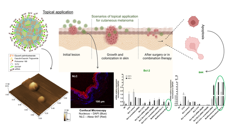
Multifunctional therapies have emerged as innovative strategies in cancer treatment. In this research article, we proposed a nanostructured lipid carrier (NLC) designed for the topical treatment of cutaneous melanoma, which simultaneously delivers 5-FU and Bcl-2 siRNA. The characterized nanoparticles exhibited a diameter of 259 ± 9 nm and a polydispersion index of 0.2, indicating a uniform size distribution. The NLCs were primarily localized in the epidermis, effectively minimizing the systemic release of 5-FU across skin layers. The ex vivo skin model revealed the formation of a protective lipid film, decreasing the desquamation process of the stratum corneum which can be associated to an effect of increasing permeation. In vitro assays demonstrated that A375 melanoma cells exhibited a higher sensitivity to the treatment compared to non-cancerous cells, reflecting the expected difference in their metabolic rates. The uptake of NLC by A375 cells reached approximately 90% within 4 hours. The efficacy of Bcl-2 knockdown was thoroughly assessed using ELISA, Western blot, and qRT-PCR analyses, revealing a significant knockdown and synergistic action of the NLC formulation containing 5-FU and Bcl-2 siRNA (at low concentration --100 pM). Notably, the silencing of Bcl-2 mRNA also impacted other members of the Bcl-2 protein family, including Mcl-1, Bcl-xl, BAX, and BAK. The observed modulation of these proteins strongly indicated the activation of the apoptosis pathway, suggesting a successful inhibition of melanoma growth and prevention of its in vitro spread.
Research Article
Bcl-2 knockdown by multifunctional lipid nanoparticle and its influence in apoptosis pathway regarding cutaneous melanoma: in vitro and ex vivo studies.
https://doi.org/10.21203/rs.3.rs-4356428/v1
This work is licensed under a CC BY 4.0 License
You are reading this latest preprint version
5-Fluorouracil
Bcl-2 siRNA
lipid nanoparticle
topical delivery
melanoma
- Development and characterization of nanostructured lipid carrier (NLC) to encapsulate 5-FU and complex efficiently Bcl-2 siRNA.
- In vitro and ex vivo skin studies in which indicated a high biocompatibility of the lipid nanoparticle with cutaneous tissue.
- The proposed nano-therapy presented a high knockdown of Bcl-2 protein and its respective mRNA, and a strong influence in other members of the Bcl-2 family, proving an efficient apoptosis induction.
Skin cancer is classified as non-melanoma or melanoma skin cancer, being the less prevalent melanoma type, although it has a worse prognosis and higher metastasis rates [1, 2]. The topical treatment presents a versatile option for cutaneous melanoma since it is less invasive and localized. However, topical treatment is indicated mainly for pre-cancer and small lesions and also to guarantee the clinical margins after surgery. Topical products are also indicated in combination with oral/intravenous chemotherapy or immunotherapy. The localized possibility of topical treatment makes this option the best choice when the lesions compromise larger areas, for facial lesions, or if the surgical procedure may cause deformation [3, 4]. Further, melanoma arises from epidermal melanocytes, and in the initial stages, the main affected layer is the epidermis. In this way, the use of topical treatment is advantageous [5, 6].
New melanoma treatment options are required because the mutations and drug resistance limit the use of existing therapies. Thus, the combination of treatments is already routine in melanoma management especially considering the complexity of the pathogenesis of cancer [7] especially considering the complexity of the pathogenesis of cancer. Combined and multifunctional therapies have been explored for melanoma management especially considering the complexity of the pathogenesis of cancer. The advantage of using a combination of approaches is the ability to affect multiple pathways. One of the combinations that have proven highly effective for several types of cancers, including skin cancer, is the combination of chemotherapeutic agents and nucleic acids [8, 9].
The delivery system, such as nanoparticles, as non-viral vectors for nucleic acids, presents promising results in cancer treatment, including melanoma [10], highlighting the co-deliver drugs and nucleic acids, such as siRNAs [1, 9].
The siRNA topical application has been studied in recent years and its topical use is considered a challenge in stability and release issues [4]. Regarding skin tissue, the use of nanoparticles as a vector of siRNA aims to protect the nucleic acid from degradation and to promote skin penetration across the human skin barrier [11]. Cationic nanoparticles, especially lipid-based ones, can be used topically, offering the permeability and skin retention ability, successfully carrying and releasing the siRNA to the target tissue [12–17].
Regarding melanoma, apoptosis and angiogenesis are critical molecular pathways that control cancer survival. The apoptosis should occur in any pathological event, but in melanoma, a high escape of this death pathway is observed. The activation of this apoptotic pathway is controlled by various down and up regulators such as the Bcl-2 proteins family [18]. The overexpression of anti-apoptotic members, such as Bcl-2, is related to multidrug resistance and inhibition of cytochrome C release, directly interfering with the caspase cascade [19]. The inhibition of a member of Bcl-2 proteins is the key to suppressing the development and survival of tumors, so it is a potential target for the knockdown gene using a specific siRNA [20]. Angiogenesis is essential to supply oxygen to a tumor site, creating a microenvironment to sustain growth [21]. Several drugs are developed to act in this survival pathway, such as 5-Fluorouracil (5-FU), which acts irreversibly, inhibiting the enzyme thymidylate synthase reducing the synthesis of DNA and RNA so that the angiogenesis process will be interrupted. This drug is mainly studied for colorectal cancer and achieved promising results but also recently has been studied for skin cancer and others [22–24]. However, regarding the several adverse effects associated with 5-FU, such as erythema, blistering, necrosis, erosions, pruritus, and burning [25], the use of a nanosystem encapsulating 5-FU has a critical role in promoting the reduction of these adverse effects and also deliver this drug in the tumor site.
Despite the existence of various multifunctional systems for the topical treatment of melanoma as reported in the literature [1, 8], our work uniquely demonstrates the topical co-delivery of 5-FU and Bcl-2 siRNA for the treatment of cutaneous melanoma. This approach introduces a multifaceted therapy that combines drug and antisense therapy, strategically targeting both angiogenesis and apoptosis pathways. This represents a novel strategy in the context of leveraging the benefits of a multifunctional therapeutic approach.
2.1. Materials
5-Fluorouracil (≥ 99% - Sigma Life Science, USA), Glyceryl palmitostearate (Precirol® ATO-5, Gattefossé, France), Caprylic/Caprylic Triglyceride (Labrafac Lipophile®, Gattefossé, France), 2-Dioleoil 3-trimetilamonium propane (DOTAP® - Avanti Polar Lipids, USA), Poloxamer 188 (P188) (Kolliphor P©, Sigma Life Science, USA), siRNAs (ThermoFisher Scientific, USA), Rhodamine 123 (ThermoFisher Scientific, USA), DAPI (Merck, Sigma Aldrich, USA), Dialysis tubing MWCO 12,000–14,000 (Fisherbrand, Fisher Scientific, USA), Lipofectamine 2000® (Invitrogen, Life Technologies, USA), Rezasurin® (G-Biosciences, USA), Propidium iodide (Sigma Aldrich, USA), Ultrapure Agarose (Invitrogen, Life Technologies, USA), Human Bcl-2 ELISA Kit (Abcam, EUA), Pierce BCA Protein Assay Kit (Thermo Fisher, #23225, Thermo Scientific, USA), Anti-BCL-2 (Abcam, EUA).
2.2. NLC Preparation
NLC was prepared by emulsification/ultrasonication technique. Briefly, the aqueous phase composed of 0.7% (w/v) P188 and 5-FU at 80°C was added in a melted lipid phase, composed of 65 mg of Precirol-ATO-5®, 25 mg of Labrafac lipophile® and 10 mg of DOTAP. Immediately the dispersion was placed in a Sonicador (Bandelin Electronic, UW2200, Berlin) at 30V for 5 minutes. The final 5-FU concentration was 500 µg/mL.
2.3. Physicochemical characterization of NLC
2.3.1. Dynamic Light Scattering (DLS) and Zeta Potential
The particle size, PdI and zeta potential were analyzed using Nano ZS instrument (Malvern, UK) equipped with a 633 nm He-Ne laser at an angle of 173° at 25°C and refractive index 1.33. The NLC was diluted 1:400, v/v in ultra-purified particle-free water and in KCl solution (0.1 nM) for Zeta potential.
2.3.2. Nanoparticle Tracking Analysis (NTA)
The number of particles was determined using NTA instrument (NanoSight NS300, Malvern, UK) equipped with 642 nm laser. NLC (1:1000, v/v) measurements (NTA 3.0 Analytical software) were performed at 25°C (n = 5).
2.3.3. Association efficiency (AE) and Drug loading (DL)
AE and DL were determined by ultrafiltration/centrifugation using Amicon® devices (50 kDa Amicon®, Millipore®). 1 mL of NLC was centrifuged (Centrifuge 5430R, Hamburg-Eppendorf, Germany) at 3000 x g for 5 minutes at 25°C. The ultrafiltrate was quantified by HPLC method previously validated at the chromatographic conditions: Shimadzu HPLC equipment using a C18 column (Phenomenex, 100 mm x 4,6 mm − 5 µm, 110 A) at 25 ºC, mobile phase composed by methanol: water (10:90, pH 4.5), flow 0.8 mL/min and 30 µL of sample injection, UV detector at 265 nm wave-length. The AE and DL was calculated using the equations 1 and 2 respectively (n = 5).
\(AE= \frac{Total amount of 5-FU - amount of 5-FU in ultrafiltrate}{Total amount of 5FU}x 100\) (Eq. 1)
\(DL= \frac{Total amount of 5-FU - amount of 5-FU in intrafiltrate}{Total amount of lipids}x100\) (Eq. 2)
2.3.4. Atomic Force Microscopy (AFM)
AFM analysis (Shimadzu Scanning Probe, SPM-9600) was performed using 5 µL NLC (1:400, v/v) deposited on a mica surface. The samples were analyzed after sample drying.
2.3.5. Complexation assay and stability of complex siRNA-NLC
The complexation of nanosystem with siRNA and the anionic competition with heparin was determined by electrophoresis according to previous studies [13, 15, 26].
2.4. Drug Release
The 5-FU release was performed using a Franz diffusion cell (Hanson Instruments, USA) with a 1.77 cm2 permeation area. The NLC 5-FU and 5-FU solution (500 µg/mL) were added on a dialysis membrane (33 mm, 23 µm, 21 in., MWCO 12,000–14,000, Fisherbrand). The receptor solution was composed of phosphate buffer (pH 7.4 ± 0.2). Franz cells were maintained at 32°C under stirring at 300 rpm. 1 mL of the permeate content (PC) was collected (n = 5) over time and analyzed in HPLC (mentioned at 2.3.3 item). This study was performed under infinite dose and sink conditions.
2.5. Skin permeation and retention studies
2.5.1. In vitro – Franz diffusion cell
In vitro permeation and retention studies were performed using a Franz diffusion cell (Hanson Instruments, USA) at the same conditions mentioned in release study. The NLC 5-FU and 5-FU solution (500 µg/mL) were studied concerning permeation and retention in porcine ear skin. The retention profile was studied by the (i) quantification of 5-FU in skin layers using tape stripping and viable epidermis and dermis (EP + D) homogenization, (ii) horizontal sectioning, and (iii) confocal microscopy (n = 5). The method was following [27] with adaptations. Confocal microscopy (Leica Microsystems Inc., USA) samples received an NLC labeled with Alexa 647 siRNA. The images were acquired with an immersion objective of 20 ×, λ = 405 nm, and λ = 638 lasers, suitable for DAPI, and Alexa 647, respectively.
2.5.2. Ex vivo - Human Organotypic Skin Explant Culture (hOSEC)
The ex vivo study was performed to observe the NLC behavior and human skin architecture after NLC application, and to quantify the amount of 5-FU retained in the human skin over time. The human skin was obtained from abdominoplasty surgery, approved by the Research Ethics Committee of Ribeirão Preto Medical School, University of São Paulo, Brazil (nº 12175/2017). The skin preparation was as follows [28]. The skin was cultivated in DMEM with 10% (v/v) SBF and 1% (v/v) antibiotic and antimycotic at 37°C, 5% CO2. The NLC 5-FU was applied daily topically, 2 µg of 5-FU per fragment (10 mm2), for seven days. The culture medium was replaced twice during the study, on the third and sixth day. The 5-FU was quantified in the cutaneous tissue and culture medium, every 24 hours in HPLC (mentioned at the 2.4.2. item).
Skin samples were analyzed in Confocal microscopy (Leica Microsystems Inc., USA) by vertical sections (n = 3, 20 µm) obtained using a cryostat (Leica, Wetzlar, Germany) at 24 and 168 hours of study. The slices were stained with DAPI and mounted with Fluoromount™. The images were acquired with an immersion objective of 20× and 40×, λ = 405 nm, and λ = 638 lasers, suitable for DAPI, and Alexa 647, respectively. The skin samples were also studied using histological analysis with hematoxylin/eosin according to (Andrade et al., 2017). The histological images were obtained in 40×.
2.6. Cellular studies
2.6.1. Culture conditions
HaCaT (human keratinocytes) and A375 (human malignant melanoma) were acquired from the Cell Bank of Rio de Janeiro and from the American Type Culture Collection (ATCC CRL-1658, USA) respectively. Both cells were cultured in DMEM with 10% (v/v) fetal bovine serum and 1% (v/v) antibiotic and antifungal solution at 37°C, 5% CO2.
2.6.2. Viability by Resazurin assay
Cell viability tests were performed in 96-well plates, 10,000 cells per well. The cells were exposed to treatments for 24 hours, and each treatment was applied at ten different concentrations. Resazurin (25 µg/mL) was added and incubated for 4 hours under the same cell culture conditions. The data acquisition was performed on a BioStack Ready microplate reader (Biotec Synergy 2, USA) under the following conditions: Excitation: 530/25, Emission: 590/35; Mirror: Top 50%, Gain: 35. The IC50 was calculated for all treatments, and the combination index (CI) was calculated to evidence a synergic effect.
2.6.3. NLC Uptake
The NLC uptake was evaluated using qualitative analysis by Confocal microscopy and quantitative analysis by flow cytometry.
a) Confocal Microscopy
The cells were seeded under a coverslip in 6-well plates, 10,000 cells per well. 2.4 x 10⁸ nanoparticles were added and incubated for 4 hours. The cells were fixed with a 1% (v/v) solution of paraformaldehyde and the nucleus and mitochondria were staining with DAPI (0.3 µg/ml) and Rhodamine 123 (1.0 µg/ml), respectively. The coverslips were mounted with Fluoromount™. Lipofectamine 2000® was used as a positive transfection control, and the Alexa 647 siRNA was used as a fluorescent probe to label both NLC and Lipofectamine 2000®. Alexa 647 siRNA naked was evaluated as a control. The images were acquired in confocal microscopy (Leica Microsystems Inc., USA) with immersion objective of 63 ×, λ = 405 nm, λ = 552 nm and λ = 638 lasers, suitable for DAPI, Rhodamine 123 and Alexa 647, respectively.
b) Flow Cytometry
The cells were seeded in 12-well plates, at a cell density of 100,000 per well, 2.4 x 10⁸ nanoparticles were added and incubated for 4 hours. The propidium iodide (10 µg/mL) was used to indicate cell viability. Quantitative cell uptake was evaluated by Flow cytometer (BD FACSCanto I).
2.7. In vitro efficacy studies - knockdown of Bcl-2 protein
The in vitro knockdown of Bcl-2 (mRNA or protein) was studied using three methods: (i) ELISA, (ii) western blotting and, and (iii) qRT-PCR. The A375 cells were seeded in 6-well plates at a cell density 100,000 cells per well for ELISA and 10,000 cell per well for western blotting and qRT-PCR. The treatments were applied, and the cells were incubated for 24 and 48 hours. The treatment concentrations applied to the cells for evaluating efficacy were chosen by viability studies that did not exhibit more than 20% death. Thus, the NLC was applied with 5-FU at 50 µM, and siBCL-2 at 60 pM (called multifunctional NLC-a) and 100 pM (called multifunctional NLC-b). The combination index (CI) was used to evaluate the synergism.
2.7.1. ELISA test
This experiment was according to Human Bcl-2 ELISA Kit (Abcam, EUA). In a nutshell, the cells were lysed and incubated with Biotin conjugated antibody followed by streptavidin-HRP and TMB substrate. The data acquisition was on a BioStack Ready microplate reader (Biotec Synergy 2, USA) in 450 nm.
2.7.2. Western Blotting
The cells were lysed with lysis buffer (150 mM NaCl, 1% Triton-X100, 0.5% sodium deoxycholate, 1% SDS, 50 mM Tris-HCl pH 8.0) plus a protease inhibitors cocktail (Sigma-Aldrich, #P8340). The lysate was centrifuged at 11,000 x g at 4°C for 15 min and the supernatant was submitted to protein quantification (Pierce BCA Protein Assay Kit). 40 µg of protein was loaded in a 12% SDS-PAGE electrophoresis gel. Immunoblotting was conducted on a nitrocellulose membrane (Sigma-Aldrich) with primary antibodies (anti-Bcl-2, Abcam; and anti-γ-Tubulin-Cell Signaling # 5886) for 1 hour; and HRP-conjugated secondary antibodies (anti-mouse W4021 and anti-rabbit W4011, Promega) for 1 hour. The image acquisition was performed (Image Quant system, Ge HealthCare - Life Science) by chemiluminescence.
2.7.3. qRT-PCR
The supernatant (culture medium and treatment) was discarded, and the RNA was purified using TRIZOL reagent (Invitrogen). RNA pellets were suspended in Diethyl pyrocarbonate (DEPC) water, and the samples were quantified at 260/280 nm (NanoVue - GE Life Sciences, England). For the quantitative RT-PCR, all RNA samples (1 µg) were converted to cDNA using the High-Capacity cDNA Reverse Transcription Kit (Applied Biosystems). The reaction was incubated at 25°C for 5 minutes, 42°C for 30 minutes, and the reverse transcriptase was inactivated by heating at 85°C for 5 minutes. For the qPCR, we used the GoTaq SYBR Green reagent (Promega), in reactions with a final volume of 12 µL containing 6 µL of GoTaq Mix, 2 µL of a sense and antisense primer mixture (400 nM of each primer in the reaction) and 3 µL of cDNA (15 ng). Amplification reactions were performed on an ABI PRISM 7500 Sequence Detection System (Applied Biosystems) using the ABI 7500 Real-Time PCR SDS 1.2 software (Applied Biosystems). 2-ΔΔCt method was used for relative quantification of mRNA expression, and the primers for the genes (Supplementary Table 1) were designed flanking intron regions to avoid the amplification of unwanted products. GAPDH was used as a reference gene.
2.8. Statistical Analysis
Data were analyzed using one-way/two-way ANOVA followed by a Tukey’s test. The results are expressed as the average ± S.D., and p values < 0.05 was considered significant in each experiment.
3.1. NLC development and characterization
NLC characterization is summarized in Table 1. The multifunctional NLC, containing both, 5-FU and Bcl-2 siRNA, presented about 250 nm and 0.2 of PdI. The zeta potential was highly positive, conferred by DOTAP, and it was neutralized when the complexation with the siRNA occurred. The number of particles did not change statistically with the loading of drug and nucleic acid. NLC 5-FU presented an AE and DL were about 45.2% (corresponding to 226 ± 17 µg FU/mL) and 4.11%, respectively. NLC multifunctional showed similar results of AE and DL despite the complexation of siRNA.
Morphological analysis by AFM suggested that NLCs have a spherical structure and no agglomerate was showed. The sizes were consistent with DLS and NTA. AMF images (Fig. 1B) can also show that there was homogeneity of the nanoparticles in the field, and the phase transitions suggested the lipids mixture. These data were consistent with the literature where hydrophilic drugs are inserted into lipid systems [29, 30].
| Characteristics | Particle size (nm) | PdI | Zeta Potential (mV) | Number NPs/mL | 5-FU AE (%) | 4-FU DL (%) |
|---|---|---|---|---|---|---|
| NLC control | 120.10 ± 2.31 | 0.196 ± 0.005 | + 39.3 ± 0.76 | 7.72 x 10¹¹ | n.a | n.a |
| NLC 5-FU | 237.30 ± 2.98 | 0.198 ± 0.004 | + 40.2 ± 0.78 | 1.04 x 10¹² | 45.2 ± 3.4 | 4.11 ± 0.31 |
| NLC multifunctional | 259.77 ± 9.96 | 0.205 ± 0.007 | -16.6 ± 0.81 | 7.80 x 10¹¹ | 46.8 ± 5.9 | 4.25 ± 0.54 |
PdI – polydispersity index; NPs - nanoparticles; n.a - not applicable, AE - association efficiency, DL – drug loading.
3.2. Complexation assay and stability of complex siRNA-NLC
The NLC with DOTAP (NLC) was able to complex siRNA as indicated by the absence of the fluorescent band (Fig. 1C). The fluorescent band stated that siRNA is free, and the distance covered by it in the gel must be the same when it detaches from the NLC, as well as there should be no presence of drag bands, as they may indicate siRNA degradation. The anionic competition using Heparin showed that siRNA was released intact from this nanosystem, as demonstrated by the fluorescent band with the same characteristics as the control (free siRNA). The absence of drag bands also confirmed the integrity of siRNA (Sato et al., 2018a; Tofani et al., 2018).
3.3. Drug Release
The release of 5-FU from NLC 5-FU showed a tendency to follow Higuchi kinetics (Supplementary Table 2) indicating a prolonged release of 5-FU. This result is compatible with the release of drugs from nanostructures. Figure 2A shows that NLC 5-FU released in 8 hours about 40% (207 µg/mL) of the content applied on the dialysis membrane. The 5-FU from solution was about 100% in PC before this time showing that free 5-FU can cross freely the membrane.
3.4. In vitro and ex vivo skin permeation and retention studies
Skin studies were accomplished to verify the capability of this nanosystem to release 5-FU through the skin as well as the delivery of this drug in skin layers. Table 2 presented the data about permeation flow, lag time, and permeation coefficient of in vitro studies in Franz cells.
Table 2. In vitro permeation and dermatokinetics parameters of 5-FU solution and NLC 5-FU in porcine ear skin. Data are presented as mean ± standard deviation (n = 5).

The NLC 5-FU presented the lowest 5-FU permeation, ending 24 hours with only 31 µg/cm2 (Fig. 2B). This result may indicate that the drug was retained in the tissue, and it did not exceed the skin layers, as a consequence of sustained release from the formulation.
The 5-FU from NLC presented maximum retention peak (Tmax) between 3 and 6 hours of study (Table 2), showing about 25 µg/cm2 of 5-FU in the viable epidermis and dermis (EP + D). This retention profile was maintained from the beginning until 12 hours (Fig. 2C). The area under curve (AUC) suggests that 5-FU from NLC was presented in EP + D five times bigger than in SC. Considering the 5-FU amounts retained in SC and EP + D, NLC provided a skin uptake of about 60% of the applied dose over time. The horizontal quantification (Fig. 2D) confirmed this retention pattern. The horizontal results evidenced that the most significant portion of 5-FU determined in EP + D belongs to epidermis, suggesting a retention profile desired for topical formulations for melanoma treatment. The confocal images (Fig. 2E) were in line with drug quantification at skin layers showing the localization of the fluorescent siRNA bonded with NLC in the epidermis and dermis.
Moving to an experimental model closer to human skin, we tested the formulations in hOSEC model, which provides a more similar condition for skin uptake because it uses explanted human skin. Advantageously, this method allows to carry out the skin permeation and retention studies for longer than 24 hours (Frade et al., 2015), so in this work, we perform this study during seven days. The NLC 5-FU presented the highest percentage of drug retention in human skin in 72 hours, that was about 60% of applied dose (Fig. 3A, 3B). Also, showed the 5-FU Cmax in 120 hours.
In Fig. 3-C we can observe the confocal microscopy images of human skin in 24 hours and 168 hours, both showing the Alexa 647 siRNA in layers of the skin. Also, we observed the skin architecture maintained over time. For the first time in this skin explant ex vivo model, it is reported that NLC protects against stratum corneum flaking compared to control, which might be suggested that occur a lipid film formation on the skin (Fig. 3-D).
3.5. Cellular studies
Viability studies showed that NLC control did not present toxicity below of 1011 nanoparticles for both cell lines A375 and HaCaT. NLC 5-FU, in both cell lines, exhibited a significant reduction of IC50 compared to 5-FU in solution. This result was expected since nanoparticles are able to increase the intracellular release of drugs. The multifunctional NLC (5-FU + Bcl-2 siRNA) presents decreased IC50 compared to NLC 5-FU and NLC Bcl-2 siRNA, suggesting a synergic action between the drug and the interfering RNA. The combination index (CI) was calculated to prove the synergism between 5-FU and Bcl-2 siRNA and presented 0.67 and 0.98 for HaCaT and A375, respectively (Fig. 4-A).
It is important to mention that Bcl-2 siRNA complexed to NLC did not influence the cell viability over 24 hours, but even in low concentrations (Used in knockdown studies – the range of 60 to 120 pM), these siRNA concentrations were enough to acts synergistically with 5-FU.
The NLC uptake was about 90% in 4 hours for A375 cells versus less than 20% for HaCaT cells (Fig. 4-C). There is no statistical difference between NLC and Lipofectamine 2000® in both A375 and HaCaT. Confocal images confirmed the results observed by flow cytometry (Fig. 4-B).
3.6. In vitro efficacy studies -- knockdown of BCL-2 protein
Figure 5. Evaluation of Bcl-2 protein knockdown, at 24 and 48 hours after insertion of treatments, using: (A) Western Blot and bands quantification by NIH Image J software (Two-way ANOVA, 95% of confidence, mean ± SD, n = 3); (B) ELISA Bcl-2 test (Two-way ANOVA, 95% of confidence, mean ± SD, n = 6. P values: a = not significant p. 0.9263; b = p. 0.0027; c = p. <0.0001; d = p. 0.0006; e = not significant p. 0.9467; f = p.<0.0001) and (C) qRT-PCR of Bcl-2 gene and related genes, Bcl-XL, MCL-1, Cyclin-2, BAX and BAK. Statistical analyses were performed using Two-way ANOVA, 95% of confidence, mean ± SD, n = 3.
qRT-PCR (Fig. 5-C) showed Bcl-2 mRNA profile in 24 hours compatible with 48 hours ELISA and Western blotting. The strong inhibition was observed for multifunctional NLC-b (containing Bcl-2 siRNA 100 pM). The CI indicated that multifunctional NLC presented a synergic effect, between 5-FU and Bcl-2 siRNA, presenting values of 0.31, 0.69 and 0.93 for western blotting (48 hours), ELISA (48 hours) and qRT-PCR (24 hours), respectively.
We studied other members of Bcl-2 protein family, and the treatments had impact on their mRNA expression. Multifunctional NLC was able to significantly increase the levels of pro-apoptotic mRNAs, such as BAX and BAK. These results suggest that the NLC multifunctional successfully induced the melanoma cells to entry in the apoptosis pathway.
The topical treatment of melanoma consists of the application of drugs directly on the skin cancer lesion and can be considered to initial stages of cancer, small injuries, and large areas in which surgical management can deform, such as the face. This option also can be adopted in association with others, such as localized radiotherapy [3]. In clinical aspects, topical management represents a treatment that is less invasive, localized, and easy to apply. However, this approach is hampered by the skin barrier conferred by the stratum corneum, which can often limit drug penetration into deeper skin layers [31].
The 5-FU is applied topically to treat skin cancer lesions as commercial products such as Efurix® (Valeant Pharmaceuticals), Fluoroplex® (Allergan), and Carac® (Dermik Laboratories). However, the application of this drug is related to several adverse effects [32] that can be overcome by delivering this drug through nanocarriers [33–35].
In this work, we proposed an NLC aiming for biocompatibility with skin and also other important features such as stability, low toxicity behavior, efficient encapsulation and release of drugs, and strong protective effect on the incorporated drugs [36]. Moreover, because of this lipid composition and positive surface charge, this NLC is capable of a co-loading of lipophilic drug, as 5-FU, and gene, as siRNA BCL-2. The characterization of NLC (Table 1) showed particle size values around 250 nm and 0.2 of PdI, and they are in accordance with literature [37–39]. AFM images showed a morphology suggesting a spherical shape (Fig. 1-A) with size in agreement with DLS and NTA analysis. The presence of siRNA, which has a negative charge, as expected, neutralized the zeta potential of NLC. Sato et al. (2018) emphasized the importance of neutralized residual charges of cationic lipid by siRNA on the toxicity decreases [40]. In the present work, this neutralization was related to the complexation process (Fig. 1-B) that occurs between siRNA and NLC containing DOTAP. The anionic competition using heparin [17, 41, 42] resulted in an efficient decomplex, where the siRNA band was intact without drag bands indicating that no degradation of siRNA occurred. In other words, this result indicates that the siRNA can be released intact inside the cell.
Concerning the co-administration of 5-FU, it is important to know the release and penetration profile of this drug. Figure 2-A indicated a Higuchi kinetic (Supplementary Table 2) that means a modified release following Fick's Law. The Higuchi kinetic model characterizes controlled release systems, such as nanoparticles. The release of 5-FU, as well as the skin permeation and retention profile, are important studies to demonstrate that NLC is appropriate for topical use. Praça et al. (2018) studied the critical parameters of skin permeation studies in several types of skin [27] and following their work, we perform the in vitro skin studies in porcine ear skin, considering all parameters, such as thickness and viability.
In order to reach the full extent of the skin, it is necessary to overcome stratum corneum (SC), the skin barrier to penetration of substances into the skin [43, 44]. 5-FU is a small molecule (130.077 g/mol), and this drug penetrates the skin layers probably by preferred paths such as pores, glands, and hair follicles crossing to the bloodstream without difficulties [8]. An association with a lipid-based nanoparticle can increase the retention in the skin layers and also guarantee that 5-FU reaches to blood as minimally as possible for adequate topical performance, being released sustained during the time and localized [16].
The permeation of 5-FU was minimal (Fig. 2-B), and its retention (Fig. 2-C) demonstrates greater retention in viable epidermis and dermis. The NLC 5-FU presented 5-FU Cmax in EP + D about 25 µg/cm2, three-fold than in SC (Table 2). The horizontal sections (Fig. 2-D) showed 5-FU in the viable epidermis, about 20 to 30 µg/cm2. These results evidenced that 5-FU was higher in EP, demonstrating the capability to overcome the SC and, which is in line with other papers for topical 5-FU to skin cancer [33, 34]. The confocal images (Fig. 2-E) were in line with the quantitative analysis that showed a fluorescence related to Alexa 647 (NLC with Alexa 647 siRNA), mostly in EP.
Although porcine skin is widely accepted for studies of skin products, the present work evaluated the behavior of the proposed NLC in human skin, using an ex vivo model, which maintains the ideal conditions for the tissue to remain alive during the time [28].
The ex vivo results indicated that NLC provided 5-FU skin delivery about 60% of the applied dose in 72 hours and, the 5-FU was detected in the culture medium only after 96 hours of application (Fig. <link rid="fig3">3</link>-A and 3-B). The siRNA retention in the skin layers is also evidenced by the confocal images (Fig. 3-C).
The in vitro and ex vivo results are in accordance, and they indicated a NLC distribution and consequently, 5-FU and siRNA releases preferential in the epidermis. Further, the NLC conferred protection to natural SC flaking, suggesting a lipid film formation. The SC flaking is a natural behavior that occurs over the time of skin culture, previously observed by authors [45]. Therefore, to our knowledge, this is the first time that lipid film formation by lipid nanoparticles was in the hOSEC model (Fig. 3-D).
Beyond the skin studies, cell toxicity and uptake of the NLC was studied (Fig. 4). The NLC and Lipofectamine 2000® did not show uptake difference in A375 cells, both presented uptakes around 90% in 4 hours. However, smaller uptake percentages were observed in HaCaT than A375.
The IC50 values indicated that NLC increased the 5-FU toxicity compared to 5-FU solution, probably due to the more effective cell internalization provided by the nanocarrier. Moreover, tumoral cells (A375) were more sensitive than non-tumoral (HaCaT). It is important to highlight that this is first time that 5-FU is applied to melanoma in combination with siRNA for Bcl-2 and this combination showed highly sensibility in A375, presenting a combination index (CI) < 1.0 for the NLC with 5-FU combined with Bcl-2 siRNA for both cell lines.
Overexpression of anti-apoptotic Bcl-2 family members is a hallmark of many neoplasms including melanoma, and the Bcl-2 siRNA has been investigated to treat this skin cancer [46]. In this paper, we evaluated the knockdown of the Bcl-2 by western blot (Fig. 5-A) and ELISA (Fig. 5-B), which showed relevant decrease (silencing) of Bcl-2 protein in 24 hours for 5-FU solution and NLC 5-FU and, in 48 hours, for treatments containing Bcl-2 siRNA. Both multifunctional treatments (Multifunctional NLC-a and -b) inhibit the Bcl-2 translation, showed synergic effect (Bcl-2 levels tended to zero) between 5-FU and Bcl-2 siRNA 100 pM (Multifunctional NLC-b). qRT-PCR (Fig. 5-C) exhibited the same pattern for the multifunctional treatments, reinforcing the synergic effect.
Our findings indicate that 5-FU enhances the reduction of Bcl-2 levels, and additionally, treatments incorporating siRNA are more effective in decreasing Bcl-2 levels compared to those relying solely on 5-FU. To further elucidate the effects of Bcl-2 inhibition by siRNA, we examined the impact on other members of the Bcl-2 protein family and related genes. Our analysis revealed that Bcl-2 knockdown significantly influences both anti-apoptotic and pro-apoptotic proteins. Notably, the co-delivery of 5-FU and Bcl-2 siRNA via NLCs led to increased levels of BAK and BAX, indicating a pronounced apoptotic response. These findings underscore the potential of our NLC formulation as a potent option for the treatment of cutaneous melanoma, demonstrating its capability to effectively trigger apoptosis.
The experiments conducted in this work have conclusively demonstrated the efficacy of nanostructured lipid carriers (NLCs) for the topical co-delivery of 5-FU and Bcl-2 siRNA. The characterization of our NLC system aligns with literature. Both in vitro and ex vivo skin studies have highlighted a consistent pattern where the NLCs are preferentially retained within the epidermis. Furthermore, these NLCs contribute to skin protection by forming a lipid film, a novel observation in the use of the human Organotypic Skin Equivalent Culture (hOSEC) model. This distinctive lipid film formation is pivotal, especially considering its alignment with natural skin composition [47] and its role in preventing desquamation, a critical aspect of lipid-based formulations. Moreover, the NLC formulation demonstrated reduced toxicity and was followed by a significant uptake in melanoma cells, leading to pronounced anti-proliferative and antisense activities. The synergy between 5-FU and Bcl-2 siRNA not only facilitated the knockdown of Bcl-2 but also implicated a broader effect on the apoptosis pathway, influencing other members of the Bcl-2 protein family. Our findings strongly suggest the potential of this NLC system to act on the apoptosis pathway, offering a promising therapeutic strategy for the treatment of cutaneous melanoma. This work underscores the value of NLCs in targeted cancer therapy, paving the way for further research and potential clinical applications.
Authors’ contribution
VIEGAS, Juliana Santos Rosa - Conceptualization, Methodology, Validation, Formal analysis, investigation, writing, visualization.
ARAUJO, Jackeline Souza – Methodology
LEITE, Marcel Nani – Methodology
PRAÇA, Fabiola Garcia – Methodology, validation
CIAMPO, Jose Orestes Del – Methodology, validation
ESPREÁFICO, Enilza Maria – Methodology, resources,
FRADE, Marco Andrey Cipriani - Methodology, resources, Writing - Review & Editing
BENTLEY, Maria Vitória Lopes Badra - Conceptualization, formal analysis, resources, Writing - Review & Editing, visualization, supervision, project administration, funding acquisition.
Acknowledgements
The development of this work was according to the framework of the National Institute of Science and Technology in Pharmaceutical Nanotechnology (INCT-Nanofarma), which is supported by Fundação de Amparo à Pesquisa do Estado de São Paulo (Fapesp, Brazil, grant #2014/50928-2) and Conselho Nacional de Pesquisa (CNPQ, Brazil, grant #465687/2014-8). Viegas, J.S.R. had a fellowship of Fundação de Amparo à Pesquisa do Estado de São Paulo (FAPESP, Brazil) grant #2017/04335. The authors thank Henrique Diniz, Fabiana Rossetto de Morais, and Eduardo Tozatto. Thanks to Gattefossé for the donation of chemicals.
Funding
Fundação de Amparo à Pesquisa do Estado de São Paulo (Fapesp, Brazil, grant #2014/50928-2) and Conselho Nacional de Pesquisa (CNPQ, Brazil, grant #465687/2014-8). Viegas, J.S.R. had a fellowship of Fundação de Amparo à Pesquisa do Estado de São Paulo (FAPESP, Brazil) grant #2017/04335.
Conflict of interest
The authors declare that they have no known competing financial interests or personal relationships that could have appeared to influence the work reported in this paper.
Consent for ex vivo studies
The protocol of Ex vivo - Human Organotypic Skin Explant Culture (hOSEC) (Section 2.5.2) using human skin was approved by the Research Ethics Committee of Ribeirão Preto Medical School, University of São Paulo, Brazil (nº 12175/2017).
Data Availability Statement
The data that support the findings of this study are available from the corresponding author, [BENTLEY, Maria Vitória Lopes Badra], upon request.
- Borgheti-Cardoso LN, et al. Nanotechnology approaches in the current therapy of skin cancer. Adv Drug Deliv Rev. 2020;153:109–36.
- Esteva A, et al. Dermatologist-level classification of skin cancer with deep neural networks. Nature. 2017;542(7639):115–8.
- Dorrani M, et al. Development of edge-activated liposomes for siRNA delivery to human basal epidermis for melanoma therapy. J Control Release. 2016;228:150–8.
- Ruan R, et al. Topical and Targeted Delivery of siRNAs to Melanoma Cells Using a Fusion Peptide Carrier. Sci Rep. 2016;6:29159.
- Jose A, et al. Effective Skin Cancer Treatment by Topical Co-delivery of Curcumin and STAT3 siRNA Using Cationic Liposomes. AAPS PharmSciTech. 2018;19(1):166–75.
- Sini MC, et al. Genetic alterations in main candidate genes during melanoma progression. Oncotarget. 2018;9(9):10.
- Brys AK, et al. Nanotechnology-based strategies for combating toxicity and resistance in melanoma therapy. Biotechnol Adv. 2016;34(5):565–77.
- Rosa J, et al. Current Non-viral siRNA Delivery Systems as a Promising Treatment of Skin Diseases. Curr Pharm Des. 2018;24(23):2644–63.
- Silvestrini AVP, et al. Nanotechnology strategies to address challenges in topical and cellular delivery of siRNAs in skin disease therapy. Adv Drug Deliv Rev. 2024;207:115198.
- Viegas JSR, Bentley MVLB, Vicentini FTMdC. Challenges to perform an efficiently gene therapy adopting non-viral vectors: Melanoma landscape. J Drug Deliv Sci Technol, 2022. 78.
- Frombach J, et al. Core-multishell nanocarriers enhance drug penetration and reach keratinocytes and antigen-presenting cells in intact human skin. J Control Release. 2019;299:138–48.
- El Moukhtari SH, et al. Lipid nanoparticles for siRNA delivery in cancer treatment. J Control Release. 2023;361:130–46.
- de Araujo MM et al. Solid Lipid-Polymer Hybrid Nanoplatform for Topical Delivery of siRNA: In Vitro Biological Activity and Permeation Studies. J Funct Biomater, 2023. 14(7).
- Silvestrini AVP, et al. Liquid crystalline nanoparticles enable a multifunctional approach for topical psoriasis therapy by co-delivering triptolide and siRNAs. Int J Pharm. 2023;640:123019.
- Viegas JSR, et al. Nanostructured lipid carrier co-delivering tacrolimus and TNF-alpha siRNA as an innovate approach to psoriasis. Drug Deliv Transl Res. 2020;10(3):646–60.
- Shastri DH. Effective Delivery Routes And Strategies For Solid Lipid Nanoparticles (Sln) And Nanostructured Lipid Carriers (Nlc). Curr Pharm Des, 2017.
- Tofani LB, et al. In Vitro TyRP-1 Knockdown Based on siRNA Carried by Liquid Crystalline Nanodispersions: an Alternative Approach for Topical Treatment of Vitiligo. Pharm Res. 2018;35(5):104.
- Schenk RL, Strasser A, Dewson G. BCL-2: Long and winding path from discovery to therapeutic target. Biochem Biophys Res Commun. 2017;482(3):459–69.
- Placzek WJ, et al. A survey of the anti-apoptotic Bcl-2 subfamily expression in cancer types provides a platform to predict the efficacy of Bcl-2 antagonists in cancer therapy. Cell Death Dis. 2010;1(5):e40.
- Samia S, et al. Recent trends and advances in novel formulations as an armament in Bcl-2/Bax targeted breast cancer. Int J Pharm. 2024;653:123889.
- Lugano R, Ramachandran M, Dimberg A. Tumor angiogenesis: causes, consequences, challenges and opportunities. Cell Mol Life Sci. 2020;77(9):1745–70.
- Cutrim ESM, et al. Preparation, characterization and in vitro anticancer performance of nanoconjugate based on carbon quantum dots and 5-Fluorouracil. Mater Sci Eng C Mater Biol Appl. 2021;120:111781.
- Mirzaghavami PS, et al. Radio-sensitivity enhancement in HT29 cells through magnetic hyperthermia in combination with targeted nano-carrier of 5-Flourouracil. Mater Sci Eng C Mater Biol Appl. 2021;124:112043.
- Nawaz A, Wong TW. Chitosan-Carboxymethyl-5-Fluorouracil-Folate Conjugate Particles: Microwave Modulated Uptake by Skin and Melanoma Cells. J Invest Dermatol. 2018;138(11):2412–22.
- Micali G et al. Topical pharmacotherapy for skin cancer: part I. Pharmacology. J Am Acad Dermatol, 2014. 70(6): p. 965 e1-12; quiz 977-8.
- Hanurry EY, et al. In vitro siRNA delivery via diethylenetriamine- and tetraethylenepentamine-modified carboxyl group-terminated Poly(amido)amine generation 4.5 dendrimers. Mater Sci Eng C Mater Biol Appl. 2020;106:110245.
- Praca FSG, et al. Evaluation of critical parameters for in vitro skin permeation and penetration studies using animal skin models. Eur J Pharm Sci. 2018;111:121–32.
- Leite MN, et al. Ex vivo model of human skin (hOSEC) for assessing the dermatokinetics of the anti-melanoma drug Dacarbazine. Eur J Pharm Sci. 2021;160:105769.
- Hawthorne D et al. Sustained and targeted delivery of hydrophilic drug compounds: A review of existing and novel technologies from bench to bedside. J Drug Deliv Sci Technol, 2022. 78.
- Li Q, Li X, Zhao C. Strategies to Obtain Encapsulation and Controlled Release of Small Hydrophilic Molecules. Front Bioeng Biotechnol. 2020;8:437.
- Viegas J, et al. Characterization of a human lesioned-skin model to assess the influence of skin integrity on drug permeability. Biomed Pharmacother. 2023;169:115841.
- Kishi P, Price CJ. Life-Threatening Reaction with Topical 5-Fluorouracil. Drug Saf Case Rep. 2018;5(1):4.
- Chinembiri TN, et al. Topical Delivery of 5-Fluorouracil from Pheroid Formulations and the In Vitro Efficacy Against Human Melanoma. AAPS PharmSciTech. 2015;16(6):1390–9.
- Petrilli R, et al. Skin cancer treatment effectiveness is improved by iontophoresis of EGFR-targeted liposomes containing 5-FU compared with subcutaneous injection. J Control Release. 2018;283:151–62.
- Sahu P, et al. pH Responsive 5-Fluorouracil Loaded Biocompatible Nanogels For Topical Chemotherapy of Aggressive Melanoma. Colloids Surf B Biointerfaces. 2019;174:232–45.
- Xu Y, et al. Surface Modification of Lipid-Based Nanoparticles. ACS Nano. 2022;16(5):7168–96.
- El-Hammadi MM, et al. Folic acid-decorated and PEGylated PLGA nanoparticles for improving the antitumour activity of 5-fluorouracil. Int J Pharm. 2017;516(1–2):61–70.
- Rajinikanth PS, Chellian J. Development and evaluation of nanostructured lipid carrier-based hydrogel for topical delivery of 5-fluorouracil. Int J Nanomed. 2016;11:5067–77.
- Safwat MA, et al. Gold nanoparticles enhance 5-fluorouracil anticancer efficacy against colorectal cancer cells. Int J Pharm. 2016;513(1–2):648–58.
- Sato Y, et al. Neutralization of negative charges of siRNA results in improved safety and efficient gene silencing activity of lipid nanoparticles loaded with high levels of siRNA. J Control Release. 2018;284:179–87.
- Krivitsky A et al. Molecular Weight-Dependent Activity of Aminated Poly(alpha)glutamates as siRNA Nanocarriers. Polym (Basel), 2018. 10(5).
- Malcolm DW, et al. The Effects of Biological Fluids on Colloidal Stability and siRNA Delivery of a pH-Responsive Micellar Nanoparticle Delivery System. ACS Nano. 2018;12(1):187–97.
- Kulchar RJ et al. Delivery of biologics: Topical administration. Biomaterials, 2023. 302.
- Louw EV, et al. Comparative study on the topical and transdermal delivery of diclofenac incorporated in nano-emulsions, nano-emulgels, and a colloidal suspension. Drug Deliv Transl Res. 2023;13(5):1372–89.
- Andrade TA, et al. Ex vivo Model of Human Skin (hOSEC) as Alternative to Animal use for Cosmetic Tests. Procedia Eng. 2015;110:67–73.
- Geng ZM, et al. Bcl-2 gene silencing by RNA interference inhibits the growth of the human gallbladder carcinoma cell line GBC-SD in vitro and in vivo. Oncol Rep. 2013;30(2):793–800.
- Zarmpi P, et al. Evaluation of chemical disposition in skin by stimulated Raman scattering microscopy. J Control Release. 2024;368:797–80.