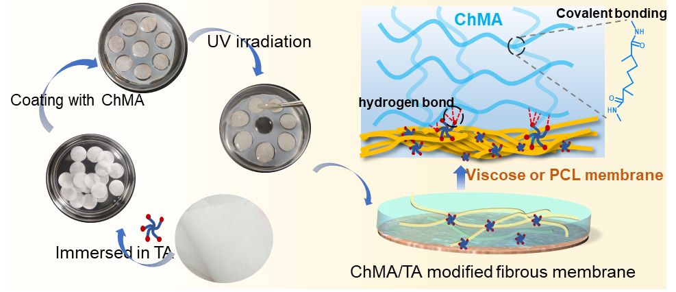
3.1 Physicochemical properties of the ChMA-modified hydrogel membranes
SEM images of the Viscose, Vis/ChMA and Vis-TA/ChMA membranes were displayed in Fig. 1a. The diameter of Viscose fibers was about 11.6 ± 0.1 μm, which was increased to 18.9 ± 0.2 μm after the modification (Fig. 1d). Fig. 1b showed the fibrous morphology of PCL membrane with a diameter of about 1.2 ± 0.1 μm, which turned to microporous structure after the modification of ChMA and TA. The pore size of the PCL nanofibrous membrane was about 2.7 ± 0.1 μm and significantly smaller than the Viscose fibrous membrane (17.8 ± 0.3 μm) (Fig. 1e), thus the dried hydrogel morphology (with pore size about 1.6 μm) was observed on the PCL-TA/ChMA sample. Fig. 1c showed the EDX spectra of the Vis-TA/ChMA and PCL-TA/ChMA membrane. Besides C and O elements, N elements from ChMA were detected on the membrane, which were comparable between Vis-TA/ChMA and PCL-TA/ChMA samples.
FTIR spectra of the Vis-TA/ChMA and PCL-TA/ChMA membranes were displayed in Fig. 2a. In the modified viscose fibrous membranes, characteristic absorption peaks of cellulose were presented, such as the stretching vibration peak of the saturated -CH bond (2921 cm-1) and the stretching vibration absorption peak of -OH (3300 – 3400 cm-1) (Zhang et al. 2023). The absorption peaks at 2948 cm-1 (-CH3, asymmetric stretching), 2864 cm-1 (-CH3, symmetric stretching) and 1723 cm-1 (C-O bond), which were infrared spectrum of PCL, could be found in spectra of PCL fibrous membranes (Li et al. 2021). New peaks were observed at 1535 cm-1 corresponding to amide II band (N-H deformation and C-H stretching) from ChMA (Xu et al. 2023b). Additionally, the peak at 1061 cm−1 was assigned to the saccharide structure of the chitosan derivatives (Xu et al. 2023b). There was no obvious new peak appeared in the spectra after TA immersed, indicating the mainly physical crosslinking between TA and ChMA (Li et al. 2023b).
The statistical results of equilibrium swelling ratio were shown in Fig. 2b. The PCL nanofibrous membrane exhibited the lowest swelling rate about 21.4 ± 1.0%. After the modification of ChMA hydrogel layer, the swelling ratio of PCL/ChMA increased to 86.0 ± 0.6%. And the addition of TA further improved the water retention of the fibrous membrane, reaching to 118.3 ± 1.5%. The Vis/ChMA had the highest swelling rate among all fibrous membranes, reaching to 203.9 ± 1.4%. Compared with the Vis/ChMA, the swelling ratio of the Vis-TA/ChMA decreased 193.4 ± 0.4%, which was attributed to the reduced pore size of the dual cross-linked network between ChMA and TA. Significantly larger swelling rate of the Vis-TA/ChMA was observed than that of the PCL-TA/ChMA group. The hydrophilicity and pore structure of the matrix are the main factors affecting the swelling rate of fibrous membranes (Shanmugapriya et al. 2018). The hydrophilic TA and ChMA as well as the porous structure of the hydrogel were the main reasons for the enhanced swelling ratios of the modified fibrous membranes. Compared with the PCL membrane, the Viscose exhibited larger pore size and higher water affinity, which led to higher swelling rate of the viscose-based membranes (Linju et al. 2023). Specially, the ChMA/TA modified viscose hydrogel fibrous membranes showed better water retention performance, which was beneficial for absorbing wound exudates.
To further study the degradation rate, the samples were soaked in PBS solution at different pH values (5 and 8) for several days. As show in Fig. 2c and d, the Viscose and PCL membrane showed similar degradation profile at different degradation solutions. And the remaining weight of all the samples was slightly larger in the basic solution than the values in the acidic solution. Specially, after the modification, the weight of the PCL-TA/ChMA was decreased to ca. 85.6 ± 2.0% by day 14 in the acidic solution, which was degraded about 2.5 times as much as the Vis-TA/ChMA (93.7 ± 0.7%) (Fig. 2d). The similar degradation pattern of the PCL-TA/ChMA was observed in basic environments (Fig. 2c). And the PCL-TA/ChMA exhibited the lowest weight ratio (83.7 ± 1.5%) at day 14 in basic environments (Fig. 2c). The PCL-TA/ChMA exhibited higher degradability than other membranes in both degradation solutions. The PCL-TA/ChMA exhibited higher degradability than other membranes in both degradation solutions, may be attributed to a less molecular interaction between PCL and TA as well as the diffuse of TA. The lack of crosslinking or molecular interaction played a major role in the degradation of fibrous membranes (Peng et al. 2012; Zhou et al. 2022b). . In addition, the degradation degree of the PCL-TA/ChMA was higher than that of the Vis-TA/ChMA, indicating that the effects of small fiber size and high porosity (high surface area) were also essential factors that required considering for fiber degradation (Peng et al. 2012). The PCL-TA/ChMA with smaller fiber diameter and high surface area, thus making the system easier to degrade.
3.2 Mechanical property of ChMA-modified hydrogel fibrous membranes
The rheology and mechanical performance of the prepared hydrogel fibrous membrane were investigated. As shown in Fig. 3a, the strain sweep assay of the Vis-TA/ChMA and PCL-TA/ChMA samples exhibited a linear viscoelastic region with a high fracture strain of about 10%. The dynamic frequency sweeps in Fig. 3b showed that G' was always larger than G'' under gradually increased frequencies, demonstrating the dominant elastic behavior of the Vis-TA/ChMA and PCL-TA/ChMA samples.
Typical tensile stress-strain curves for the ChMA-modified fibrous membranes at wet state were presented in Fig. 4a-f. From Fig. 4a-c, we can see that the tensile strength and elongation at break of the Viscose were about 2.3 ± 0.2 MPa and 55.1 ± 0.9%, respectively, which were slightly decreased to 1.6 ± 0.2 MPa and 42.5 ± 5.4%, respectively, after the modification of the ChMA/TA hydrogel. Fig. 4d-f showed the typical strain-stress curves, tensile strength and elongation at break of the PCL, PCL/ChMA and PCL-TA/ChMA at wet state. And the tensile strength and elongation at break of PCL were about 3.6 ± 0.5 MPa and 585.8 ± 21.1%, respectively, which were slightly decreased to 3.2 ± 0.4 MPa and 618.2 ± 18.2%, respectively, after the modification of the ChMA-TA hydrogel (Fig. 4ef). No significant difference in mechanical properties among the PCL, PCL/ChMA and PCL-TA/ChMA samples. Moreover, the tensile strength and elongation at break of ChMA-modified PCL membranes were significantly larger than the values of the ChMA-modified viscose membranes. Although the tensile strength and elongation at break of the PCL and Vis membranes were slightly decreased after modification, the mechanical property of all the samples were still in the range of nature skin tissue (Gao et al. 2023).
3.3 Antibacterial property of ChMA-modified hydrogel fibrous membranes
Bacterial infections pose a huge threat to human health globally, which is the necessity for designing clinic dressings with excellent antibacterial property (Zhou et al. 2023). As show in Fig. 5, the antibacterial activity of ChMA-modified fibrous membranes was evaluated by both qualitative and quantitative methods. Fig. 5a showed the inhibition zone against E. coli and S. aureus of the ChMA-modified fibrous membranes by the qualitative methods. The Viscose and Vis/ChMA fibrous membranes without loading TA did not show obvious inhibition zone, but the Vis-TA/ChMA showed inhibition zone with diameter of 1.8 ± 0.1 and 1.9 ± 0.2 cm against E. coli and S. aureus, respectively (Fig. 5a). Similarly, the PCL-TA/ChMA showed inhibition zone with diameter of 1.6 ± 0.1 and 1.6 ± 0.3 cm, respectively. There was no significant difference in the inhibition zone between the two dressings. In addition, as shown in Fig. 5b, the bacterial survival rates of E. coli and S. aureus were quantitatively characterized. The antibacterial rates of Vis/ChMA against E. coli and S. aureus were 38.0 ± 7.4 and 43.6 ± 2.1%, respectively, while the antibacterial rates of PCL/ChMA against the two bacteria were 33.2 ± 2.6% and 39.8 ± 3.4%, respectively. Additionally, the Vis-TA/ChMA exhibited the significantly larger antibacterial rate of 95.8 ± 0.2% against E. coli and 98.5 ± 1.0% against S. aureus. The antibacterial rates of PCL-TA/ChMA against E. coli and S. aureus increased to 90.0 ± 1.4% and 98.9 ± 0.6%, respectively. The statistics of the number of bacterial colonies (Fig. 5c) in the treated bacterial solution were consistent with the above pattern. The great antibacterial performance of ChMA and TA modified fibrous membranes was attributed to the positive charge of chitosan as well as the rich phenolic hydroxyl groups of TA drugs (Zhou et al. 2022a). The antibacterial property of the TA encapsulated membrane was achieved by membrane destruction mechanisms of TA, which could destruct the bacterial cell wall structure and change cell membrane permeability (Li et al. 2023b). Wu et al. achieved great antibacterial effects by increasing the content of TA solution (5-20 wt %) and assisting with photothermal methods (Wu et al. 2023). Herein, modified hydrogel membranes loading with 9% TA, exhibited excellent antibacterial rate (>90% bacterial death upon). Therefore, the ChMA and TA modified fibrous membrane exhibited excellent antibacterial properties, especially against S. aureus (>95%), which were beneficial for the wounds repair.
3.4 Antioxidant property of ChMA-modified hydrogel fibrous membranes
The antioxidant activity of ChMA/TA modified hydrogel membranes was evaluated by the reduction of DPPH free radicals (Fig. 6a). After receiving electrons or hydrogen atoms, DPPH radicals were neutralized, and the color of the supernatant changed from purple to yellow (Selvaraj et al. 2017). As show in Fig. 6b, the purple color of the supernatant was faded in the ChMA/TA-modified hydrogel fibrous membrane. And the Vis-TA/ChMA and PCL-TA/ChMA groups showed almost no purple color, indicating that free radicals were almost completely captured (Fig. 6b). The radical scavenging rates of the Vis/ChMA and Vis-TA/ChMA were enhanced to 39.2 ± 1.4% and 89.9 ± 0.5%, respectively, comparing with the Viscose (Fig. 6a). Moreover, the radical scavenging rates of the PCL/ChMA and PCL-TA/ChMA were increased to about 60.4 ± 8.7% and 91.0 ± 0.4%, respectively, comparing with the PCL. And no significant difference of the radical scavenging values between the PCL-TA/ChMA and Vis-TA/ChMA. Antioxidant activity of the ChMA-modified hydrogel fibrous membrane was significantly improved. And the capture rates of DPPH radicals by Vis-TA/ChMA and PCL-TA/ChMA were over ca. 90%. The enhanced DPPH free radical scavenging ability of the ChMA-modified fibrous membrane was attributed to the free amino groups on ChMA with electron transfer ability, which could react with free radicals and exhibit antioxidant activity (Xu et al. 2023b). Meanwhile, the abundant pyrogallol and catechol groups in TA endow it with the ability to inhibit free radicals through electron transfer (Xu et al. 2023a). Moreover, the free radical scavenging ability of TA was also relevant with its concentration (Xu et al. 2023a). Given that the content of TA was identical in both viscose and PCL samples, there was no significant difference of the radical scavenging values between the PCL-TA/ChMA and Vis-TA/ChMA. The synergies of ChMA and TA enabled the ChMA-modified hydrogel fibrous membrane to maintain excellent antioxidant capacity.
3.5 Hemolysis and cell culture
Hemocompatibility was particularly important for the application of wound healing materials (Lu et al. 2023a; You et al. 2022). As shown in Fig. 7a., the supernatant in modified membranes exhibited similar color (nearly colorless) to that of the PBS group. However, the control group of deionized water showed a uniform red solution, which was caused by complete rupture and hemolysis of red blood cells. In addition, quantitative analysis of blood compatibility showed that the hemolysis rates of all samples did not exceed 3% (Fig. 7a). Therefore, the ChMA/TA modified hydrogel fibrous membranes possessed good blood compatibility. The in vitro cell compatibility of ChMA/TA modified fibrous membranes was evaluated through co-incubation with fibroblasts. The results of the cytotoxicity test were shown in Fig. 7b. Compared with the pure Viscose group, ChMA/TA modified membranes did not exhibit negative effects on cell survival. There was no significant difference in cell viability among the groups. ChMA/TA modified hydrogel fibrous membranes exhibited excellent cell compatibility, which was benefited from the good biocompatibility of raw materials and scientific preparation processes.
3.6 In vivo wound healing
The in vivo wound healing capacity of ChMA-modified hydrogel fibrous membranes were evaluated by a full thickness defect wound model. As shown in Fig. 8a-c, on day 3, the wound contraction of the ChMA-modified viscose and PCL membrane groups reached to about 21.6% and 20.1%, respectively. And on day 7, the wound contraction of the ChMA-modified viscose and PCL membrane groups was improved to about 73.2% and 77.3%, respectively, significantly larger than the Viscose and PCL groups. On day 14, the wounds treated by the Vis TA/ChMA and PCL-TA/ChMA groups were almost closed, with wound contraction ratios of 95.6% and 98.1%, respectively, whereas the wounds of pure Viscose or PCL group remained macroscopically non-healing. The detail of the wound healing was further evaluated by the H&E staining on day 14 (Yang et al. 2024; Mirhaj et al. 2022). As shown in Fig. 8e, compared with the viscose and PCL groups, the ChMA/TA-modified groups showed more fibroblasts (blue arrow) and less inflammatory cells (red arrow) around the impaired region. Moreover, a layer of epithelium was formed in the Vis-TA/ChMA and PCL-TA/ChMA group (Fig. 8d). And the dermal tissue and skin appendages were regenerated in the ChMA/TA-modified groups on day 14. Granulation tissue thickness of the Vis-TA/ChMA and PCL-TA/ChMA groups were measured about 759.1 μm and 454.3 μm, respectively, which were larger than the value of the viscose (407.4 μm) and PCL group (437.9 μm). Simultaneously, more regular hair follicles were observed in the ChMA/TA-modified groups. No significant difference between the ChMA/TA-modified PCL or viscose membrane. These results evidenced that the ChMA/TA-modified hydrogel fibrous membranes with spatially designed structure displaying excellent wound healing behavior. The treatment effects of the ChMA/TA-modified hydrogel fibrous membranes were attributed to the spatially designed structure and the composition (Yang et al. 2024; Mirhaj et al. 2022). Firstly, the porous structure of ChMA hydrogel maintained proper wound pus absorption effect, and providing a moist environment that was benefit for wound repair. Besides, TA exhibited an antibacterial effect by reducing S. aureus growth and alleviate the inflammatory immune response induced by bacterial infection and accelerate the wound healing process. In summary, ChMA/TA-modified hydrogel fibrous membranes exhibited large wound contraction, thick granulation tissue and superior wound healing effectiveness in vivo. Considering the ease of accessibility of viscose as well as the simplicity and cost-effectiveness of the current approach, ChMA/TA-modified viscose membrane would be utilized as highly promising wound dressings.