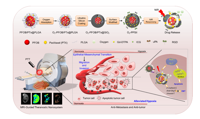
3.1 Facile synthesis and Characterization of O2-PPSiI Nanosystem
The O2-PPSiI nanosystem was chemically synthesized by a two-stage self-assembly process, which was the PFOB core as the oxygen carrier, and then encapsulated into an ultrathin-walled silica shell (Fig. 1A). In the first stage, the PFOB core (i.e., the core without the silica coating) was synthesized via an emulsion–(solvent-evaporation) method[35], and PTX was mixed into PLGA to form the PFOB/PTX@PLGA (abbreviated as PP) nanocapsule. And then the PP nanocapsule was used to absorb O2 as a carrier. In the second stage, the ultrathin-walled silica shell was synthesized by hydrolysis and condensation of TEOS. The silica shell was heterogeneous in nature, and thus biocompatible DLmenthol was used to prevent oxygen release from the nanosystem. The loading efficacy of PTX in O2-PPSil was about 20 %.
Transmission electronic microscopy (TEM) pictures presented the morphology of O2-PPSi which was a typical spherical shell/core structure (Fig. 1B) with an average diameter of 203 nm (Fig. 1C). Compared to PP (− 8.8 mV), the zeta potential of O2-PPSi elevated to + 22.9 mV (Fig. 1D), which is due to the formation of the aminated silica shell on the PP surface. The elemental mapping images of O2-PPSi nanoparticles showed the presence of Si and F elements (Fig. 1E), which further verified the loading of PFOB and coating of the silica shell. Benefiting from the rich amino and silicon hydroxyl groups on the surface, the tumor-targeting ligands uPA and RGD were covalently modified to the surface of O2-PPSi nanoparticles; photothermal agent ICG and MR contrast agent Gd-DTPA (Gd) were also added by electrostatic interaction. The morphology of the nanosystem (named O2-PPSiI) was also observed by TEM and scanning electron microscopy (SEM). Figures 1F and 1G show uniform O2-PPSiI with an average diameter of 203 nm, and the thickness of silicon shell with the decoration on its surface was about 8 nm (Fig. 1H).
The X-ray photoelectron spectroscopy (XPS) and Fourier transform infrared spectroscopy (FTIR) results further proved that the O2-PPSiI was successfully synthesized. The silicon and the gadolinium peaks were seen in the XPS survey spectra for the surfaces of O2-PPSiI, suggesting that the silica shell and Gd-DTPA were successfully coated and modified on the shell of O2-PPSiI (Fig. 1I). The high resolution Gd 4d XPS spectra for O2-PPSiI inserted in Fig. 1I revealed that the characteristic peaks centered at 142.8 eV and 153.7 eV could be attributed to Gd 4d5/2 and Gd 4d3/2, suggesting the existence of Gd (III), which derived from Gd-DTPA[36]. A satellite peak at 148.2 eV in the Gd 4d XPS spectra may derived from the coordinate bond of Gd (III) and DTPA. The XPS spectrum of C 1s is shown in O2-PPSi (Figure S1A), and the peak components at 284.5 eV and 286.6 eV were appointed to the C-C and C-N groups, respectively. These were attributed to APTES on the surface of O2-PPSi. After ICG and Gd-DTPA grafting on the surface of O2-PPSi, two new peaks at an energy of 284.7 eV and 288.3 eV were seen and could be attached to the C = C and C = O group, respectively (Figure S1B). The chemical structure of O2-PPSiI was analyzed using FTIR. Versus the FTIR spectrum of O2-PPSi, the broad band at ≈ 1600 cm− 1 resulted from the C = N band and the peaks at 1414 cm− 1 that are represented by the vibrational stretching of the C = C groups (Fig. 1J), which are from the ICG. The spectrum of DTPA exhibited the characteristic peak of the asymmetric and symmetric carbonyl (C = O) stretch of anhydride at 1738 cm− 1. Meanwhile, a characteristic UV absorption spectra peak of ICG emerged at 808 nm after the ICG was grafted on the surface of O2-PPSi (Figure S2), which matched the wavelength of the NIR laser applied for PTT. Versus the O2-PPSi, the color change of O2- PPSiI indicated that the ICG was coated on the surface (Figure S3).
We also evaluated the magnetic properties of O2-PPSiI using T1-weighted imaging (T1WI) and T1 mapping MRI, and the results showed that the T1WI signal of nanosystem was linear and concentration-dependent. The T1 relaxivity (r1) of O2-PPSiI to be 27.812 mM− 1/s− 1 (Figure S4), and it was dramatically superior than the free Gd-DTPA (4–5 mM− 1/s− 1), indicating a favorable T1WI contrast effect of O2-PPSiI. These results demonstrated that O2-PPSiI could enhance T1 positive contrast. Therefore, we further examined the accumulation of O2-PPSiI in vivo by MRI. The accumulation of O2-PPSiI in the tissue was quantitatively evaluated by the decrease of T1 value (longitudinal relaxation time, normalized as △T1 to the base). The injected O2-PPSil was selectively accumulated in the tumor and reached its maximum at 8 h (△T1 26.28%) after the systemic administration in vivo (Fig. 1K). However, the accumulation of Gd-DTPA had no significant selectivity between the tumor and normal muscle tissue. It reached the maximum at 30 min (△T1 18.45%) but recovered to baseline after injection for 2 h (Fig. 1L). These results demonstrated that O2-PPSiI could enhance T1 positive contrast of Gd-DTPA and tumor targeting ability against TNBC in vivo.
3.2 Photothermal Ability of O2-PPSiI Nanosystem
The photothermal ability of O2-PPSiI was then examined in this study. The absorption of O2-PPSiI at 750 − 850 nm showed obvious concentration dependence (Fig. 2A). The extinction coefficient of O2-PPSiI at 808 nm for different concentrations was 82.16 Lg− 1 cm− 1 (Figure S5), indicating that the O2-PPSiI nanosystem has high photothermal-conversion performance due to ICG modification. The photothermal-conversion efficiency (η) of the O2-PPSiI nanosystem was determined as 45.45% based upon the data analysis in Figure S6 and Fig. 2B. To estimate the photothermal-conversion performance of O2-PPSiI, it was stimulated by an 808 nm NIR laser with increased power densities (0.5, 1.0, 1.5, and 2 W cm− 2) when the concentration was locked at 300 µg mL− 1. As shown in Fig. 2C, the temperature elevation of O2-PPSiI increased by 35°C (reached 61°C) over 3 min of irradiation (1.0 W cm− 2), and the thermal pictures of this process were documented with an IR thermal camera (Figure S7), demonstrating that O2-PPSiI had an excellent photothermal-conversion performance. The packaging of ICG in O2-PPSiI can contribute to the improvement of photostability versus free ICG, as proved with a reducible photothermal effect under repeated laser cycling for five cycles[37] (Figure S8).
3.3 NIR-triggered O2 Release and Drug Delivery
The system has NIR-induced photothermal effects from ICG and active oxygen release in PFOB [38]. We encapsulated the oxygen reservoir and antitumor agent into the nanocore and developed an NIR-responsive on-demand drug releasing nanomedicine system to overcome the drug leakage of conventional nano-based drug delivery system (NDDSs). We measured the O2 concentration change of O2-PPSiI in deoxygenated water under a nitrogen atmosphere and found that the O2 concentration increased significantly in the O2-PPSiI solution; many bubbles appeared with longer NIR irradiation times (Fig. 2D and Figure S9). Subsequently, ultrasound imaging was used to visualize the NIR-triggered O2 release and drug delivery in vitro. Figure 2E and Figure S10 show an apparent enhanced echo intensity in the O2-PPSiI solution after NIR irradiation. This was further confirmed via successful development of the NIR-responsive on-demand drug releasing nanomedicine system in this study. Meanwhile, the O2 concentration of the O2-PPSiI solution without NIR irradiation at room temperature showed no obvious change within 24 h (Fig. 2F), suggesting that the stability of O2-PPSiI was favorable. This proved that the O2-PPSiI has higher O2storage ability than other O2delivery nanosystems. The temperature elevation of O2-PPSiI with NIR laser irradiation was the key contributor of oxygen release from O2-PPSiI. Figure 2G and Figure S11 show that the morphology of O2-PPSiI swelled and then gradually collapsed at 60 ℃ and 70 ℃ when the reaction temperature reached 50 ℃. This indicates that the O2 release from O2-PPSiI was accompanied with the structural collapse of the SiO2 shell.
Subsequently, the photothermal-conversion performance of O2-PPSiI and the O2 release from O2-PPSiI nanosytem in vivo were further investigated via an IR thermal camera and ultrasound imaging. The outward temperature of tumor treated with O2-PPSiI gradually increased from 35 to 60°C along with the laser irradiation time came to 3 min (808 nm, 1 W cm− 2; Fig. 2H). This temperature increase could ablate the tumor. Moreover, the ultrasound images recorded the O2 release process under the laser irradiation (Fig. 2H and Figure S12). These results demonstrated that O2-PPSiI offered NIR-responsive on-demand drug-release in vitro and in vivo.
3.4 Distribution of O2-PPSiI Nanosystems In Vivo
We first used MRI to monitor the distribution of the O2-PPSiI nanosystem in vivo at various time points. Figure 3A shows that intravenously injected O2-PPSiI was selectively accumulated in the tumor region and came to its maximum at 8 h. Meanwhile, the fluorescence imaging in vivo was used to further monitor the distribution of the O2-PPSiI nanosystem in the tumor by capturing the ICG signal. The fluorescence images also verified the injected O2-PPSiI accumulating in the tumor; it reached its maximum at 8 h and then had long-term retention in the tumor region until 72 h, which corresponded to the MRI data (Figure S13). Therefore, dual modal imaging can effectively and precisely monitor the distribution of the O2-PPSiI nanosystem in vivo and confirm the best time point of NIR irradiation.
Transporting the drugs to the deep tumor tissue is limited owing to abnormal angiogenesis and irregular tumor blood flow. Currently, functional nanosystems have provided new strategies to enhance tumor penetration and achieve effective and successful antitumor therapy[39]. Here, intravoxel incoherent motion diffusion-weighted imaging (IVIM-DWI) was used to evaluate the in vivo tumor vascularity. As a non-contrast-based functional MRI sequence, IVIM-DWI is an attractive approach to assess not only the tumor cellularity but also the micro-perfusion with no need to use exogenous contrast agents. This allows it to be repeatedly used as a method of quantitatively monitoring the therapeutic response in vivo [40]. In this work, we applied IVIM-DWI to evaluate the microperfusion of the baseline tumors, and then drew two orthogonal lines in the Dorsal/Ventral (Do/Ve) and Medial/Lateral (Me/La) directions crossing the tumor center. This divided each radius into 10 segments with equivalent lengths (each segment was then appointed with a figure of 1–10, i.e., the outermost segment was appointed with 1 and the innermost segment of 10 at a radial position). We then confirmed the heterogeneous vascularity of TNBC and found that the intratumor blood flow at the normalized radius ≥ 6 in both the Ve/Do and La/Me direction was significantly decreased versus the outermost segment (Fig. 3B and Fig. 3C), which was also observed by Gaustad [41]. Based on these results, we further divided the entire tumor into peripheral (the outermost segment 1 to segment 5) and central (segment 6 to the innermost segment 10) zones. The injected O2-PPSiI infiltrated deep into the central region with no significant difference in △T1 versus the peripheral zone (Fig. 3D). The Gd-DTPA mainly accumulated in the peripheral region and presented a lower △T1 than the central zone (Fig. 3E). These results confirmed a favorable intratumor penetration of O2-PPSiI, and the potential mechanism may be associated with the cell-to-cell transport through the active targeting of uPAR in the tumor and stromal cells [42].
We further investigated the biodistribution of injected O2-PPSiI in the tumor-bearing nude mice. The T1 MRI data presented that, except for the intratumor accumulation, the injected O2-PPSiI was mainly collected by the kidney within 24 h (Figure S14A). It had a fluctuating yet increasing trend in △T1 after 24 h (Figure S14B), which may reflect the urinary excretion of O2-PPSiI. In addition, the injected O2-PPSiI was also taken up by the liver and spleen that showed a fluctuating change in △T1 (Figure S15C–D). Moreover, the fluorescence images showed most of the injected O2-PPSil accumulated in the intestinal system within 8 h, and then vanished gradually after 12 h excepted for the retention in the tumor region. These results indicated a urinary and intestinal excretion of the O2-PPSiI, further confirming its favorable biodegradability in tumor theranostics.
3.5 Antagonizing Tumor Hypoxia Statues by NIR-triggered Oxygen Release In Vivo
Hypoxia offers multiple problems in the treatment of cancers[43]. Antagonizing tumor hypoxia statues is important for tumor therapy[44, 45]. Therefore, the efficacy of relieving tumor hypoxia for O2-PPSiI nanosystems was further investigated with MDA-MB-M231 tumorbearing nude mice. First, based upon the paramagnetic effect of deoxyhemoglobin, we used blood oxygenation level-dependent magnetic resonance imaging (BOLD-MRI) to monitor the realtime improvements in tumor hypoxia as well as the efficacy of NIR-triggered oxygen-shuttle nanomedicine in promoting tumor oxygenation. Improvements in tumor hypoxia were evaluated quantitatively using the decreased R2* values of BOLD-MRI [46]. Figure 3F shows that the tumors treated with NIR-triggered O2-PPSiI and O2-PSiI (without PTX, 808 nm, 1 W cm− 2) demonstrated a reduction in R2* (normalized as △R2* to the base) versus the baseline in both the central and peripheral zones. These results proved a satisfactory tumor oxygenation level in the local tissue. Versus the saline group, the tumors treated with a single NIR laser, PTX, and O2-PPSiI showed no significant difference in △R2*, while the NIR-triggered O2-PPSiI and O2-PSiI treatment groups had a significant decrease in △R2* at the endpoints of the treatment in both the central and peripheral zone (Fig. 3G). The tumor treated with NIR-triggered PPSiI was considered as a negative control to verify the contribution of O2 release for the tumor hypoxia relieving effect. Versus the NIR-triggered PPSil group, the tumor treated with NIR-triggered O2-PPSil showed a significantly decreased △R2* in both the central and peripheral zones, which further proved the improvements in tumor hypoxia statues by NIR-triggered oxygen release[47]. Furthermore, we performed HIF-1α immunofluorescence staining to confirm the efficacy of NIR-triggered O2-PPSiI in promoting tumor oxygenation (Fig. 3H). Consistent with the results of BOLD-MRI, the tumor treated with NIR-triggered O2-PPSiI showed a decreased expression of HIF-1α. But then, tumors treated with single NIR, PTX, O2-PPSiI, and NIR-triggered PPSiI exhibited high expression of HIF-1α with strong green immunofluorescence in different levels. The BOLD-MRI and immunofluorescence staining data suggested that NIR-triggered O2-PPSiI could mitigate the hypoxia microenvironment of TNBC in vivo, especially to the central tumor that presented as an avascular region with poor blood flow.
3.6 Synergistic Therapeutic Efficacy of NIR-Triggered O2-PPSiI
The in vitro cytotoxicity of the O2-PPSiI nanosystem was investigated with MDA-MB-231 cells by MTT [48]. Figure S15 shows that the viability of the cell treated with NIR irradiated O2-PPSiI reduced to 17.8%. Meanwhile, the MDA-MB-231 cells treated with PTX and NIR irradiated PPSiI and O2-PSiI exhibited higher cell viabilities of 55.9%, 35.1%, and 48.2%, respectively. The excellent cancer cell killing efficiency for NIR irradiated O2-PPSiI nanosystem in vitro could be attributed to the hyperthermia of ICG and the chemotherapeutic effect of PTX releasing from the O2-PPSiI nanosystem.
To further investigate the cancer cell killing mechanism of O2-PPSiI in vitro, we measured the ROS and 1O2 generated by O2-PPSiI[49]. Versus PTX and NIR-triggered PPSiI, O2-PPSiI under NIR laser irradiation showed more high-efficiency ROS and 1O2 generation in solution and MDA-MB-231 cells (Figure S16), indicating an important role of O2 release in the production of 1O2 and ROS [50]. Subsequently, the in vivo synergistic antitumor effect of the NIR-triggered O2-PPSiI nanosystem was further investigated with MDA-MB-231-bearing nude mice. Based on the precise monitoring for the distribution of O2-PPSiI with T1 mapping MRI and fluorescence imaging in vivo (Fig. 3A and S11), the NIR irradiation was triggered at 8 h after the injection of PPSiI, O2-PSiI, or O2-PPSiI. The relative changes of tumor volume and weight are revealed in Fig. 4A–4C. Over 21 days of treatment, the relative tumor volume (normalized as △Volume to the base) of mice treated with only saline, O2-PPSiI, and NIR irradiation groups increased significantly (Fig. 4A). The mice treated with PTX and NIR-triggered O2-PSiI (without PTX) presented a slightly increased tumor volume compared to day 0, demonstrating the moderate tumor inhibition of the single chemotherapy and phototherapy against TNBC. However, the tumor volume of mice treated with NIR-triggered PPSiI showed no significant difference at 14 days and 21 days compared to day 0, while the NIR-triggered O2-PPSiI group showed significantly decreased tumor volumes (Fig. 4B), and the volume as well as the tumor weight at the endpoint of both the groups were significantly lower than the PTX and NIR-triggered O2-PSiI groups (Fig. 4C), demonstrating an obvious inhibition and regression effect of the synergistic chemo-phototherapy. After treatment for 21 days, the picture of separated tumors in each group also presented similar results as the tumor volume and weight revealed above (Figure S17). Moreover, the tumor volume and weight of the mice treated with NIR-triggered O2-PPSiI were significantly lower than those treated with NIR-triggered PPSiI after 21 days coincided with the results of 3D-CUBE T2WI at 21 days (Fig. 4D). The excellent antitumor efficiency in vivo for the NIR-triggered O2-PPSiI nanosystem could be ascribed to an enhanced synergistic chemo-phototherapy efficiency induced by O2 release triggered by NIR laser irradiation.
To further investigate the antitumor effect of the NIR-triggered O2-PPSiI in vivo, the parameters of IVIM-DWI (D and f value) were applied to track the tumor cellularity and perfusion during the therapy[51]. Figure 4E and Figure S18 showed the △D value (normalized as △D to the base) in both the central and peripheral regions of the NIR-triggered O2-PPSiI group increased significantly compared the PTX, NIR-triggered O2-PSiI, and PPSiI groups, which demonstrated a reduced cellular density and activity of the tumor in vivo [52]. The results suggest that the enhanced synergistic chemo-phototherapy efficacy induced by tumor hypoxia improvement under NIR laser irradiation could effectively inhibit the tumor cell proliferation especially to the avascular central tumor. Moreover, tumor progression was suppressed by the synergistic chemo-phototherapy which was confirmed with microscopic analysis of tumor tissues stained by hematoxylin and eosin (H&E). Figure 4F shows that O2-PPSiI under NIR laser irradiation could effectively decrease the cell viability in tumor regions which offering an additional support for the antitumor efficacy of the nanosystem in vivo. Terminal deoxynucleotidyl transferasemediated dUTP nick endlabeling (TUNEL staining assay) was also completed to further determine the cell proliferation and apoptosis in tumor tissue at treatment of 21 days. As displayed in Fig. 4G, the green and blue fluorescence of the NIR-triggered O2-PPSiI (G7) group had excellent overlap confirming DNA fragmentation and large apoptosis areas of tumor tissue.
3.7 The Suppression of Tumor Metastasis
The metastasis of the tumor is a major concern in the clinic. It has been reported that the hypoxic microenvironment in the tumor could influence the progression of metastasis[53]. The combination of O2-PPSiI and the NIR laser could release the O2 to relieve tumor hypoxia due to the rupture of the silica shell, which might antagonize hypoxia-induced tumor metastasis. Therefore, we first used the wound-healing migration assay together with the transwell assay to evaluate the suppression effect of the tumor metastasis in vitro of O2-PPSiI under NIR laser irradiation. Figure 5A compares the PTX group and the PPSiI + NIR laser group. The O2-PPSiI under NIR laser irradiation could effectively suppress the migration of MDA-MB-231 cells at the same concentrations. Meanwhile, the invasive ability of MDA-MB-231 cells treated by different group was also detected and monitored in real-time. Versus the control (refer to the Saline and Laser group), the invasive ability of MDA-MB-231 cells treated by PTX, PPSiI + NIR laser, or O2-PPSiI + NIR laser was obviously decreased (Fig. 5B and Fig. 5C). Certainly, the combination of O2-PPSiI and NIR laser showed more effective suppression than PTX, PPSiI, and NIR laser. These results proved a synergistic effect of NIR triggered O2-PPSiI nanosystem on suppressing migration and invasion of MDA-MB-231 cells, and suggested a positive role of the NIR-triggered oxygen release and tumor hypoxia improvement.
In the tumor hypoxic microenvironment, signaling pathways that facilitate cell survival and metastasis are activated to give the tumor cell the ability to migrate and invade via the epithelial-mesenchymal transition (EMT) [54, 55]. EMT is a biological process that promotes the transformation of immotile epithelial cells to motile mesenchymal cells, involving a reduction of epithelial markers and the increase of mesenchymal markers in the tumor [56]. To further explore the effect of NIR-triggered O2-PPSiI on suppressing the tumor metastasis in vivo, the epithelial-specific marker E-cadherin together with the mesenchymal-specific markers vimentin and Snail-Slug were chosen to perform immunohistochemistry microscopy [57]. Figure 5D and Fig. 5E show that the expression of epithelial-specific marker E-cadherin in the tumor treated with NIR-irradiated O2-PPSiI was much higher than that of the single PTX group, O2-PSiI + Laser group, or PPSiI + Laser group. In addition, the expression of mesenchymal-specific markers Snail-Slug and vimentin in the tumor treated with NIR-irradiated O2-PPSiI was much lower than that of the single PTX group, O2-PSiI + Laser group, or PPSiI + Laser group. These results demonstrated that the NIR-irradiated O2-PPSiI could inhibit the process of EMT in the tumor and decrease its migration and invasion due to the tumor hypoxia improvement induced by the oxygen release under NIR laser irradiation and synergistic chemo-phototherapy. These effects were further verified by the results of correlation analysis presented in Figure S19. In addition, the uPA/RGD dual-targeting molecules of O2-PPSiI may play a positive role in this process as indicated by significantly downregulating the expression of mesenchymal-specific markers vimentin and Snail-Slug in the O2-PPSiI group compared to the Saline [57, 58]
Furthermore, the partial activation of EMT program was considered a major driver of tumor progression from initiation to metastasis. Particularly, zinc finger E-box binding homeobox 1 (ZEB1) and transforming growth factor β (TGF-β) play essential roles in the proliferation, migration, and invasion of tumor cells. We next examined whether O2-PPSiI could inhibit ZEB1 and TGF-β levels in orthotopic MDA-MB-231 tumor-bearing mice. Immunofluorescence for the ZEB1 verified that O2-PPSiI combined with NIR laser could efficiently reduce the expression of ZEB1 withintumor in comparison with the saline group suggesting that this combined strategy suppressed the activation of cell motility and stemness (Fig. 6A and Fig. 6D). Versus the mice in PPSiI་Laser group, the O2-PPSiI combined with NIR laser distinctly inhibited the expression of ZEB1, which confirmed that relieving tumor hypoxia could effectively abrogate the hypoxia-induced EMT. Meanwhile, the EMT was also reduced by TGF-β downregulation due to the synergistic effects of NIR-irradiated O2-PPSiI (Fig. 6B and Fig. 6D).
Matrix metalloproteinase-2 (MMP2) is a member of the zinc-binding endopeptidase family and plays an essential role in the invasion and metastasis of cancer cells. Upregulation of MMP2 expression can also promote tumor metastasis[59]. Therefore, we further detected the expression of MMP2, proving that O2-PPSiI combined with NIR laser could inhibit tumor metastasis. Figure 6C and Fig. 6D show that O2-PPSiI under NIR laser irradiation could effectively downregulate the MMP2 expression. The negative correlation analysis of E-cadherin, vimentin, Snail-Slug and ZEB1, TGF-β, and MMP2 demonstrated that the O2-PPSiI + NIR laser group could effectively influence the EMT (Fig. 6E and Figure S20). Therefore, these results indicated that O2delivering strategies could alleviate hypoxia of the tumor tissue and abrogate the hypoxia-induced EMT to inhibit tumor metastasis.
3.8 Biosafety Evaluation of NIR-triggered O2-PPSil In Vivo
The in vivo biosafety for O2-PPSiI was systematically inspected via body weight monitoring, blood biochemical analysis, and H&E staining. Figure S21 shows that the body weight (normalized as △Body weight to the base) of the mice in NIR-triggered O2-PPSiI group increased during treatment for 21 days. This was significantly higher than the baseline and the saline group at 21 days. The body weight of PTX-treated mice declined significantly compared to the baseline over 9 days after injection. The blood biochemical analysis of mice treated with PTX also indicated an impaired metabolic function of the liver with significantly elevated Alanine transaminase (ALT) and Aspartate aminotransferase (AST) in the blood (Fig. 7A). This was further verified by the presence of patchy lymphocyte infiltration and hepatocyte swelling in liver H&E staining (Fig. 7B); no obvious hematological and histological abnormalities were observed in the other groups (Fig. 7A and Fig. 7B). These results indicated the hepatotoxicity of single PTX treatment and proved the safety of O2-PPSiI for clinical translation.