3.1. Characterization of prepared carriers
The crystallographic data of the prepared SiGe hybrid was addressed by X-ray diffraction analysis, as indicated in Fig. 1. The appeared XRD pattern shows a broad peak at 2θ = 15–35 ° which is coressponded to formation of amorphous phase of the silica according to JCPDSn PDF data (29–0085) [25], as well as amorphous structure of gelatin [26].
The chemical properties and formation of prepared carriers was evaluated using FTIR analysis, as indicated in Fig. 2. The broad band around 3400 cm− 1 in all spectra is due to stretching vibrations of the O‒H or N‒H bonds [27]. In the FTIR spectra of the SiGe, and SiGe/BSA-FA, the observed bands around 1100, 470 and 800 cm− 1 are related to Si − O−Si stretching, Si − O stretching, and Si − O bending mode of silica network, respectively [28]. Also, the appeared bands around 1670 cm− 1 (C = O stretching or amide-I), 1540 cm− 1 (amide-II), and 1240 cm− 1 (amide-III ) in FTIR spectra of BSA-FA, and SiGe/BSA-FA show characteristic bonds of gelatin and BSA [15, 21]. Futhermore, the observed bands around 2920 cm− 1 in all spectra are due to stretching vibrations of C − H bonds [29].
The surface area, mean pore size and total pore volume of the prepared samples measured using BET and BJH methods were reported in Table 1. Accordingly, the Bet surface area of the SiGe, and SiGe/BSA-FA hybrid nanocarriers were measured about 374, and 378 m2/g, respectively, indicating proper surface area of prepared porous nanocarriers. However, loading of 5FU into the pores of the SiGe/BSA-FA caused in significant decrease of SBET, confirming the successful encapsulation of the 5FU into the SiGe/BSA-FA.
Table 1
The surface area, mean pore size and total pore volume of the prepared samples
| Samples | Surface area (m2/g) | Mean pore size (nm) | Total pore volume (cm3/g) |
|---|
| SiGe | 374 | 5.1 | 0.74 |
| SiGe/BSA-FA | 378 | 6.5 | 0.67 |
| SiGe/BSA-FA@5FU | 271 | 8.7 | 0.78 |
Figure 3 displays the nitrogen adsorption-desorption isotherms and pore size distribution of SiGe, SiGe/BSA-FA, and drug loaded nanocarrier. Regarding the IUPAC classification, the adsorption isotherms of the prepared nanocarriers were in line with type-IV, confirming the formation of mesoporous structure in all samples [30, 31]. Furthermore, the appeared hysteresis loops in these isotherms shows the occurrence of capillary condensation in mesoporous structure of nanocarriers. Here, the type of hysteresis loops refers to type H-2, which indicates interconnected pore networks along with a partially agglomeration in structure [27, 32].In addition, the pore size distribution in Fig. 4.b shows a narrow and uniform distribution in the range of 2–40 nm.
The morphology and surface properties of the SiGe, and SiGe/BSA-FA were investigated using FE-SEM micrographs. The results in Fig. 4 clearly exhibited a porous structure with spherical shape for SiGe in the range of 200–300 nm. However, systematic changes due to surface modification of the SiGe nanocarrier with BSA-FA was observed in the SEM image of the SiGe/BSA-FA system, so that an approximately spherical and porous particles along with a rough surface is visible for SiGe/BSA-FA sample. Similar SEM results were also observed by Maheswari, when the surface of the calcium ferrite spherical nanoparticles was modified with BSA [21].
3.2. Entrapment efficiency and release study
The prepared SiGe/BSA-FA carrier was used to evaluate the entrapment property and delivery behavior of 5FU. Here, the content loading (LC) and entrapment efficiency (EE) of 5FU, measured using UV–Visible spectroscopy method, were obtained at about 70% and 29%, respectively. The high entrapment efficiency of 5FU is mainly thought to be related to penetration of drug molecules and chemical interactions between hydrophilic drug of the 5FU and proper functional groups in the SiGe/BSA-FA structure [33].
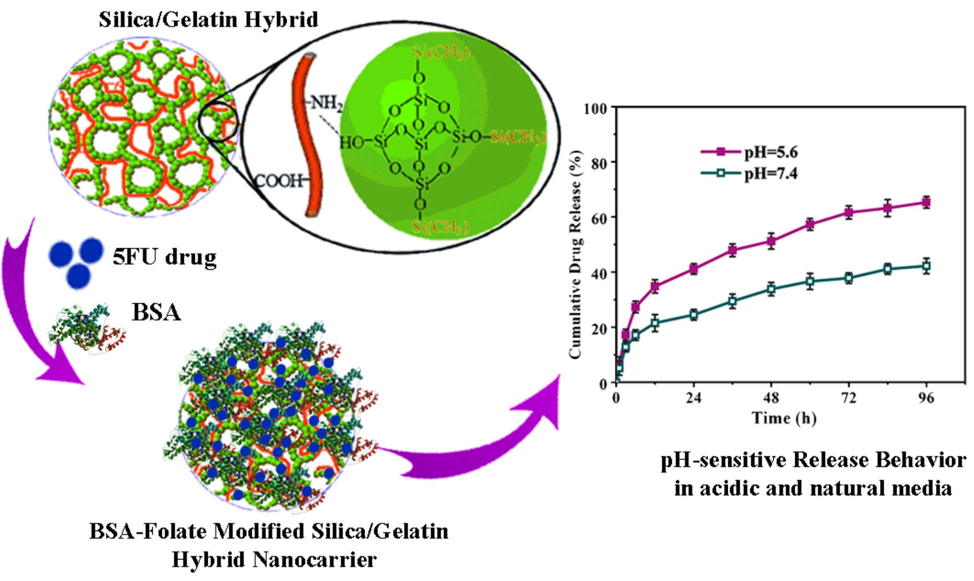
In cancerous tissue, changes in the body's metabolic conditions and the uncontrolled proliferation of cancer cells lead to an acidic microenvironment [21]. Based on these properties of cancer conditions, stimuli-responsive drug delivery platforms such as pH-sensitive systems are of great interest for targeted delivery [34]. Here, the pH-responsive release behavior of the drug from the SiGe/BSA-FA@5FU nanocarrier was investigated under two different pH media of 5.6 and 7.4, similar to the acidic condition of cancer tissue and the normal physiological condition of the body. It should be noted that the in-vitro drug release under acidic condition was carried out to evaluate the pH-responsive behavior of nanocarrier, effect of acidic microenvironment of cells on release profile. Figure 5 indicates the cumulative 5FU release from the SiGe/BSA-FA@5FU hybrid nanocarrier under two different PBS solution during 96 h and temperature of 37°C. The results showed a partial burst release from the nanocarrier at the early times (less than 12 h) for both release media, and a sustained and gradual release profile until 96 h. The chemical interactions between 5FU and main matrix of the SiGe/BSA-FA nanocarrier can control the diffusion of drug towards PBS solution and create a sustained and gradual release behavior after 12 h [21, 35]. However, the rapid release rate of 5FU duration of first 12 h can be due to high surface area of the carrier, as well as the dissolution of drugs deposited at surface layers [36]. In addition, it was observed that the amount of cumulative drug release was enhanced as the pH of the medium increased, which confirms the pH-responsive behavior of the developed nanocarrier. The maximum amount of 5FU release from the SiGe/BSA-FA@5FU nanocarrier was obtained at about 42.2% at natural pH of 7.4 after 96 h. Whereas, maximum 5FU release was reached to 65.44% at a pH of 5.6, which is thought to be related to the partial solubility of the carrier structure in this acidic condition after 96 h.
3.3. Kinetic models and mechanism study
The Higuchi, and Korsmeyer-Peppas kinetic models were used to describe the release mechanism and investigate the behavior of drug release, as presented in Fig. 6. According to the results, Higuchi's kinetic model is able to adequately describe the cumulative release data of fluorouracil from the SiGe/BSA-FA@5FU nanocarrier. These results show that the diffusion mechanism mainly controls the drug release from the carrier. It has been reported that the release of poorly soluble or insoluble drugs from silica mesoporous carriers often follows the Higuchi model [37, 38].
The details related to the estimated kinetic parameters of Higuchi and Korsmeyer-Peppas kinetic models were reported in Table 2. According to the results, the value of n estimated from Korsmeyer-Peppas model was found to be less than 0.5, which indicates the effect of both penetration and swelling mechanisms in controlling the fluorouracil release process.
Table 2
Results of Higuchi and Korsmeyer-Peppas kinetic models for SiGe/BSA-FA@5FU carrier in acidic and basic media
| Sample | Higuchi | Korsmeyer-Peppas |
|---|
| R2 | KH | R2 | n |
|---|
| SiGe/BSA-FA@5FU (pH = 5.6) | 0.988 | 7.36 | 0.965 | 0.45 |
| SiGe/BSA-FA@5FU (pH = 7.4) | 0.989 | 4.68 | 0.962 | 0.41 |
3.4. Toxicity Studies
One of the most important issues encountered in chemotherapy is acute side effects of the cytotoxic drugs that occur after treatment [39]. This issue can be solved to a great extent by using bioactive materials in smart drug delivery systems and encapsulating cytotoxic drugs in these systems [40, 41]. Within this realm, the biocompatibility and interactions between the developed carriers with the living cells are of significant importance [42]. Here, MTT assay as a non-animal approach was applied to detect the toxicity of 100 µg/mL of SiGe/BSA-FA@5FU and drug-free carrier against HDF normal cells and OVCAR-3 human ovarian cancer cells after 24 and 48 h, as reported in Fig. 7. Each reported data is the average of three performed test. For both HDF and OVCAR-3 cells, a slight difference from control samples was observed in viability of cells treated with SiGe/BSA-FA. These results support the low toxicity and biocompatibility of designed carrier [43]. Considering the non-toxicity and biocompatibility of silica, gelatin and BSA, these findings seem to be logical [44, 45]. However, a toxic response was obtained by exposure the SiGe/BSA-FA@5FU carrier to both cancer and normal cells, which is due to the presence of 5FU. In addition, the drug-loaded carrier showed less cell viability against OVCAR-3 cells than HDF cells after 24 and 48 h, at about 69 and 51%, respectively, which confirmed more cytotoxic effect of SiGe/BSA-FA@5FU against cancerous cells compared to the normal cells [27]. In addition, increasing exposure time from 24 to 48 h, was led to less cell viability from 69% down to 51% in cancerous cells, and from 81–73% in normal cells, respectively.