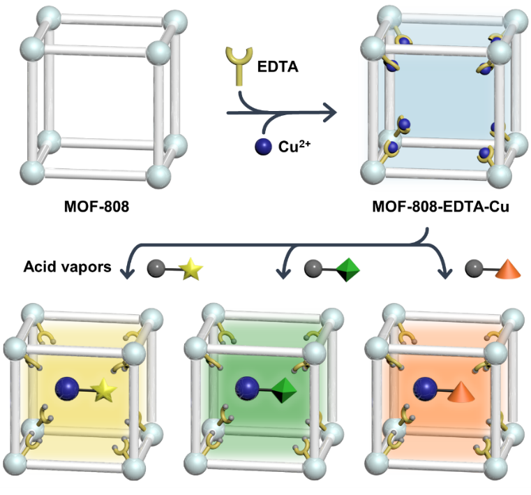
Preparation and characterization of MOF-808-EDTA-Cu
To develop an acid vapor decoder system featuring a fully exposed colorimetric decoder, EDTA-coordinated MOF-808 (designated as MOF-808-EDTA) was prepared following a previously reported method29–30 (Supplementary Figs. 1 and 2). The colorimetric center Cu2+ was introduced by soaking MOF-808-EDTA in a 100 mM Cu2+ solution for 24 h, affording a cyan-colored powder. The Cu-ion-incorporated MOF-808-EDTA was denoted as MOF-808-EDTA-Cu (Fig. 1a). The X-ray powder diffraction (XRPD) patterns of MOF-808-EDTA-Cu confirmed that the framework structure was maintained even after post-synthetic treatments (Fig. 1b). Inductively coupled plasma-atomic emission spectrometry (ICP-AES) confirmed that the Cu2+ ions capable of chelating up to 82% of EDTA were successfully incorporated into MOF-808-EDTA-Cu. Concurrently, the Fourier transform-infrared (FT-IR) spectrum of MOF-808-EDTA-Cu showed the absence of peaks associated with NO3− constituting the Cu precursor, ruling out the possibility of the physical inclusion of Cu precursors into the MOF pores (Supplementary Fig. 3)31–32. Upon the inclusion of Cu ions into MOF-808-EDTA, the ultraviolet visible near-infrared (UV-vis-NIR) spectrum revealed a new absorption peak at 13140 cm− 1, corresponding to the d-d transition 2Eg → 2T2g of an octahedral six-coordinated Cu ion, which is the same as in the previously reported [Cu(EDTA)(H2O)]33, suggesting the presence of Cu-chelated EDTA in the MOF-808-EDTA-Cu system (Supplementary Fig. 4). X-ray photoelectron spectroscopy (XPS) of MOF-808-EDTA-Cu (Supplementary Fig. 5) revealed a significant shift in the Zr 3d binding energy compared to MOF-808, with the monocarboxylate ligand removed, indicating that the EDTA molecule chelating the Cu ion remained grafted to the Zr6 cluster of MOF-808-EDTA-Cu29,34. Nitrogen sorption measurements of MOF-808-EDTA-Cu showed a decrease both in surface area and pore size of larger cavity (1127 m2/g and 0.79 nm) compared to that of the pristine MOF-808 (2108 m2/g and 1.30 nm), indicating that Cu chelated EDTA, the colorimetric center, exists in accessible internal pores of MOF-808 rather physical mixed (Fig. 1c and Supplementary Fig. 6). Therefore, MOF-808-EDTA-Cu containing fully exposed Cu-EDTA within its accessible pores, was successfully prepared as a colorimetric sensor via a simple post-synthetic modification.
Colorimetric response of MOF-808-EDTA-Cu to HCl vapor
An interesting naked-eye detectable color change from cyan to yellow was observed in MOF-808-EDTA-Cu within 20 s of exposure to HCl vapors evaporated from a concentrated HCl solution (Fig. 1e). Remarkably, this transition occurred without any structural changes in the MOF-808 framework, suggesting that the eye-detectable color shift did not originate from structural decomposition (Fig. 1b). Further analysis confirmed the incorporation of chlorine into MOF-808-EDTA-Cu after exposure, as evidenced by scanning electron microscopy coupled with energy-dispersive X-ray spectroscopy (SEM-EDS) (Fig. 1d) and the XPS spectra of Cl 2p (Supplementary Fig. 7). The emergence of a Cu–Cl stretching vibration peak at ~ 289 cm− 1 in the Raman spectra (Supplementary Fig. 8) indicated the formation of new bonds between Cu and Cl 35. Upon exposure to HCl, UV–vis–NIR spectroscopy showed new absorption peaks at 34364 and 25316 cm⁻¹, suggesting that the coordination environment of the Cu2+ ion is changed (Fig. 1f). Interestingly, these peaks align with the LMCT characteristics reported for tetrahedrally structured CuCl42 − 2,36−37, implying that free Cu2+ ions de-chelated from EDTA forming a new yellow Cu-Cl complex (Fig. 1f). Importantly, MOF-808-EDTA-Cu demonstrated selectivity towards acid gas, maintaining its colorimetric response unchanged when exposed to potentially interfering air gases such as N₂, O₂, and CO₂, as well as variations in humidity and temperature within a substantial range (Supplementary Fig. 9). This highlights its potential as a reliable acid-vapor-selective sensor for practical applications.
An intriguing colorimetric response was observed upon exposure of MOF-808-EDTA-Cu to HCl vapor, which was detectable even at concentrations as low as 120 ppm, although a longer detection time was required. Cu-EDTA and Cu(CH₃COO)₂, characterized by non-porous and densely packed structures, displayed gradual color changes after acidic exposure for 1 h at 120 ppm. Conversely, MOF-808-EDTA-Cu exhibited a visible color shift within 5 min, underscoring the significance of grafting Cu-EDTA onto MOF-808 and directly expose it to external acidic vapors for efficient acid sensing (Supplementary Fig. 10). Upon immersion in water, the yellow color of the HCl-exposed MOF-808-EDTA-Cu immediately changed to cyan and was recovered as a cyan powder through filtration (Fig. 1e). The recovered MOF exhibited the same UV-Vis-NIR spectrum as that of MOF-808-EDTA-Cu (Supplementary Fig. 11). This intriguing regeneration of the sensor is attributed to the characteristics of EDTA, which is known to efficiently chelate various metal ions even at low concentrations in aqueous solutions30,38–39. Consequently, the EDTA-decorated MOF-808 demonstrated an ability to re-chelate approximately 80% of Cu²⁺ ions during the regeneration process, as confirmed by ICP-AES analysis. Furthermore, during three cycles of alternating exposure to HCl and water, MOF-808-EDTA-Cu continued to exhibit reversible cyan-yellow color variations, highlighting the reusability of the sensor (Fig. 1e).
Acid-triggered colorimetric decoding mechanism
To reveal the underlying mechanism of the formation of yellow Cu-Cl complexes from highly stable cyan-colored Cu-EDTA complexes, a series of controlled experiments were conducted with MOF-808-EDTA-Cu. First, to elucidate the formation conditions of the Cu-Cl complexes from MOF-808-Cu-EDTA, three types of aqueous solutions containing the same 4 M Cl− ions were prepared: two neutral (NaCl and KCl) and one acidic (HCl) solution. In the 4M Cl− solution, a portion of the introduced free Cu2+ ions formed a yellow Cu-Cl complex, leading to a color change from blue to greenish-yellow (Supplementary Fig. 12a). Notably, MOF-808-EDTA-Cu turned greenish-yellow only in the acidic HCl solution, while retaining its cyan color in the other solutions, indicating the generation of free Cu2+ ions from Cu-EDTA only under acidic conditions (Supplementary Fig. 12b). This unique acid-condition-selective color transformation stems from alterations in the functional groups of EDTA, as evidenced by the FT-IR spectra of MOF-808-EDTA-Cu before and after exposure to HCl (Fig. 1g). Compared to MOF-808-EDTA-Cu, the HCl-exposed MOF-808-EDTA-Cu showed decreased intensity of the peak at 1566 cm− 1, corresponding to the νas,COO− of EDTA40–43, while the emergence of a new peak at 1719 cm− 1 was attributed to the νC=O of carboxylic acids, indicating the protonation of the carboxylate in EDTA27,44. XPS spectra of the HCl-exposed MOF-808-EDTA-Cu compared to MOF-808-EDTA-Cu revealed new peaks at 401.6 and 533.1 eV corresponding to the N 1s of the protonated amine (-HN+-) and O 1s of the carboxylic acid in EDTA45–47, respectively, further supporting the protonation of EDTA which hardly chelates the Cu2+ ion, resulting in the release of free Cu ions (Fig. 1h and Supplementary Fig. 13). Therefore, both Cl− and H+ are essential for the colorimetric decoding of acid vapors by MOF-808-EDTA-Cu. These unique acid-selective color transition mechanism demonstrates the potential of MOF-808-EDTA-Cu as a true colorimetric acid sensor that can selectively react with anions in acidic environments.
MOF decoder to visualize exposed acid vapors
Building on the unique acid-triggered and anionic participation in the colorimetric sensing mechanism, we explored the potential of MOF-808-EDTA-Cu as a colorimetric sensor for the visualization of colorless hydrohalic acid vapors. The MOF-808-EDTA-Cu sensor distinctly visualized the exposed hydrohalic acid vapors, namely HF, HBr, and HI, as white, dark purple, and brown, respectively (Fig. 2a). Interestingly, these observed color changes were aligned with the expected colors resulting from the interaction of free Cu ions with halide ions, suggesting that the de-chelated Cu2+ ions from the protonated EDTA in MOF-808-EDTA-Cu reacted with hydrohalic acids (Supplementary Fig. 14). Specifically, the UV-vis-NIR spectra of MOF-808-EDTA-Cu after exposure to HF and HBr were consistent with those of CuF2 and CuBr42−, respectively48, implying the formation of white CuF2 and purple CuBr42− within the MOF sensor following acid exposure (Supplementary Figs. 15 and 16). Furthermore, exposure to HI resulted in the formation of white CuI(s) and brown I2(aq), as confirmed by PXRD and UV-vis-NIR spectra, suggesting that the brown color of the HI-exposed MOF-808-EDTA-Cu originated from I2(aq) rather than CuI(s) (Supplementary Fig. 17). The exceptionally strong chelation of EDTA in MOF-808 renders it versatile and enables the incorporation of various metal ions into the MOF-808 sensor. To further explore our methodology, we prepared Fe3+-chelated MOF-808-EDTA (referred to as MOF-808-EDTA-Fe) as a colorimetric sensor, which exhibited a distinct color change from ivory to yellow and orange upon exposure to HCl and HBr, respectively (Fig. 2b and Supplementary Fig. 18).
Furthermore, the strong chelation capability of EDTA allowed MOF-808-EDTA to effectively integrate multiple metal ions (Cu2+ and Co2+) with atomic-level dispersion, thereby expanding the scope of visually-identifiable acid vapors within a single-domain sensor (Fig. 2c). MOF-808-EDTA-Cu/Co, featuring Cu-chelated EDTA and Co-chelated EDTA, exhibited a unique ability to differentiate between six acidic vapors within a single-domain sensor (Fig. 2d). This capability arises from the co-presence of Cu-chelated EDTA, adept at decoding hydrohalic acid and Co-chelated EDTA, proficient at decoding nitric acid and trifluoroacetic acid (TFA). Such findings demonstrate the versatility and resilience of our sensor platform, providing visual identification of a variety of acid vapors with a single sensor domain.
To exploit the outstanding properties of the MOF-808-EDTA-Cu platform for the visualization of colorless acid vapors, the fabrication of miniaturized portable acid vapor sensors that could be used for real-time on-site monitoring was explored (Fig. 3a). For transformation into a portable acid vapor decoding sensor, a MOF sensor-based ink was fabricated by combining MOF-808-EDTA-Cu with polyvinylidene fluoride (PVDF) in a dimethylformamide (DMF) solution, which can be applied to various substrates, including foil, paper, fabric, and glass (Supplementary Fig. 19). When exposed to HCl vapor evaporating from a concentrated HCl solution (approximately 15,500 ppm)49–50, the MOF-808-EDTA-Cu portable sensor underwent a distinct color shift from cyan to yellow, which was detectable by the naked eye and recorded by a camera sensor (Fig. 3b). This color change was further translated into RGB channel values, allowing the quantification of the color changes and 24-hour real-time monitoring. Notably, when exposed to low concentrations of HCl where the color change is not saturated, the sensor exhibited a reduced transition from cyan to yellow within the same exposure timeframe, suggesting its potential as an acid–gas concentration analyzer (see Fig. 3c). This transition can be precisely quantified using the equation |dB|/B0, where |dB| denotes the absolute value of change in the blue channel value from the initial blue channel value B0. Correlation of the |dB|/B0 ratio with different HCl vapor concentrations can establish a linear range spanning from 120 to 740 ppm, providing experimental validation of the portable sensor as a colorimetric sensor capable of quantifying the concentration of exposed HCl vapor. Additionally, when exposed to an atmosphere with high relative humidity (RH) 85%, no color change was detected both with the naked eye or even with RGB values, demonstrating its practical use as a portable sensor capable of selectively visualizing acidic vapors, even in the presence of humidity interference (Fig. 3d).
Based on the obtained results, we extended our investigation on the color changes of sensors upon exposure to various acid vapors. In experiments with hydrohalic acid vapors, including HF, HBr, and HI, the color change of the MOF-808-EDTA-Cu portable sensor was not complete until 25 min, however, changes detectable by the naked eye appeared within 10 min (Supplementary Fig. 20). Interestingly, when monitoring the color shifts of the portable sensor via the RGB channel values, distinct trends in the RGB channel values depending on the exposed acid vapors were observed, even in the early stages when they were barely detectable by the naked eye (Fig. 3e). Furthermore, these acid-dependent distinct color alterations enable the statistical validation of exposure to hydrohalic acid vapor within 2 min by applying principal component analysis (PCA) and hierarchical cluster analysis (HCA) methods. As shown in Fig. 3f, the 12 datasets of dR, dG, and dB obtained from three repeated 2-min exposure experiments with four different hydrohalic acids formed distinct clusters that were well-spaced apart, implying efficient identification. HCA-based data classification using Ward's method revealed that when the closest data points were clustered, three points originating from the same acid vapor exposure experiments were successfully grouped together, confirming the ability of the sensor to discriminate between acids (Fig. 3g). Moreover, this versatile portable sensor platform can incorporate various transition metals, such as Co and Fe, to broaden the decoding range of acid vapors to up to six types or to adjust the detection color (Supplementary Fig. 21), providing experimental validation of its applicability to diverse industrial requirements.