Synthesis and Crystallography
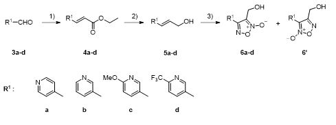
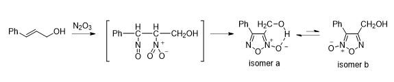
Totally, thirty nine novel oxadiazole-2-oxide derivatives were synthesized following Scheme 1 and Scheme 2. The structures of these compounds were detailed in Table 1. During synthesis of the important intermediate 6, two isomers were inevitably formed when the oxadiazole-N-oxide ring was constructed by sodium nitrate-mediated cyclization. Gasco and co-workers reported [19] that intramolecular hydrogen bond played a crucial role on the formation of the main isomer during cyclization and could stabilize the structure of the isomer a (Scheme 3).
For our case, two isomers 6 and 6' were obtained (Scheme 1) and one of them was the main product after cyclization. To confirm the position of N-oxide moiety, is the main product 6 or 6'? The crystal of the main product was obtained. It was interesting that the X-ray confirmed that the main product had a similar conformation with the isomer a in Scheme 3, and specifically, the main product was 6a (Scheme 1). The crystal structure showed that there were two independent molecules in an asymmetric unit (Figure 3a). One molecule’s nitrogen atom was labeled with N1, N2 and N3, and the other molecule’s nitrogen atom was labeled with N4, N5 and N6. However, intermolecular hydrogen bonds instead of intramolecular hydrogen bonds were found in the crystal of 6a. The molecular labeled N1, N2 and N3 of nitrogen atoms formed a hydrogen bonds helix chain (Figure 3b), in addition, the molecular labeled N4, N5 and N6 of nitrogen atoms formed a hydrogen bonds zigzag chain (Figure 3c). This result also indicated that after the pyridine replaced the benzene, the intramolecular hydrogen bond showed in Scheme 3 might not be necessary for the formation of main isomer 6a-d.
Figure 3: (a) Crystal structure of 6a; (b) Hydrogen bond helix chain formed by molecules labeled N1, N2 and N3 of nitrogen atoms; (c) Hydrogen bond zigzag chain formed by molecules labeled N4, N5 and N6 of nitrogen atoms.
Activity assessments of TGR
With furoxan as the positive control, oxadiazole-2-oxides (6, 7, 9) were evaluated for their inhibitory activities against both rSjTGR-Sec and SWAP containing wtSjTGR under different concentrations (Table 1). As shown in Table 1, all intermediates bearing hydroxy (6a-6d) showed better inhibitory activity against both two enzymes than furoxan. The pyridine ring in the structure of compound 6a had a stronger electron withdraw effect on the oxadiazole-2-oxide ring than compound 6b, and 6a (IC50 70nM of SWAP containing wtSjTGR, IC50 36nM of rSjTGR-Sec) had better inhibitory activity on both two enzymes than 6b (IC50 0.14µM of SWAP containing wtSjTGR, IC50 0.39µM of rSjTGR-Sec). Compared with 6b, the electron-donating methoxy group at the ortho position of pyridine (like compound 6c) decreased the inhibitory activity (6c, IC50 1.1µM of SWAP containing wtSjTGR, IC50 2.4µM of rSjTGR-Sec). Notably, electron-withdrawing group trifluoromethyl made 6d exhibited excellent inhibitory effect (IC50 7.5nM of SWAP containing wtSjTGR, IC50 55.8nM of rSjTGR-Sec). These data indicated that the electron-withdrawing substituents very possibly provided potency enhancements of activity.
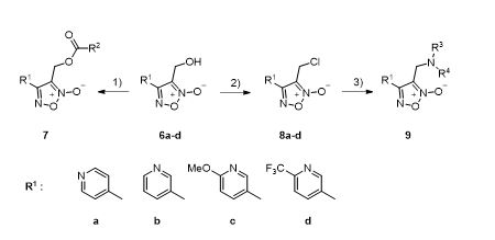
To further investigate the relationship between the structure and bioactivity, modification of hydroxyl group into the ester group and amine, respectively afforded compounds 7 and 9. It was found that the ester group could not effectively improve the inhibitory effect, except the ethyl ester (7ca), chloracetyl ester (7ae, 7be), benzoyl ester (7af, 7cf) and acryloyl ester (7bj) exhibited inhibitory effects similar to furoxan. Only 7ca (IC50 34.77µM of SWAP containing wtSjTGR) showed inhibitory effect against SWAP containing wtSjTGR, but not as good as furoxan. Among chloracetyl esters, maybe is because the pyridine ring of 7ae had a stronger electron-withdrawing effect, 7ae (IC50 4.23µM of SWAP containing wtSjTGR) had a slight advantage over 7be (IC50 16.40µM of SWAP containing wtSjTGR, IC50 14.82µM of rSjTGR-Sec). However, 7ae did not show an effective inhibitory effect on rSjTGR-Sec. As for phenyl derivatives, 7af (IC50 8.87µM of SWAP containing wtSjTGR, IC50 3.23µM of rSjTGR-Sec) and 7cf (IC50 12.51µM of SWAP containing wtSjTGR) did not differ significantly in inhibiting SWAP containing wtSjTGR, but 7cf did not show an inhibitory effect on rSjTGR-Sec. Among acrylate compounds, only 7bj showed inhibitory activity against SWAP containing wtSjTGR (IC50 5.80µM).
Some amine derivatives (9aa, 9ab, 9bd, 9be, 9da) showed inhibitory activity, among which 9bd had an inhibitory effect against SWAP containing wtSjTGR at the nanomolar level (IC50 = 42nM). Compared with phenyl amino derivative 9aa, 9ba, 9ca and 9da, the pyridine with strong electron-withdrawing ability improved the activity, such as, compounds 9aa (IC50 21.57µM to SWAP containing wtSjTGR) and 9da (IC50 11.62µM to SWAP containing wtSjTGR, IC50 3.73µM to rSjTGR-Sec) showed better inhibition effect than 9ba (IC50>50µM) and 9ca (IC50>50µM). Similar to phenyl amino derivatives, 4-trifluoromethroxyphenyl amino derivative 9ab (IC50 0.62µM to SWAP containing wtSjTGR, IC50 1.52µM to rSjTGR-Sec) showed better inhibitory effect than 9bb (IC50>50µM both to SWAP containing wtSjTGR and rSjTGR-Sec). However, among piperazine substituted derivatives (9ac, 9ad and 9bd), 9bd exhibited good inhibitory activity against SWAP containing wtSjTGR (IC50 0.042µM). Interestingly, compound 9be is an intermediate of a synthetic target molecule, but it displayed much better inhibitory effects (IC50 0.63µM to SWAP containing wtSjTGR, IC50 0.22µM to rSjTGR-Sec) than furoxan Perhaps the sulfamide or Si-O-N moieties in molecule 9be has some influence on enzyme inhibition ability, this result might provide a new direction for more compounds design.
Docking studies
To rationalize the obtained bioactivity data and to understand how the synthesized inhibitors interact with schistosomal proteins, the selected compounds with high activity 6d, 7af and 9ab were docked to the available crystal structure of SjTGR (thioredoxin glutathione reductase from Schistosoma japonicumi, PDB ID:4LA1). The crystal structure of SjTGR is a homodimer, and we retained chain B for docking studies. The results were displayed in Figure 4 and Figure 5.
Compound 6d formed four hydrogen bonds with Asp433, Ser117 and Thr153, and a π-cation interaction with Arg393(Figure 4a) and two strong hydrogen bonds existed between the hydroxyl moiety of 6d and SjTGR. These results could explain the highest activity of 6d. Compound 7af formed two hydrogen bonds with Cys154 and Asp433, and a π-cation interaction with Arg393(Figure 4b), while the pyridine ring and oxadiazole ring didn’t form any type of interactions with the residues of Arg393. Compound 9ab formed one hydrogen bond with Ser276, a π-cation interaction with Arg393 and a π-π stacking interaction with Tyr296. It’s noticed that the oxadiazole ring participated in the formation of chemical bonds, to constitute more interaction bonds. The distance between the oxadiazole ring and other ring -shaped conjugated structures should be taken into consideration. After superimposing three compounds (6d,7af and 9ab) together (Figure 4d), each of them formed one π-cation interaction with Arg393, therefore, Arg393 was speculated to play a role of anchor during the binding process.
Figure 4: In (a), (b) and (c), the protein (SjTGR) is shown as ribbons, the synthesized inhibitors are shown in orange sticks, and the residues that can interact with inhibitors are shown as green sticks. (a) Compound 6d forms a π-cation interaction (red dashed line) with Arg393 and hydrogen bonds (yellow dashed lines) with Ser117, Thr153 and Asp433. (b) Compound 7af forms a π-cation interaction with Arg393 and hydrogen bonds with Cys154, and Asp433. (c) Compound 9ab forms a π-cation interaction with Arg393, a π-π stacking interaction (blue dashed line) with Tyr296, and a hydrogen bond with Ser276. (d) Superposition of docking poses of compounds 6d (blue), 7af (purple), 9ab (green) in the binding pocket of S. japonicum TGR.
On the other hand, the hydrophobic effect was showed by red arcs with spokes radiating toward the atoms involved (Figure 5). The numbers of the residues that had hydrophobic contacts with the inhibitors were associated negatively with the enzyme inhibitory activities. Among three compounds (6d, 7af and 9ab) compound 7af had the most of hydrophobic contacting residues and it had the lowest activity, compound 6d had the least of hydrophobic contacting residues and it exhibited the highest activity.
Figure 5: LigPlot+ generated two-dimensional schematic overview of molecular interactions between SjTGR and compound 6d, 7af and 9ab. Hydrogen bonds are indicated by green dashed lines with corresponding distances between the atoms given in Å. Hydrophobic contacts are shown by red arcs with spokes radiating toward the atoms involved.