EXPERIMENTAL METHODS
Ethics. An informed consent was obtained from each patient concerning diagnostic procedures, treatment to remove salivary stones and the subsequent use of these specimen for research purposes, including data analysis. The study was approved by the University´s ethical review committee (186_19 Bc).
Patients and clinical interventions. All patients of our study cohort with symptomatic sialolithiasis (n = 37) received a clinical examination, ultrasound imaging and a subsequent sialendoscopy to ensure the diagnosis. Diagnostic and therapeutic interventions were carried out in a tertiary referral center specializing in salivary gland diseases (FAU Medical School, Department of Otolaryngology, Head and Neck Surgery, University of Erlangen-Nuremberg, Erlangen, Germany), obtaining stones from submandibular (n = 30) and parotid glands (n = 7).
Submandibular stones were removed by transoral surgery which allowed to obtain a complete unfractionated sialolith. Parotid stones were removed either in total during an open surgical approach in which the stone was removed as a whole, or by sialendoscopically assisted basket extraction (when the concrement was small enough to evacuate it through the duct). In cases of larger parotid concrements which could not be evacuated in total due to their size or which were not obtained during an open surgical approach, stone fragments were collected after sialendoscopically controlled intraductal pneumatic lithotripsy and subsequent basket extraction.
Chemicals and Biochemicals. If not stated otherwise, we obtained all chemicals and biochemicals from Sigma-Aldrich/Merck (Darmstadt, Germany).
Salivary stones storage and processing. Sialoliths were stored at room temperature (for µCT, calcium staining) or dry at -20°C (immunofluorescence) until analysis. Some stones were embedded in methacrylate and cut into 2 µm sections with a rotary microtome (RM2265, Leica, Wetzlar, Germany); others were fixed for 4-12 hours with paraformaldehyde and decalcified in Teitel buffer (140 g EDTA free acid; 90 ml NH3 [30%] in 500 ml H2O; pH = 7.2; adjusted with NH4OH) for up to 4 weeks. The stones used for dye penetration assays were incubated with trypan blue (100 µg/ml), propropidium iodide (10 µg/ml), or acridin orange (10 µg/ml) at ambient temperature for 10 minutes and subsequently ground with a diamond file. By mechanical grinding with ceramic or diamond tools we obtained gravel of sialoliths, and material from the surface, the intermediate and the center of the stones was received by selectively skimming off the layers.
Micro-computed tomography. For µCT scanning of the calculi we used a cone-beam desktop microcomputer-tomograph (µCT 40, Scanco Medical AG, Bruettisellen, Switzerland). The samples were positioned, fixed, and scanned (voxel size 9 µm, 45 kVp). µCT images were processed and analyzed quantitatively with Photoshop CS5 64 Bit (Adobe, Munich, Germany).
Digital reconstruction of volumetric µCT data. Based on the volume data generated in CT, a physically based volume rendering algorithm using a Monte Carlo path tracing method simulated the complex interactions of photons (emission, absorption, scattering) within the scanned specimens 67, 68. Cinematic Rendering generated photorealistic images by calculating realistic lighting by light transport simulation along hundreds or thousands of photons paths per pixel, using a stochastic process. Thus, even complex effects such as ambient occlusion or tissue density could be modeled.
Macrophotography. Macro images were taken with a Nikon 700 camera (Nikon, Tokyo, Japan) with a CMOS sensor in FX format (36.0 x 23.9 mm, 12.87 million pixels) and two different objectives (Nikon AF-S Nikkor 60mm/2.8G ED; Nikon AF-S Nikkor 105mm 1:2,8G VR). For illumination, we employed white light.
Microscopy. We used a BZ-X700 automated video microscope for fluorescence microscopy, equipped with Z-stack and stitching technology to increase the depth of field and the size (Keyence Corporation, Osaka, Japan). For bright field microscopy, oblique illumination was available. We used Photoshop CS5 or CC2018 (Adobe, Munich, Germany) for post-processing of pictures and morphometry. Areas for morphometry were selected by a blinded investigator.
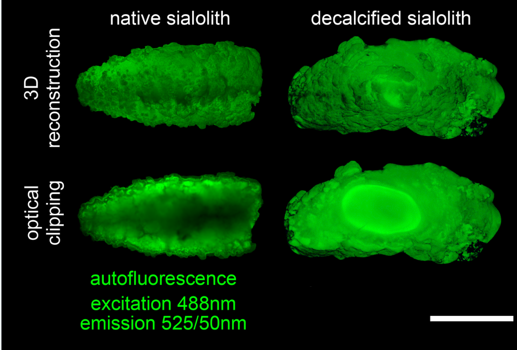
Optical clearing and Light sheet fluorescence microscopy (LSFM) of submandibular sialoliths. We fixed and decalcified submandibular sialoliths with paraformaldehyde and Teitel buffer, respectively, as described above. DNA of both decalcified and non-decalcified sialoliths was stained with propidium iodide (10 μg/ml) in PBS for 5 days at ambient temperature. We performed optical clearing according to established protocols 69. Briefly, samples were dehydrated in ethanol series of 50%, 70%, and twice 100% (v/v). Each dehydration step was carried out at room temperature for 2 days in gently shaking 5 ml tubes. After dehydration, samples were transferred to ethyl cinnamate, optically cleared for one day at ambient temperature and imaged with a LaVison BioTec Ultramicroscope II including a LaVision BioTec Laser Module (LaVision BioTec GmbH, Bielefeld, Germany), an Olympus MVX10 zoom body (Olympus Germany, Hamburg, Germany), and an Andor Neo sCMOS Camera (Andor, Belfast, UK) with a pixel size of 6.5 μm. We used detection optics with fourfold optical magnification and NA 0.5. Autofluorescence was excited by a 488 nm optically pumped semiconductor laser (OPSL) and detected at 525/50 nm. For propidium iodide excitation, a 561 nm OPSL and a 620/60 nm emission filter were employed. For 3D reconstruction and optical clipping of LSFM data we used Imaris software (Version 9.1, Bitplane AG, Zurich, Switzerland).
Von Kossa staining for mineralized areas. We rinsed methacrylate sections several times with distilled water, incubated them with a 5% aqueous silver nitrate solution in the dark for 20 minutes and finally exposed them to UV-light until a black precipitate had formed. The remaining ionic silver was removed by incubation with 5% sodium thiosulfate for 5 minutes and extensive rinsing with distilled water.
DNA staining. We stained the extracellular DNA of stones and histological samples with 10 µg/mL and 1 µg/mL propidium iodide in isotonic PBS (both from Thermo Fisher Scientific, Waltham, USA) for at least 10 minutes at ambient temperature. Digestions with DNase 1 (1U/ml; 10mM Mg2+; 5mM Ca2+) were performed for 120 minutes at 37°C to confirm extracellular DNA as the origin of the observed propidium iodide signals.
Immunostaining for neutrophil markers. Histological sections and gravels from sialoliths were analyzed for extracellular DNA, neutrophil elastase and citrullinated histone H3. Samples were fixed with 4% paraformaldehyde for 10 minutes and blocked for 18 hours at 4°C in PBS containing 10% FBS (Merck Millipore, Billerica, Waltham, USA). Primary antibodies detecting neutrophil elastase or citrullinated histone H3 (Abcam, Cambridge, UK; ab21595 and ab1503, respectively) were used following manufacturer´s recommendations. AffiniPure Cy5-conjugated Goat-Anti-Rabbit IgG (H+L) secondary antibodies (Jackson Immuno Research Labs, West Grove, USA) were co-incubated with 1 µg/ml Hoechst stain or propidium iodide. Slides were embedded in DAKO fluorescent mounting medium (Agilent Technologies, Santa Clara, USA) according to manufacturer´s recommendations.
Neutrophil elastase activity in human sialoliths. 1 mg of sanding dust from sialoliths (diamond file, grain size 300) was resuspended in 500 µl of PBS. 25 µL of this material (or H2O as controls) were added to 175 µl of PBS and 25 µl of 1 M fluorogenic substrate MeOSuc-AAPV-AMC (Santa Cruz Biotechnology, Heidelberg, Germany; sc-201163). Samples were continuously analyzed in black 96-well plates (Thermo Fisher Scientific, Waltham, USA; 137101) every 10 minutes for up to 12 hours at 37°C in a thermostated fluorescence reader (TECAN Infinite 200 Pro, Tecan Trading AG, Switzerland; excitation 360 nm; emission 465 nm).
DNA extraction, sequencing and microbiome analysis. To remove DNA contaminations we washed the surfaces of the sialoliths extensively with at least 3 changes of > 20-fold volumes of PBS. We ground the stones with a gentleMACS Dissociator (Miltenyi Biotec, Bergisch Gladbach, Germany) using M-tubes and TE buffer (100 mM Tris-Cl, 10 mM EDTA, pH = 8.0). After phase separation, we recovered the sialoliths-borne DNA by adding ammonium acetate to a final concentration of 2 M to the aqueous phase, followed by 0.6 volumes of isopropanol and overnight incubation at -20°C. The pellet was redissolved in 300 mM sodium acetate and reprecipitated by the addition of 2 volumes of absolute ethanol (12 hours at -20°C). An aliquot of the DNA samples was run on an agarose gel.
The 4 samples with the highest DNA content were analyzed by 16S-based metagenomic sequencing as described elsewhere 70. The V3-V4 region of the bacterial 16S rRNA gene was amplified by PCR (35 cycles; 98°C for 15 seconds, 58°C for 20 seconds, 72°C for 40 seconds; NEB Next Ultra II Q5 Master Mix, New England Biolabs, Ipswich, USA) using template DNA and region-specific primers (containing barcodes and Illumina flow cell adaptor sequences; Eurofins Genomics, Munich, Germany). The amplicons were purified with Agencourt AMPure XP Beads (Beckmann Coulter, Brea, USA), normalized and pooled. Sequencing was performed with an Illumina MiSeq device using a 600-cycle paired-end kit and the standard Illumina HP10 and HP11 sequencing primers. The read Fastq files were bioinformatically processed (merging, demultiplexing, quality filtering, dereplication, chimera removal) using the 64-bit version of Usearch 10 according to the Uparse pipeline 71. Operational taxonomic units (OTUs) were selected at a threshold of 97% similarity and taxonomically classified by comparing the representative OTU sequence to the reference file of the ribosomal database project (RDP version 16).
Statistical analysis. One Way ANOVA was used to calculate significances of the differences between independent groups with the SPSS Software (IBM, Armonk, USA, version 24). p-values ≤ 0.05 were considered statistically significant.