Patients and samples
Forty-one patients with a total of 84 specimens were enrolled from Sir Run Run Shaw Hospital, College of Medicine, Zhejiang University and Fudan University Shanghai Cancer Center in this study. As shown in Figure 1, four specimens were excluded because of insufficient tumor content, and two specimens were excluded due to high content of necrotic tissue in the samples; thus, six corresponding patients were excluded. Thirty-five MPMTs from 73 specimens met all quality control criteria and were analyzed by the 90-gene expression assay. Based on the invasion site of the tumor, 73 specimens were sorted into 12 types, including tumors in the colorectum, gastroesophagus, lung, ovary, endometrium, breast, liver, kidney, urinary, prostate, head & neck and thyroid. Table 1 presents the demographics of the 35 MPMT patients. Among these patients, 30 harbored synchronous MPMTs, and five harbored metachronous MPMTs. Twenty-one patients were male, and fourteen patients were female. The median age at diagnosis was 62.5 (range 33-77) for the first cancer, 63 (range 33-77) for the second cancer and 68 (range 53-70) for the third cancer. The most common invasion sites of the first, second and third tumors were the colorectum, gastroesophagus and colorectum, respectively. Of the 73 specimens, tumors were most frequently located in the colorectum (24.7%, 18 of 73), gastroesophagus (16.4%, 12 of 73) and lung (12.3%, 9 of 73). The distribution of tumor locations is shown in Figure 2. The most common stages of the first, second and third cancers were II, I and II, respectively. Thirty-two of 35 patients (91.4%) underwent surgery, and 62.9% (22/35) underwent chemotherapy.
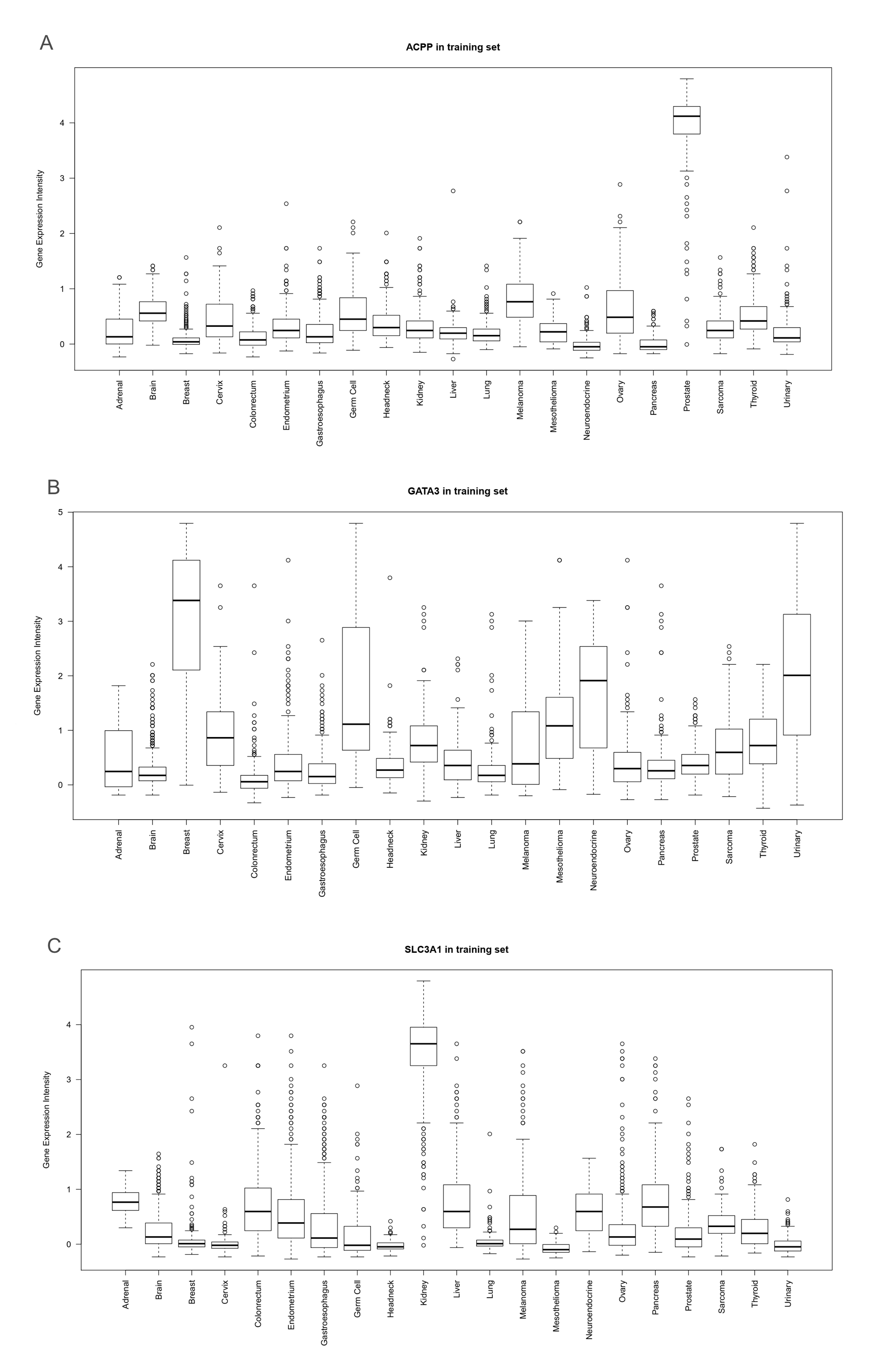
Performance of the 90-gene expression assay in MPMTs
The overall workflow for the 90-gene expression assay is shown in Figure 3. The concentrations of total RNA from 73 samples ranged from 3.3 ng/μL to 255.5 ng/μL, with a median of 66.6 ng/μL. The median A260/A280 ratio (purity of RNA) was 1.98 (range 1.74-2.04).
With the 90-gene expression assay, 20 specimens were classified as colorectal tumors, 11 as gastroesophageal tumors, 7 as lung tumors, 6 as breast tumors, 6 as ovary tumors, 5 as endometrium tumors, 4 as urinary tumors, 4 as kidney tumors, 3 as liver tumors, 3 as head & neck tumors, 2 as prostate tumors and 2 as thyroid tumors. For the entire cohort, the 90-gene expression assay showed an overall accuracy of 93.2% (68 of 73, 95% confidence interval (CI): 0.84-0.97). As shown in Table 2, the sensitivity of 90-gene expression assay were 100% for classifying tumors from the colorectum, ovary, endometrium, breast, kidney, urinary tract, prostate, head & neck and thyroid . Furthermore, the 90-gene expression assay correctly classified 83.3% of the gastroesophageal cancer cases, 77.8% of the lung cancer cases and 75.0% of the liver cancer cases. As shown in Table 3, 5 out of 73 specimens had discordant molecular classifications compared with the pathological diagnosis, including two lung tumors, two gastroesophageal tumors and one gallbladder tumor.
The performance of 90-gene expression assay stratified by histopathological features was further investigated. Among 73 specimens, 40 (55%) were well-moderately differentiated tumors, 25 (34%) were poorly or undifferentiated tumors, and 8 (11%) were squamous cell carcinoma. Regarding to the predictions of 90-gene expression assay, the overall accuracy was 95.0% (38 of 40; 95% CI, 0.82-0.99) for well-moderately differentiated tumors and 92.0% (23 of 25; 95% CI, 0.82-0.99) for poorly or undifferentiated tumors, with no statistically significant difference (p-value > 0.5). In addition, the overall accuracy was 87.5% (7 of 8; 95% CI, 0.47-0.99) for squamous cell carcinoma.
Specific case
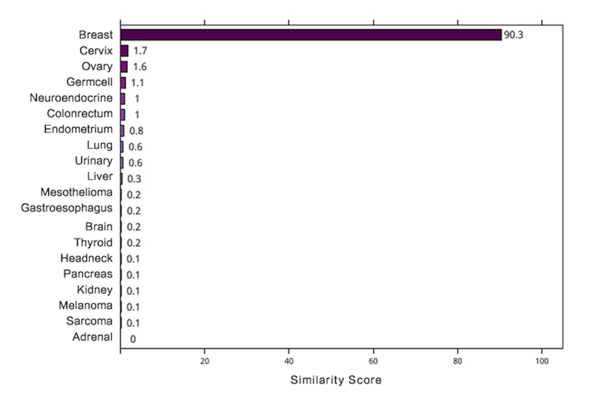
A 77-year-old man noticed chest tightness, shortness of breath, nausea/vomiting and fever. He underwent endoscopic biopsies in the gastroesophageal region (29-35 cm from the incisors) and was diagnosed with squamous cell carcinoma (SCC). In the meantime, a chest computed tomography (CT) scan found a lesion in the left upper lung, and the lung biopsy was diagnosed as SCC. Through comprehensive clinical and pathology examinations, the clinician confirmed that the patient had multiple synchronous primary tumors according to the Warren and Gates criteria [14]. FFPE tumor tissues taken from the esophagus and lung were analyzed by the 90-gene expression assay, and the predictions showed that the two specimens were gastroesophageal cancer and lung cancer (Figure 4).
SCC comprises a wide range of tumors originating from diverse anatomical locations that share a common histomorphology and expression of squamous cell differentiation markers, making it difficult to distinguish whether the subsequent SCC is a primary tumor or metastatic lesion. This patient had two simultaneous lesions on the esophagus and lung. Histopathologically, esophageal SCC often metastasizes to the lung. Pathological diagnosis could only confirm the two lesions in the esophagus and lung as SCCs. It was challenge to determine whether the two lesions were synchronous SCCs or represented SCC metastasis based on immunohistochemistry (IHC) and morphology assessment. The clinical outcome of synchronous esophageal and lung SCCs is better than that of metastatic cancer, and the diagnosis of the tumor will directly affect the treatment options. If the new lesion is a second primary tumor, surgical resection supplemented by chemotherapy or radiotherapy has been the preferred therapeutic regimen instead of palliative treatment. Thus, for patients highly suspected of having metastatic cancer, the 90-gene expression assay can be useful to identify the tissue of origin more quickly when imaging and IHC examinations are ineffective, and support the choice of precise treatment.