Cell and Bacterial culture
J774a.1 cells were obtained from the Cell Culture Center, Xiehe Medical University (Beijing, China). The cells were cultured in a humidified incubator at 37°C with 5% CO2 in DMEM (Hyclone, Logan, UT, USA) supplemented with 10% FBS (Gibco, Grand Island, NY, USA), 100 μg/ml streptomycin and 100 U/ml penicillin (Gibco). Mouse bone marrow monocyte-derived macrophages (BMDMs) were isolated from the femurs of 6 to 8-weeks-old female C57BL/6 mice, and cultured in six-well plates (Corning) for 7 days in RPMI1640 (Hyclone) supplemented with 10 ng/ml M-CSF (Pepro Tech), 10% FBS, 100 microgram/ml streptomycin and 100 U/ml penicillin (Gibco). Virulent M. bovis Beijing strain number C68004 was provided by the China Institute of Veterinary Drug Control (CVCC, China), and grown in 7H9 Middlebrook media (Difco) supplemented with 10% albumin-dextrose-catalase (ADC) enrichment solution, 2 mg/L sodium pyruvate and 0.05% Tween–80 at 37°C under biosafety conditions level 3 (BSL3).
Mice and M. bovis infection
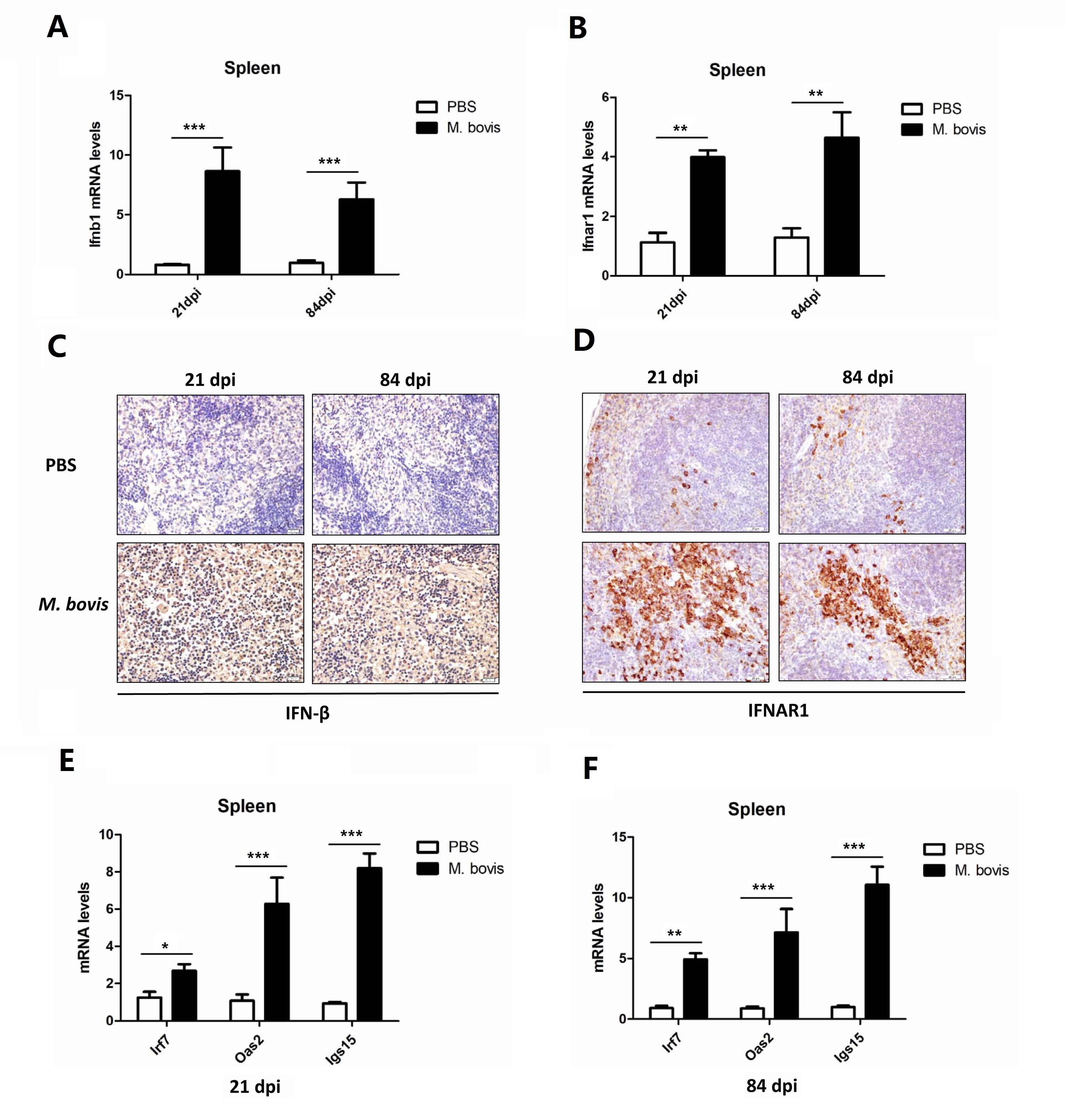
C57BL/6 female mice (6–8 weeks of age) were obtained from Vital River Laboratories (Beijing, China). Mice were maintained in a strict biosecurity measures in BSL-III laboratory of China Agricultural University, under the protocols of the Laboratory Animal Ethical Committee of China Agricultural University (Protocol 20110611–01). Our experimental protocols (CAU20180212–2–4) were approved under Authorization for Experimentation on Laboratory Animals by Animal Ethical Committee of China Agricultural University. Mice were housed in groups as many as 5 mice per cage with free access to feed and water under sterilized condition. In the current study, we infected mice with M. bovis at 100 CFU (Colony-forming units) or PBS through intranasal route as previously reported [21]. Briefly, for intranasal challenge mice were anaesthetized by intraperitoneal injection of Zoletil 50 (50mg/ml; Virbac, France) diluted in PBS. One day post infection (p.i), about five mice were selected randomly and sacrificed for collecting lung tissues to enumerate viable M. bovis bacilli.
In vivo IFNAR1 blocking antibody treatments
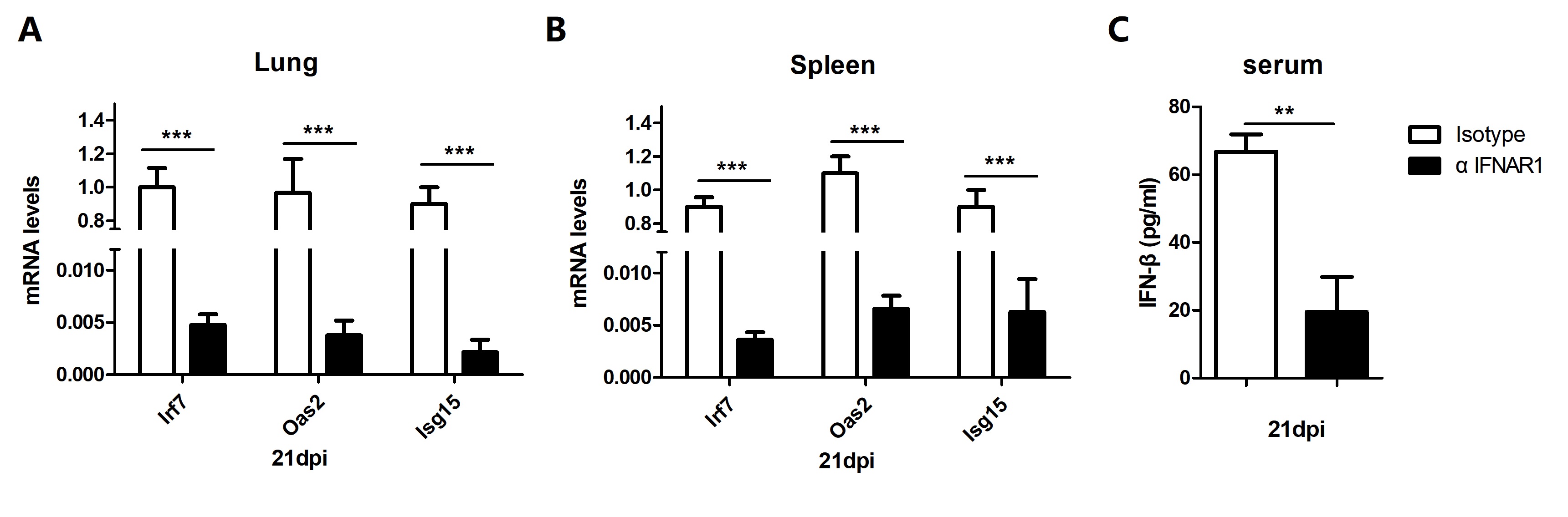
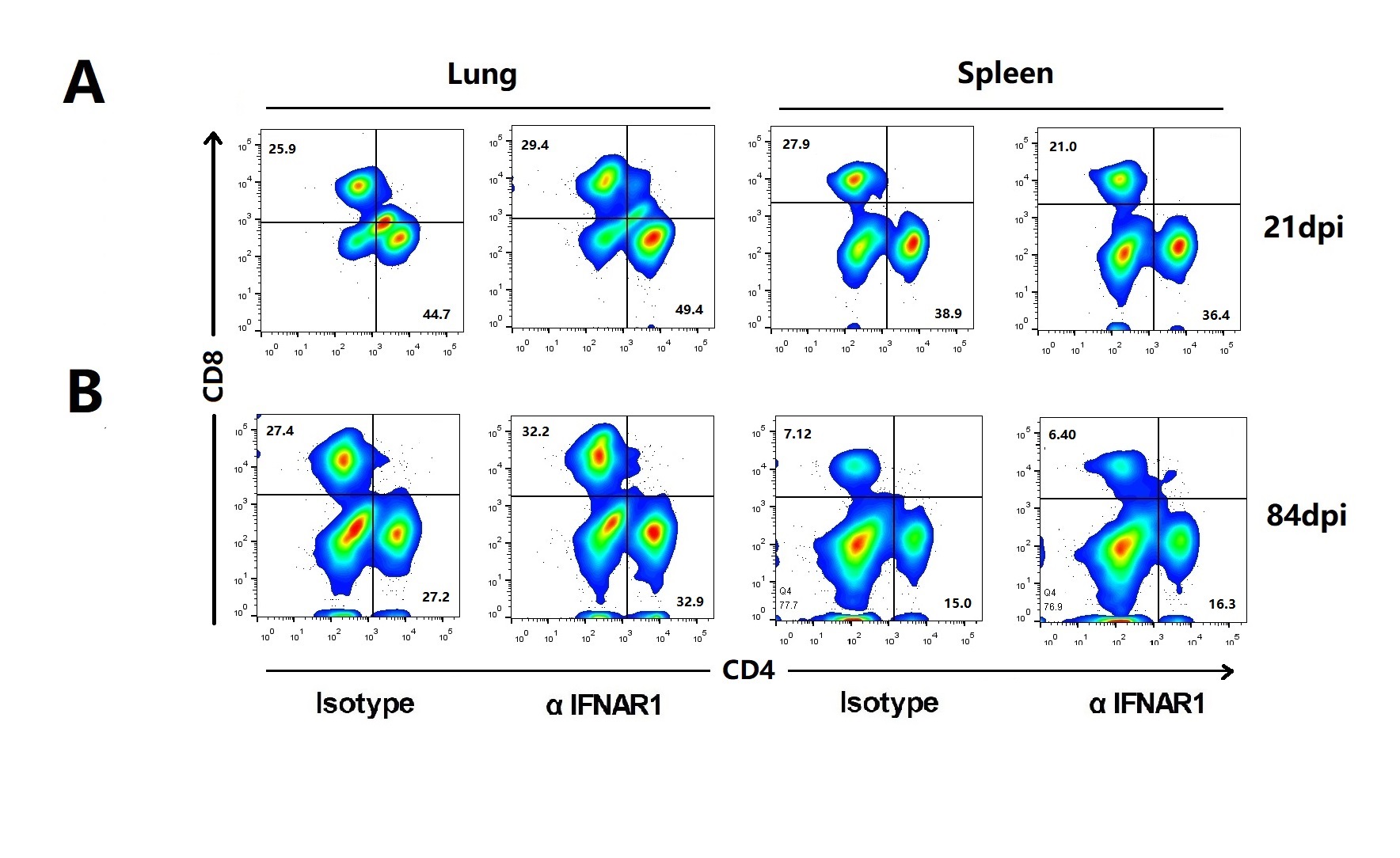
Each groups 12 mice C57BL/6 mice were treated with 500µg of anti-IFNAR1 antibody (clone MAR1–5A3; Biolegend San Diego, CA, USA) or mouse IgG1 (500µg) isotype control (clone MOPC21; Bio X Cell) one day before to M. bovis infection and at 6 weeks post infection (500µg) through intraperitoneal (i.p) injection. Six mice from each groups were sacrificed at 21 and 84 days post infection. For histopathology analysis, animals were euthanized by cervical dislocation method as previously describe [22]. The cervical dislocation method was carried out because it minimizes the effect on immune system [22]. This method preferably brings quick loss of consciousness, does not leave tissue residues, and it is also safe and effective. This choice was approved by the Ethic Committee for Animal Care and Use, China Agriculture University. Lung and spleen tissues were collected aseptically as soon as possible and stored at –80°C till further experiments.
Determination of M. bovis load in mice
For the enumeration of total viable bacilli, lung and spleen tissues were collected aseptically and lysed with small ceramic beads in phosphate buffered saline (PBS), in a tissue homogenizer apparatus (WKT technology) in accordance with the guidelines of manufacturer. An appropriate tenfold serial dilution was prepared in sterilized PBS. The dilutions were separately plated on Middlebrook 7H11 agar plates in triplicates supplemented with ampicillin (10 μg/ml) and sodium pyrovate (2–4mg/liter). After 2–3 weeks of incubation at 37°C colonies were counted for M. bovis.
IFNAR1 blocking antibody treatment in vitro
To investigate the inhibitory effect of type I interferon signaling in vitro, Mouse bone marrow derived macrophages (BMDMs) were treated with 1 µg/ml of an IFNAR1 blocking antibody (Biolegend) for 2 hours. Then the cells were washed three times with warm PBS and infected with M. bovis at multiplicity of infection (MOI) of 10 and incubated at 37oC for 24 hours.
IFN-β production assay
The level of IFN-β production in the serum samples were calculated by using ELISA assay as previously described (Cusabio, Wuhan, Hubei, China) according to the manufacturer’s protocols [23]. Briefly, 100 µl of standards and samples were added in respective wells of 96 wells antibody coated plate. After incubation and washing steps, conjugated secondary antibodies were added for 1 hour followed by same washing steps. After washing, substrate solution was added in each well followed by addition of stop solution. A standard curve was obtained by using 2-fold dilutions of the standard for each independent experiment. The concentration of cytokines was calculated using a standard curve.
Multiplex cytokine assays
The lung tissues were homogenized in PBS 0.05% v/v Tween 20 with a supplementation of protease inhibitor cocktail (Roche). The detection of multiple cytokines was carried out by using multiplex bead-based immunoassay kits (Millipore) according to the manufacturer’s protocols. MAGPIX® instrument (Luminex, USA) was used to perform multiplex bead-based immunoassay for the detection of multiple cytokines.
RNA quantification
The cDNA synthesis from total RNA (50 ng) was performed by using the Revert Aid first-strand cDNA synthesis Kit (Thermo Fisher Scientific, MA, USA) according to the manufacturer’s protocol. For the quantitative analysis of mRNA, real time-PCR (qRT-PCR) was performed by using AceQ qPCR SYBR Green Master Mix kit (Vazyme Biotech, Nanjing, China) according to the manufacturer’s instructions. β-actin was used as a house keeping gene for data analysis. The sequences of primers used in the current study are mentioned in Table 1. Thermal cycling conditions were 95°C for 5 minutes then 40 cycles at 95°C for 10 seconds and 60°C for 30 seconds. All fold changes were analyzed by using the ΔΔCt method. Amplifications were performed with the 700 Fast Real-Time PCR Systems (ViiA7 Real-time PCR, ABI). Samples were measured in triplicate from three independent experiments.
Histopathology
For histopathology analysis the left lung lobes were selected from mice of all experimental groups. 10 % formalin buffer was used as a tissue fixative solution, after fixation the lung tissues were sectioned at 5 μm of thickness, followed by H&E or Ziehl-Neelsen staining methods. The inflammatory changes in the lung tissue was measured by observing the H&E stained lung sections under light microscopy at magnifications at low (x10) and high (x40) magnifications. The superior lobes of the left lung were stained with H&E to assess the severity of inflammation; minimum 10 microscopic fields were selected for each section. The level of inflammation in the lung sections were quantified by measuring the aria of lesion out of the total area of the section by using ImageJ software (National Institutes of Health, USA).
Immunohistochemistry
The lung tissues that were already fixed in formalin buffer (10%) and parafinized tissue were sectioned for immunohistochemical analysis. Briefly after deparafinization and antigen retrieval the lug sections were blocked with BSA for 15 minutes at 37oC. After blocking the sections were incubated at 4oC with anti-mouse IFNAR1 (Biolegend) or anti-mouse IFN-β (Santa Cruz Biotechnology, CA, USA) overnight followed by HRP-labeled secondary rabbit anti-mouse IgG antibodies (Proteintech, Wuhan, China). The enzymatic activity was revealed by using 3, 3’-Diaminobenzidine (DAB). Digital images were collected on Olympus microscope fitted with DS-Ri2 camera. To quantify the intensity of IFNAR1 and IFN-β, 3 lung sections from independent animals of each group were visualized under low and high power of magnifications. The stained area compared with the total tissue area was determined by using Image-J software.
Lung cell isolation
Right lung lobes were washed with PBS before excision, and minced on the ice. The minced lung tissues were incubated with DMEM containing 50μl Collagenase 1 (1mg/m1) (Solarbio, Beijing, China) and 50μl DNase 1 (150U/m1) (Roche Biochemicals) for 1 hour at 37 °C and shaked once every 15 minutes. After that tissue homogenates were passed through a cell strainer of 70μm pore size (BD Falcon, NY, USA) to make single cell suspension. Lung cells were centrifuged in complete DMEM for 5 minutes at 1000 rpm for obtaining leukocytes from cell suspension. After centrifugation the supernatants were removed and the cell pellets were resuspended, and the lysis of erythrocytes, Red Blood Cell Lysis Buffer (MultiSciences, Hangzhou, China) was added to the cell suspension and keep at room temperature for 5 minutes. About 10 ml of DMEM containing 10% FBS was added to stop the lysis reaction, and after centrifugation the supernatant was removed. After that the cells were resuspended in PBS and the number of cells was counted.
Flow cytometry
For differential quantification of innate immune cells, isolated leukocytes for both lung and spleen tissue were stained with antibodies against Ly6G PerCP-Cyanine5.5 (eBioscience, California, USA), CD11b-APC, CD11c-FITC, Ly6C-PE, CD11c-PE, MHC-II-FITC, CD40-FITC, CD80-FITC, CD86-FITC CD4-FITC and CD8-APC (all from eBioscience). For leukocytes analysis, single-cell suspensions from lungs were stimulated with 10 μg/ml of EAST–6 in the absence or presence of 4 μg/ml brefeldin A (Sigma, St. Louis, USA) for 6 h at 37°C. Antibodies were used for detection of the following lymphocyte subsets and intracellular cytokines: CD206-PE (eBioscience).
Statistical analysis
The data was analyzed by using GraphPad Prism 5 software USA. Student t test was applied for comparing between two groups. One way or two way ANOVA was applied for the comparison of multiple groups.