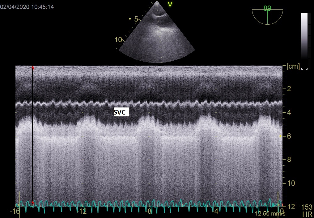
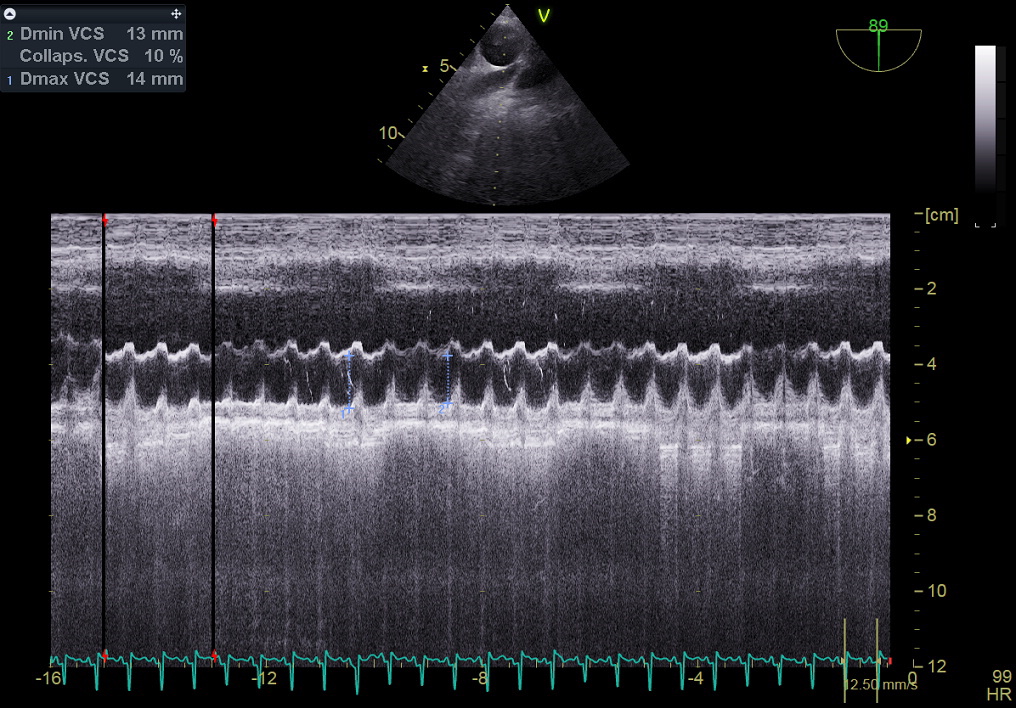
To the best of our knowledge, we report here the first case series of echocardiographic patterns in critically ill patients admitted for COVID-19. This study was enabled by our systematic and protocolized use of CCE for more than 25 years. Among the 79 patients admitted during the study period, 65 underwent CCE during the first week, in 39 cases because they required catecholamine infusion and in 26 cases systematically. The most frequent echo pattern was vasoplegia (≈ 38%), followed by ACP (≈ 28%), LV systolic dysfunction (≈ 25%), and hypovolemia (≈ 8%). Changes in echo patterns were observed when CCE was performed during D3 − 7, with more patients exhibiting ACP. Interestingly, all patients had a decrease in pulmonary acceleration time (PAAcT).
The pattern of ACP is probably the one which deserves the most discussion. It was the most frequent pattern during D3 − 7. We found thrombus in the pulmonary arteries in 67% of cases when CTPA was performed. Significant coagulopathy with clinical consequences has been reported in COVID-19 [16]. In an autopsy series of 12 patients, Wichmann et al. reported a high incidence of deep venous thrombosis and pulmonary embolism [5]. In 337 COVID-19 patients who underwent CTPA, Poyiadji et al. reported pulmonary embolism in 22% of cases [17]. Another explanation for ACP is distal microthrombosis of the pulmonary circulation due to extensive inflammation in the lung. This was noted in 7 lungs from patients who died from COVID-19 by Ackermann et al., who especially reported alveolar capillary microthrombi and severe endothelial injury [18]. The fact that our patients had a decrease in pulmonary artery acceleration time, even without ACP, suggests that most of them had a certain degree of pulmonary vascular dysfunction with pulmonary hypertension. In more “usual” ARDS, RV failure has also been suspected to be related to respiratory settings and alveolar overdistension. It is unlikely that such a mechanism could act in this specific situation as COVID-19 patients have near-normal lung compliance and are easily ventilated, at least during the first days [6] but larger studies are needed.
The other echo patterns are probably less specific to COVID-19 and are already well-described in septic shock [8]. They are probably due to the cytokine storm which injures the cardiovascular system [19]. Two different mechanisms of LV systolic dysfunction can be discussed in COVID-19. The first is much closer to septic cardiomyopathy [20], which is described as an acute injury of the heart related to cytokines [21]. In our population, LV systolic dysfunction, when present, did not involve LV dilatation, suggesting an acute injury. The second is related to the Kawasaki-like syndrome described in children and adolescents [22]. In our population, we had 2 young women, respectively 17 and 24 years of age, who developed such a syndrome with shock and severe LV failure.
Our study has some limitations. First, it was not a prospective study. However, our systematic and protocolized use of CCE for more than 20 years led us to record off-line and retrospectively most of the important information, so as to describe the echo patterns. While 14 patients did not undergo CCE, all patients who required catecholamine infusion did, at the time catecholamines were started. Second, it is a pure descriptive study, but this is the first case series reporting echocardiographic patterns in COVID-19 patients. Third, we decided not to report any longitudinal data and to limit the analysis to the first week. Indeed, due to the high ICU length of stay in these patients, we considered that after 1 week the disease progression was no longer very specific to COVID-19, but more related to the usual clinical course of critically ill patients, as ventilator-associated pneumonia or bleeding. Finally, we were unable to perform CTPA in all patients with ACP. This was mostly due to their instability. However, among those who underwent CTPA, 67% had thrombus in the pulmonary circulation.