The details of technique, methods, reliability, search for published evidence and references are provided in the online supplement (ESM supplement; website link…please insert when ready). The commonly used parameters for hemodynamic monitoring in critically-ill children are measurements of: blood pressure, central venous pressure (CVP), central venous oxygen saturation, cardiac output, serum lactate, pulmonary artery catheter, transpulmonary dilution, clinical signs, near-infrared spectroscopy, fluid responsiveness, microcirculation and role of ultrasonography A brief summary of the evidence related to each sub-section has been summarized below.
1. Clinical signs
Pediatric resuscitation courses (such as ETAT WHO, APLS) teach initial assessment of the shocked child well. Most caregivers will be familiar with the clinical signs and symptoms that help assess the hemodynamic status in children, including heart rate (HR); blood pressure; respiratory rate (RR); state of consciousness; diuresis; core and peripheral temperature; capillary refill time and peripheral perfusion. Some of these parameters are age-dependent and some can be altered by ambient temperature, pain, anxiety and many other factors. The mission of the primary resuscitation team is to identify the shocked child in need for urgent intervention and treatment, usually with fluids and then inotropes or vasopressors in some combination.
All recommendations reached a high level of agreement, both in identifying children in need for treatment and in the limited value of clinical signs to guide hemodynamic treatment. There is a significant variability in clinicians’ abilities to assess hemodynamic clinical parameters at the bedside. Early signs of hemodynamic decompensation may be subtle and can be easily missed by the clinicians [14]. For these reasons, the frequent and trend evaluation of clinical signs are more important than a single specific determination. A combination of vital signs can be more useful to evaluate hemodynamic state than individual parameters [15].
Disappointingly, there is no good correlation between clinical assessment and invasive hemodynamic parameters, which only indicates that clinical parameters and invasive parameters do not measure the same compartment [16]. Hence, in hemodynamically unstable patients apart from frequent meticulous assessment, monitoring trends of several measurable clinical, biochemical and monitoring parameters should be used to guide the therapy timely and accurately.
Table 1: Recommendations on use of clinical examination and blood pressure measurement in hemodynamic monitoring in critically ill children.
|
Sr No
|
Recommendation
|
Level of agreement
|
|
Clinical signs
|
|
1.
|
There is no single clinical parameter that allows to evaluate the global hemodynamic status in children and, therefore, we recommend to analyze several parameters and make frequent assessments.
|
Strong agreement
|
|
2.
|
We recommend to perform a clinical assessment as the initial evaluation in all patients for the detection of hemodynamic alterations and to evaluate clinical signs periodically together with hemodynamic monitoring parameters in unstable patients.
|
Strong agreement
|
|
3.
|
We do not recommend to titrate hemodynamic therapy or fluid loading solely based upon clinical signs or a reduced urine output alone in unstable patients with the exception of the initial resuscitation phase.
|
Strong agreement
|
|
Arterial blood Pressure
|
|
4.
|
We recommend the use of intra-arterial blood pressure (IBP) over oscillometric blood pressure (OBP) measurement when a reliable blood pressure (BP) measurement is of importance or when fast changes in blood pressure need to be detected.
|
Strong agreement
|
|
5.
|
In children over 12 years of age we recommend a target blood pressure of ³ 65 mmHg MAP (according to adults surviving sepsis guidelines) unless in children known to have prior hypertension.
|
Strong agreement
|
|
6.
|
We recommend not to use BP as the only therapeutic target in unstable children. The hemodynamic state should be evaluated integrating several clinical and hemodynamic parameters.
|
Strong agreement
|
|
7.
|
We recommend IBP monitoring in children in shock not responsive to initial fluid therapy or requiring vasopressor treatment, and hypertensive emergencies to control the effect of continuous invasive hypotensive drugs.
|
Strong agreement
|
- Arterial blood pressure
Blood pressure (BP) measurement is one of the most commonly used hemodynamic parameters for diagnostic and therapeutic decisions in critically-ill children, not least due to ease of utilization and, if invasive, the additional benefit of arterial blood sampling, as well as continuous data sampling. Both a low and a high BP on admission are related to an increased mortality [17]. Accurate measurement of BP is considered essential for the diagnosis and treatment of hypertension as well as of hypotension, including various categories of hemodynamic shock [18, 19]. BP can be measured invasively but also by using several less reliable non-invasive methods [20].
The committee strongly agreed on the use of intra-arterial blood pressure (IBP) over oscillometric blood pressure (OBP) measurement when there is a need for reliable BP measurement in children with shock not responding to initial fluid therapy or requiring inotropes or vasoactive medication; in patients with intracranial hypertension and intracranial pressure monitoring to measure cerebral perfusion pressure; during major surgery and in children with malignant hypertension or other hypertensive emergencies and to monitor the effect of continuous intravenous vasoactive medications or inotropes. However, the clinical value of BP in guiding hemodynamic therapy was not appreciated equally among the panel members. Nevertheless, there was strong agreement that BP should not be used as the only therapeutic target in unstable children, so the hemodynamic state should be evaluated integrating BP with several clinical and additional hemodynamic parameters [21].
Optimal values for BP in healthy and critically-ill children, including therapeutic thresholds, should be related to the clinical condition, age, sex and body size [20-25]. There was only weak agreement concerning BP values in children under 12 years of age. In children over 12 years of age generally we strongly recommend a target mean arterial pressure (MAP) of ³ 65 mmHg, although in specific situations the targeted BP may be higher such as when managing raised intracranial pressure.
- Central venous pressure (CVP)
The committee shared a strong common opinion regarding CVP. CVP should be measured as accurately as possible and be evaluated only as part of multi-modal hemodynamic monitoring to assess intravascular volume and cardiac function [26, 27]. Isolated CVP measurement is of limited value but trends of CVP, both the value and the wave morphology, or change in CVP in response to fluid or vasoactive therapy may provide useful information about overall hemodynamic status and cardiovascular physiology in critically-ill children. Specifically, a rise or high levels of CVP should be avoided [28]. The committee agreed that CVP is not of great value in the initial treatment of critically ill children but it can deliver important additional information in children with shock refractory to initial hemodynamic treatment, however, use of CVP requires a good understanding of its limitations and pathophysiology of underlying disease process. For example, CVP should not be used as a sole parameter to guide fluid therapy [29-32].
- Central venous oxygen saturation measurement
Central venous oxygen saturation (ScvO2) approximates but does not equal mixed oxygen saturation (SmvO2). The normal ranges for ScvO2 and SmvO2 are 70-80% and 60-70%, respectively, in the setting of a normal aortic saturation [33, 34]. Trends between ScvO2 and SmvO2 are often interchangeable, although SmvO2 values are generally around 7-10% lower than ScvO2. A low ScvO2 typically indicates a mismatch between oxygen supply and utilization. Conversely, a normal, or high ScvO2 value, does not necessarily signify supply-demand adequacy, as tissue dysoxia (which may occur in sepsis) may cause an artificial elevation (or normalization) of ScvO2. Lastly, ScvO2 in isolation cannot be considered a surrogate of cardiac index / cardiac output [35]. However, there is some evidence that resuscitation in sepsis might be more beneficial when ScvO2 is incorporated in the treatment strategy [36].
The committee agreed that ScvO2 is an important parameter in unstable patients not responding to first treatment and that its trend is helpful in hemodynamic management. However, we recommend that hemodynamic therapy should not be targeted solely based upon ScvO2 levels.
- Volume resuscitation and fluid responsiveness
Volume resuscitation is one of the most commonly used therapeutic options. Nevertheless, excessive fluid administration may impair tissue perfusion even further by promoting edema and third-space fluid accumulation [6, 7, 37]. A rise in cardiac output (or stroke volume) as a result of volume resuscitation is called fluid responsiveness. To prevent unnecessary fluid administration, it could be beneficial to predict fluid responsiveness before the fluids are delivered. Unfortunately, there is no clear, simple and proven method to predict fluid responsiveness in children. Static measures, mostly CVP, are not appropriate to test fluid responsiveness [29, 31]. The published evidence suggests that respiratory variation in aortic blood flow peak velocity {(ΔVPeak / velocity time integral (VTI)} is the most reliable indicator of fluid responsiveness, but only in ventilated children that fulfil various criteria [38]. Other dynamic methods, like passive leg raising test and liver pressure, have not been adequately assessed in children of all ages [39].
Due to the lack of simple bedside available methods to determine fluid responsiveness and the risk of fluid overload with aggressive approach, the committee recommended the following: recurrent smaller fluid boluses (maximal 5-10 ml/kg) in a short time interval in patients with hemodynamic instability while tracking changes in cardiac output, blood pressure and CVP to confirm or assess fluid responsiveness. Furthermore, we strongly agreed to recommend withholding fluid therapy in patients with an increasing CVP and no significant increase in blood pressure or cardiac output as a result of previous fluid therapy. No specific recommendations regarding estimating fluid responsiveness can be made in patients with raised intracranial pressure or extracorporeal life support (ECLS).
Table 2: Recommendations on use of measurement of CVP, SCVO2, and prediction of fluid responsiveness in hemodynamic monitoring in critically ill children
|
Sr No
|
Recommendation
|
Level of agreement
|
|
Central venous pressure
|
|
1.
|
We recommend to place the tip of a central venous catheter at the junction of the superior caval vein (SCV) and the right atrium to obtain an optimal central venous pressure (CVP) measurement or ScvO2 sample.
|
Strong agreement
|
|
2.
|
We recommend to measure CVP in all unstable patients refractory to initial hemodynamic treatment.
|
Strong agreement
|
|
3.
|
We recommend against the use of CVP to predict fluid responsiveness; Fluid loading should not be started solely based upon a low CVP.
|
Strong agreement
|
|
4.
|
An isolated CVP measurement is of limited value in clinical practice. However, trends in CVP may provide important information regarding changes in cardiovascular pathophysiology such as evolving right heart failure and an abrupt elevation in CVP upon fluid administration should raise suspicion of significant cardiac dysfunction.
|
Strong agreement
|
|
Central venous oxygen saturation measurement
|
|
5.
|
We recommend to measure central venous oxygen saturation (ScvO2) in unstable patients not responding to the initial treatment. ScvO2 < 65% suggest a possible hemodynamic alteration, however, in sepsis a normal or high ScvO2 may reflect mitochondrial dysfunction and mask hemodynamic alterations.
|
Strong agreement
|
|
6.
|
ScvO2 is not an adequate marker of cardiac index (CI).
|
Strong agreement
|
|
7.
|
We recommend against targeting hemodynamic therapy solely based upon ScvO2.
|
Strong agreement
|
|
Volume resuscitation and fluid responsiveness
|
|
8.
|
We recommend to observe the patient’s clinical situation, physical exam and various perfusion indicators suggesting an inadequate CO (or oxygen transport) caused by hypovolemia before considering fluid loading.
|
Strong agreement
|
|
9.
|
In delivering a bolus of fluid, we recommend to administer a small bolus of fluid in a short time period while tracking changes in cardiac output, blood pressure and CVP, and when possible or available, to confirm fluid responsiveness before commencing fluid loading therapy.
|
Strong agreement
|
|
10.
|
We recommend alternative therapeutic strategies for hypotension management in fluid non-responders.**
|
Strong agreement
|
|
11.
|
We recommend to withhold fluid therapy in patients with an increasing CVP and no significant increase in blood pressure or cardiac output as a result of previous fluid therapy.
|
Strong agreement
|
|
12.
|
We recommend fluid therapy (with boluses 5-10 ml/kg) as part of early resuscitation in unstable patients guided by the effect on blood pressure and / or cardiac output.
|
Strong agreement
|
|
**Non-responders defined cases who had no rise in cardiac output (or stroke volume) as a result of volume resuscitation.
|
- Echocardiography / Ultrasonography
Cardiac ultrasound or functional echocardiography is non-invasive and easily available at the bedside in the intensive care setting, and it allows rapid evaluation of hemodynamic status in real time. It can be used to assess cardiac function and preload, to estimate cardiac output and fluid responsiveness, to measure pulmonary artery systolic pressure and serial assessment allows to evaluate response to therapy [10, 40]. Hence, it can be used, as an adjunct to other parameters, in children with hemodynamic instability to gain additional information required for making accurate clinical decisions such as myocardial failure, pulmonary hypertension or cardiac tamponade [10]. It can help in understanding the pathophysiology of shock in children, and it can help in selecting timely, targeted specific and right intervention [10, 41, 42].
The committee strongly recommended the use of cardiac ultrasound for hemodynamic evaluation of infants and children with hemodynamic instability. Since cardiac ultrasonography does not provide continuous measurements and is limited by a wide intra and inter-observer variability [40, 43] we strongly recommend using serial assessments. However, we do not consider cardiac ultrasound as a tool for the routine hemodynamic monitoring in the intensive care setting.
- Cardiac output monitoring and transpulmonary indicator dilution
Cardiac output (CO) is the product of heart rate and stroke volume. Stroke volume depends on preload, contractility and afterload. The physical examination and simple commonly used hemodynamic parameters are the surrogate markers of cardiovascular well-being but they do not provide direct assessment of the cardiovascular hemodynamic status of the patient and clinical estimation of cardiac output has showed to be mostly inaccurate [14]. Hence, there seems to be a need of advanced hemodynamic monitoring to titrate therapies specifically when volume expansion or vasoactive drugs are delivered in order to improve cardiac output or systemic vascular resistance [44, 45]. In patients with refractory shock, when an effective and accurate measurement of CO is needed, the following methods may be used depending upon available resources and expertise: measurement of CO using transthoracic ultrasound (echocardiography) and transpulmonary dilution (TPD) [44-46]. Ultrasonography is non-invasive, easily available and can provide a fairly accurate and serial estimation of cardiac output at the bedside to monitor the initial response to therapy [40, 47]. However, it requires specific skills and it is operator-dependent. Despite being the most reliable clinical method to measure cardiac output, application of TPD in the clinical practice may be challenging because of resources, technical difficulties or lack of expertise. It is invasive and not suited to emergency resuscitation. Only two methods are available in children below 40 Kg: transpulmonary thermodilution (TPTD) by PiCCO (Pulsion Medical Systems, Germany) and transpulmonary ultrasound dilution (TPUD) by CO status (Transonic, USA) [44-46, 48]. Neither are in frequent use in ICU due to their intricate set-up, and particularly for PICCO the risk to children’s vessels from relatively large femoral arterial catheter required. Moreover, because of their intermittent measurement technique TPD methods are not suitable for the detection of rapid, frequent, changes in hemodynamic status, as required in some critically-ill children.
In the clinical practice, we recommend to use cardiac output ultrasound/Doppler methods for estimating CO in stable patients and the initial assessment of unstable patients. The committee did not reach to a strong agreement on the methods to estimate CO in children with refractory shock needing escalation of treatment and hemodynamic monitoring. There was a strong agreement that TPD methods are the most reliable but whether their use should be advised in situations needing escalation only reached a weak agreement. TPD methods also measure blood volumes and lung water but the committee recommended against using these parameters for targeting hemodynamic goals. Measuring CO using PAC is not recommended.
Cardiac output can also be estimated at the bedside using other non-invasive methods like bioimpedance and bioreactance, pulse contour and Doppler. Validation of these methods in critically-ill children is sparse and these methods are therefore not consensually viewed as accurate enough to estimate absolute values of CO in the intensive care setting in children [49]. However, they might provide a trend over time. In general, the committee strongly agreed that at the current time no recommendations regarding these methods can be given due to the limited experiences in critically-ill children.
- Pulmonary artery pressure (PAP)
The pulmonary artery catheter (PAC) can provide continuous measurement of right atrial, PAP, measurement of CO and pulmonary arterial occlusion pressure (wedge pressure). However, because of invasiveness and size it is not used or recommended in intensive care clinical practice in children [50]. Similarly, left atrial pressure can be measured using surgically inserted left atrial catheters (LAC) [51, 52]. Still, alternative less invasive techniques are being used in children to estimate left atrial pressure in unstable patients and LAC or PAC are rarely used in today’s intensive care clinical practice [53, 54].
Because of the above, the committee recommends not to use a PAC in children to measure cardiac output or PAP in the ICU. Instead, transthoracic ultrasound echocardiography can be easily used to estimate PAP at the bedside non-invasively and it can provide serial assessment to monitor the response to therapy or disease process (as above). However, it should not be used to estimate PAP in patients with right ventricular (RV) failure [55]. For precise measurement of PAP, we recommend using the PAC only at the cardiac catheterization laboratory.
Table 3: Recommendations on the use of cardiac ultrasound and other methods to estimate cardiac output for hemodynamic monitoring in critically ill children
|
Sr No
|
Recommendation
|
Level of agreement
|
|
Echocardiography / Cardiac ultrasound
|
|
1.
|
We recommend to use cardiac ultrasound as an adjunct to gain additional information required for making accurate clinical decisions in infants and children with hemodynamic instability but not as a tool for routine hemodynamic monitoring in intensive care setting.
|
Strong agreement
|
|
2.
|
Cardiac ultrasound can help in diagnosing pulmonary hypertension and assessing severity of pulmonary hypertension, and in detecting cardiac tamponade.
|
Strong agreement
|
|
3.
|
We recommend monitoring of pulmonary artery pressure (PAP) using ultrasound with refractory shock states to exclude pulmonary hypertension. Cardiac ultrasound may help in identifying underlying pathophysiology of shock and choosing the right intervention based upon deranged hemodynamic physiology (preload, afterload or cardiac function).
|
Strong agreement
|
|
4.
|
Cardiac ultrasound may help in assessing fluid responsiveness and we recommend using velocity time integral (VTI) across aortic valve for assessing fluid responsiveness rather than inferior vena cava collapsibility in mechanically ventilated infants and children.
|
Strong agreement
|
|
5.
|
We recommend using serial longitudinal assessments to assess response to therapy in patients with significant hemodynamic instability.
|
Strong agreement
|
|
Cardiac output measurement and transpulmonary indicator dilution
|
|
6.
|
We recommend to use ultrasound/Doppler based methods of estimating CO in stable patients, for the initial assessment of unstable patients and to decide if a more invasive method is required. When reliable absolute measurements of CO are deemed necessary, thermodilution (TPD) is the method of first choice.
|
Strong agreement
|
|
7.
|
In patients with a refractory shock when an accurate measurement of CO is needed, we recommend to use transpulmonary thermodilution (TPTD) or semi-invasive transpulmonary ultrasound dilution (TPUD).
|
Weak agreement
|
|
8.
|
We recommend to use invasive (and if possible continuous) CO monitoring in unstable post-operative patients after major (cardiothoracic) surgery, multiple trauma injuries or burns or patients with complex cardiopulmonary interactions.
|
Strong agreement
|
|
9.
|
We recommend against targeting fluid therapy based upon blood volumes measured with TPD or targeting hemodynamic therapy based upon lung water measurement to assess pulmonary oedema in critically ill children.
|
Strong agreement
|
|
10.
|
Because of their intermittent measurement technique, TPD methods are not suitable for the detection of fast changes in CO unless used in conjunction with continuous trend monitoring using pulse contour analysis, calibrated by transpulmonary indicator dilution technology.
|
Strong agreement
|
|
Pulmonary artery pressure
|
|
11.
|
We do not recommend to use pulmonary artery catheter (PAC) to measure CO in children. However, monitoring of left atrial pressure only in selected cardiac surgery patients or patients after lung transplant using a surgically inserted catheter can be helpful
|
Strong agreement
|
- Lactate measurement
Determination of blood lactate concentration is a cheap, fast and easy bedside parameter that has demonstrated utility to predict the outcome or to trigger the need to intensify medical treatment [56]. The committee showed some variation in their approach to the use of lactate in children since 5 out of 10 recommendations needed a revision.
In critically-ill patients or children with shock, early and serial lactate blood sampling from a reliable site such as a central venous or arterial indwelling catheter is recommended, though peripheral venous sampling with tourniquet time < 60 secs is possible [57]. This is specifically recommended when the initial capillary lactate value is > 3.0 mmol/L [57-59]. Studies report an association between failure to normalize lactate levels to a certain threshold (3.0±1.0 mmol/L) during the first 12 to 24 hours of ICU admission, and adverse outcomes regardless of the reason for ICU admission [60, 61]. Experts could not agree on the use of lactate as part of a goal-directed approach and only weakly agreed on the approach to a persistent high lactate level. In the latter, lactate levels should always be used in conjunction with other clinical indicators of poor systemic perfusion and monitoring parameters. Persistently elevated lactate levels may reflect other mechanisms rather than those derived from poor tissue perfusion in shock, and instead reflect aerobic glycolytic mechanisms including catecholamine administration or endogenous release [62].
- Near-infrared spectroscopy
Near-infrared spectroscopy (NIRS) is a non-invasive, bedside technique to estimate regional capillary-venous hemoglobin saturation (rSO2). The mean baseline cerebral rSO2 is >70% in healthy children. Infants and children with cyanotic heart disease may have a cerebral rSO2 between 46-57% [63-67]. Moreover, practitioners should be mindful about a considerable variability in NIRS values between commercially available devices. It has been observed that values measured in both monitors INVOS 5100-C® (Medtronic; Boulder, CO, USA) and Foresight Elite® monitor (CAS Medical Systems; Branford, CN, USA) are not interchangeable [68]. Although NIRS is mainly used to measure rSO2 in the brain, there are also reports of its use on other organs. In a study by Dabal et al. [69], it appears that renal NIRS and inferior vena cava desaturations precede rScO2 changes in the prediction of serious cardiovascular adverse events in patients after stage 1 Norwood palliation. Trend in NIRS values may provide valuable physiological information in children with hemodynamic instability although clear (cut-off) values and evidence of benefit are lacking.
The committee strived to define recommendations with regard to this subject and 6 out of 7 recommendation had to be redefined. As a result, the only strong recommendation was to advise against routine use of NIRS in all children with hemodynamic instability. Moreover, the committee agreed not to make recommendations regarding the use of NIRS while treating children in shock, post-cardiac arrest, post traumatic brain injury and infants with hypoxic-ischemic encephalopathy. Lastly, there was no agreement on the clinical usefulness of a decline of cerebral rSO2 under 40-50% or a change in baseline of more than 20% [70 ].
- Microcirculation
Microcirculatory assessment by videomicroscopy using side-stream or incident dark field is expensive, and not widely available. Currently it does not allow for assessment of rapid circulatory changes during resuscitation [71]. No studies have defined the normal values of microcirculation in children outside the neonatal period but do report that vascular density seems to decrease with age [72]. So far, published studies have not defined target values of microcirculatory parameters in critically ill children [72-77]. At this point in time, the committee recommends its use only for research purposes.
The committee also states that many routinely used parameters like capillary refill, peripheral temperature, lactate, may reflect aspects of the hemodynamic condition but do not adequately reflect the microcirculation and cannot be used as such. Although central venous to arterial CO2 difference could provide additional insight into the microcirculatory condition, we currently recommend against its use to guide resuscitation in critically ill children.
Table 4: Recommendations on use of serum lactate, near infrared spectroscopy (NIRS) and microcirculation assessment for hemodynamic monitoring in critically ill children
|
Sr No
|
Recommendation
|
Level of agreement
|
|
Serum lactate measurement
|
|
1.
|
We recommend to obtain a repeat blood sample from a reliable site when the lactate value of a capillary sample is higher than 3.0 mmol/L and to closely follow up patients and intensify treatment until lactate values at least drop below 3.0 mmol/L, especially if other concerns regarding tissue hypoxia are present.
|
Strong agreement
|
|
2.
|
We recommend to interpret lactate levels always in conjunction with clinical indicators of poor systemic perfusion and monitoring parameters.
|
Strong agreement
|
|
Near infrared spectroscopy
|
|
3.
|
Trend in NIRS values may provide valuable physiological information in children with hemodynamic instability but routine use in all children with hemodynamic instability is not recommended.
|
Strong agreement
|
|
4.
|
Near infrared spectroscopy (NIRS) can be useful during the peri-operative period after surgery for congenital heart defects, however we recommend against the routine use of NIRS during non-cardiac surgery.
|
Weak agreement
|
|
Microcirculation
|
|
5.
|
Many routinely used parameters like capillary refill, peripheral temperature, lactate, NIRS etc. reflect aspects of the hemodynamic condition but they do not adequately reflect the microcirculation. Although central venous to arterial CO2 difference could provide additional insight into the microcirculatory condition, we recommend against its use to guide resuscitation in critically ill children
|
Strong agreement
|
|
6.
|
We recommend against routine microcirculation evaluation by video microscopy in stable children except those in clinical studies
|
Strong agreement
|
Limitations
We acknowledge the limitations of these recommendation: 1) The most important limitation is the lack of high-quality evidence. These recommendations are based upon expert consensus and review of the published literature including experts’ opinions, which can involve subjective value judgments, 2) Both lower and upper limits of age, from term infant >37 weeks and postnatal age > 4 weeks to 18 years, are artificial - to avoid overlapping with neonatal and adult population specific guidelines, and 3) some of these recommendations may not be appropriate for low-resources settings and may not be applicable in all settings requiring hemodynamic monitoring in children because of their limited availability or expertise.
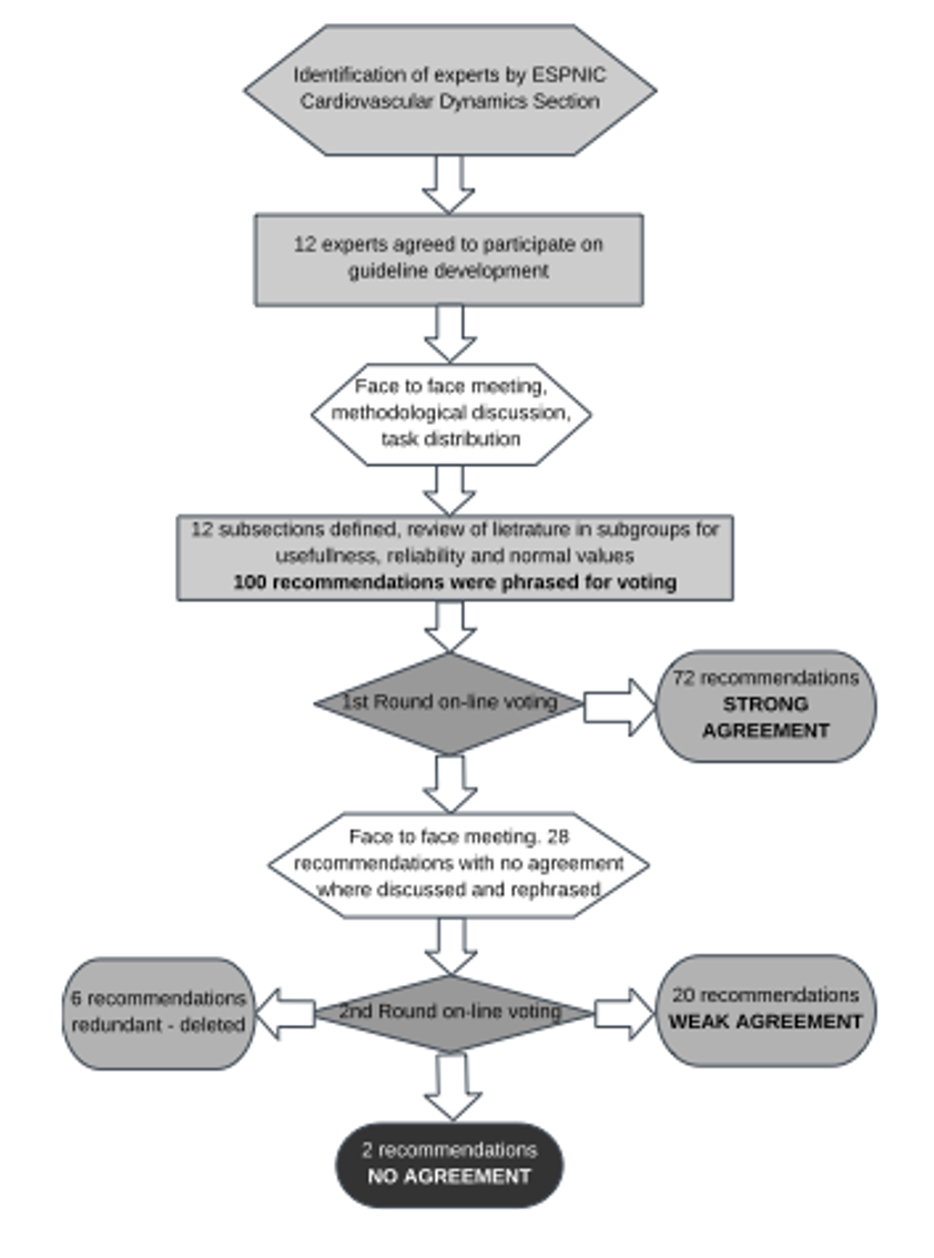
Nevertheless, despite these limitations the committee members believe that these are consensual expert recommendations based upon literature review and rigorous standardized process of developing expert consensus – followed DELPHI approach, a well-established standardized approach (DELPHI approach) – to reach consensus in such circumstances of limited published evidence to develop evidence-based guidelines.
Future directions
The committee recognize that there is an important lack of knowledge and evidence concerning hemodynamic monitoring in children. There is a great need for: 1) studying the relationship between measured parameters and end-organ perfusion, and 2) evaluating the clinical efficiency and patient outcome when therapy is guided by specific monitoring technologies.