Reagents
Sodium L-lactate (71718), sodium pyruvate (792500), streptozotocin (STZ, S0103) and aprotinin (A6106) were purchased from Sigma-Aldrich (Missouri, USA). lnsulin (2018283062) was purchased from Novo Nordisk (Bagsværd, Denmark). Glucagon (HY-P0082) and FSK (HY-15371) were purchased from MedChemExpress (New Jersey, USA). Glucose (20171108) was purchased from Sinopharm Chemical Reagent (Beijing, China). A blood glucose meter (06656919032) and test strips (1072332990) were purchased from Roche (Basel, Switzerland). Sulfo-Cyanine7 NHS ester (Cy7, GY1058) was purchased from Goyoo Biotechnology (NanJing, China). Dulbecco’s modified Eagle medium (DMEM, high-glucose, D5796), protease inhibitor cocktail I (20-201) and a dipeptidyl peptidase-4 (DPP4) inhibitor (DPP4-010) were purchased from Millipore (Massachusetts, USA). Low-glucose DMEM (31600-500), glucose-free DMEM (90113-500) and L-alanine (A8210) were purchased from Solarbio (Beijing, China). Fetal bovine serum (FBS, A3160901) was purchased from Gibco (New York, USA). LipofectamineTM 2000 (11668-027) was purchased from Invitrogen (Massachusetts, USA). Chow (HD1001) and a HFD (HD001) were purchased from BiotechHD (Beijing, China). cAMPS-Rp triethylammonium salt (151837-09-1) was purchased from Tocris Bioscience (Bristol, England). Collagenase IV (2091) was purchased from BioFroxx (Einhausen, Deutschland). PerfectStartTM Green qPCR SuperMix (AQ601) and TransScript® One-Step gDNA Removal and cDNA Synthesis SuperMix (AT311) were purchased from TransGen Biotech (Beijing, China). NucleoZOL (740404) was purchased from MACHEREY-NAGEL (MN, Düren, Deutschland). A mouse insulin ELISA kit (PI602) and BCA protein concentration determination kit (P0012) were purchased from Beyotime (Shanghai, China). A mouse glycated plasma protein kit (80420) was purchased from Crystal Chem (Washington, USA). Mouse aspartate transaminase (AST, 200218), alanine transaminase (ALT, 191230), triglyceride (TG, 200224) and cholesterol (CHO, 200224) biochemical test kits were purchased from Ruierda Biological Technology (Beijing, China). A cAMP assay kit (ab133051) and PKA Kinase Activity Kit (ab139435) were purchased from Abcam (Illinois, USA). An Amplex™ Red Glucose/Glucose Oxidase Assay Kit (A22189) was purchased from Invitrogen (Massachusetts, USA).
Antibodies
Anti-α-tubulin (T6074, 1:5,000 dilution) and anti-Flag (A8592, 1:5,000 dilution) antibodies were purchased from Sigma-Aldrich (Missouri, USA). Anti-glucagon (ab92517, 1:2500 dilution), anti-insulin (ab6995, 1:200 dilution), anti-CREB-phospho S133 (ab32096, 1:1000 dilution), and anti-CREB (ab32515, 1:1000 dilution) antibodies were purchased from Abcam (Illinois, USA). An anti-GP73 antibody (F-12, sc-393372, 1:200 dilution) was purchased from Santa Cruz (Texas, USA). An anti-His antibody (KM8001, 1:1000 dilution) was purchased from Taihua Lekang Biotechnology (Beijing, China). Anti-phospho-Akt (Thr308, 13038, 1:1000 dilution), anti-Akt (9272, 1:1000 dilution), anti-phospho-PKA-C-α (Thr197, 5661, 1:1000 dilution), anti-phospho-PKA substrate (RRXpS/T, 9624, 1:1000 dilution), and anti-PKA-C-α (5842, 1:1000 dilution) antibodies were purchased from Cell Signaling Technology (Danvers, USA). Anti-rabbit HRP-IgG (ZB-2301, 1:5000 dilution) and anti-mouse HRP-IgG (ZB-2305, 1:5000 dilution) secondary antibodies were purchased from ZSGB-BIO (Beijing, China). An anti-GP73 monoclonal antibody (mAb) for the blocking experiment was custom made. Isotype-matched IgG (A7028) was purchased from Beyotime (Shanghai, China).
Plasmids and cell culture
Mammalian expression vectors encoding Flag-tagged human, mouse and rat GP73 were constructed by inserting the corresponding PCR-amplified fragments into pcDNA3 (Invitrogen, Massachusetts, USA). The HepG2 (CRL-10741), Vero E6 (CRL-1568), HK-2 (CRL-2190), 293T (CRL-3216) and L6 (CRL-1658) cell lines were obtained from the American Type Culture Collection (ATCC, Rockville, MD, USA). The Huh-7 (0403) cell line was obtained from the Japanese Collection of Research Bioresources. All cell lines were tested for mycoplasma contamination and were incubated in DMEM at 37°C in a humidified atmosphere with 5% CO2. LipofectamineTM 2000 was used for transfection following the manufacturer’s protocol.
To knock out human GP73 in Huh-7 cells, two small guide RNAs (sgRNAs) targeting GP73 were designed and inserted into the LentiCrispr v2 vector to construct transfer plasmids. 293T cells were transfected with pMD2. G, psPAX2 and the corresponding transfer plasmid to produce lentivirus. A total of 108 Huh-7 cells were infected with lentivirus at a multiplicity of infection (MOI) of 2.0 and selected with 4 μg/mL puromycin for two weeks to ensure proper selection.
The following sgRNA sequences were used:
sgRNA-1: 5’-CACCGCACACACAGAGGTGCCACAA-3’
sgRNA-2: 5’-CACCGACCAGTTAAAGACCCTGCAG-3’
control- 5’-CACCGCGCTTCCGCGGCCCGTTCAA-3’.
PMHs were isolated and purified using a modified two-step collagenase perfusion method. Cells were resuspended in low-glucose DMEM containing 5% FBS and seeded on 15-cm dishes at 80% confluence. Five hours later, the cells were washed and cultured in serum-free medium overnight. For gluconeogenesis-related assays, the medium was replaced with glucose- and phenol-free DMEM the next day in the presence of 10 mM pyruvate sodium and 10 mM sodium lactate, and the cells were treated with the indicated concentrations of rmGP73 or rrGP73, 200 mM cAMPS-Rp, or 2 μM glucagon. Then, 64 nM GP73 was used for 10 min for the cAMP and PKA assays and for 1 h for phosphoproteomics. The results were normalized to the protein content.
Sample acquisition from COVID-19 patients
The Ethics Committee of Huoshenshan Hospital approved the study (HSSLL036). Given the urgency of the COVID-19 pandemic, the need for informed consent forms was waived by the ethics boards of the hospitals. Basic information and serum biochemical test results were collected from 136 COVID-19 patients at Huoshenshan Hospital (Wuhan, Hubei province, People’s Republic of China) from January 11 to March 11, 2020. Diagnosis was based on chest computed tomography (CT) manifestations and/or reverse transcription-polymerase chain reaction (RT-PCR) according to the criteria of the New Coronavirus Pneumonia Prevention and Control Program (5th edition) published by the National Health Commission of China. According to these criteria, COVID-19 patients were classified into mild, moderate, and severe COVID-19 subgroups. Data were excluded if the subject was younger than 18 years or older than 75 years, had incomplete medical records, acute lethal organ injury (e.g., acute myocardial infarction, acute coronary syndrome, acute pulmonary embolism, or acute stroke), or decompensated or end-stage chronic organ dysfunction (e.g., decompensated cirrhosis, decompensated chronic renal insufficiency, or severe congestive heart failure), were pregnant or had malignancy (Table 1). Human blood samples were collected from the COVID-19 patients analyzed in this study before intervention. Fifty patients were classified as having mild COVID-19, 65 patients had moderate COVID-19, and 21 patients had severe COVID-19 (Table 2).
Sample acquisition from healthy and diabetic patients
Human blood samples from the healthy and diabetic patients used in this study were obtained from individuals admitted to the Third Medical Center of the Chinese PLA General Hospital. The detailed characteristics of the recruited subjects are described in Table 3. All individuals in this study provided a signed statement of consent. The Committee for Ethics in Human Studies from the Third Medical Center of the Chinese PLA General Hospital approved this study (KY2021-009).
Infection with authentic SARS-CoV-2
The SARS-CoV-2 strain (2019-nCoV BetaCoV/Beijing/AMMS01/2020) used in the present study was isolated from the lung lavage fluid of an infected patient and preserved at the State Key Laboratory of Pathogen and Biosecurity at Beijing Institute of Microbiology and Epidemiology. SARS-CoV-2 infections were performed in the BSL-3 Laboratory of the Beijing Institute of Microbiology and Epidemiology. The infectious virus titer was determined as plaque-forming units in Vero E6 cells and was used to calculate the MOI. Cells were infected with SARS-CoV-2 at the indicated MOIs for the indicated times in glucose-free medium for the production assay. The results were normalized to the protein content. Cells were infected with SARS-CoV-2 at the indicated MOIs for the indicated time, and the supernatant was harvested for the GP73 assay and viral load assay. Viral RNA was extracted from the supernatants using the QIAamp Viral RNA Mini Kit (52906, Qiagen) according to the manufacturer’s instructions. Viral RNA was analyzed using qRT-PCR and a One-Step PrimeScript RT-PCR Kit (RR064B, TaKaRa) using SARS-CoV-2-specific primers in an Applied Biosystems 7500 Real-time PCR System. The following sequences of the SARS-CoV-2 probes were used:
SARS-CoV-2 open reading frame 1b (ORF1b):
Forward: 5’- CCCTGTGGGTTTTACACTTAA-3’
Reverse: 5’- ACGATTGTGCATCAGCTGA-3’.
Probe: 5’-FAM- CCGTCTGCGGTATGTGGAAAGGTTATGG-BHQ 1-3’.
SARS-CoV-2 nucleocapsid (N):
Forward: 5’- GGGGAACTTCTCCTGCTAGAAT-3’
Reverse: 5’- CAGACATTTTGCTCTCAAGCTG-3’.
Probe: 5’-FAM- TTGCTGCTGCTTGACAGATT-BHQ 1-3’.
The number of copies per microliter (μl) was determined using a synthetic RNA fragment to amplify the target region.
Recombinant GP73 protein purification
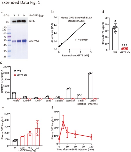
Human, mouse, and rat GP73 cDNAs, each with a six-amino-acid His tag on the N-terminus, were cloned into the pCDNA3.1 vector. Ni-NTA His-Bind column-bound His-GP73 protein from 293T cells transfected with the above plasmids was further purified using size-exclusion columns and polymyxin B-based endotoxin-depletion columns after extensive washing. The final His-GP73 proteins used in all recombinant protein experiments were >90% pure (endotoxin<=2 EU/ml) and stored at -80°C.
Animals, intervention and monitoring
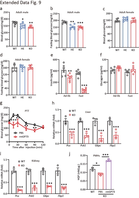
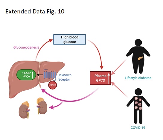
Male C57BL/6N WT mice (8 to 10 weeks old) were purchased from SPF Biotechnology (Beijing, China). GP73 KO mice (T20200316-18[D25]) were generated by Southern Model Biotechnology (Shanghai, China). Male C57 BLKS/J db/db and BKS control mice (8 weeks, 36-40 g) were purchased from GemPharma Tech Co. Ltd. (Jiangsu, China). All mice were group-housed conventionally on a 12-h light/dark cycle for 3 days before any experiments. All animal experiments were performed at the AMMS Animal Center (Beijing, China) and were approved by the Institutional Animal Care and Use Committee. For single injections, mice were injected i.v. with 0.1 mg/kg recombinant His-tagged GP73, and plasma was collected at the indicated times via tail bleeding for insulin and glucose level measurements. The ITT, GTT, PTT, and ATT were performed using standard procedures. A 0.75 U/kg insulin bolus was used for the ITT, a 1.5 g/kg glucose bolus was used for the GTT, a 1.5 g/kg pyruvate bolus was used for the PTT, and a 0.6 g/kg alanine bolus was used for the ATT. For immunological sequestration experiments, mice were injected i.v. with 15 mg/kg custom-made anti-GP73 mouse mAb or an equivalent dose of IgG (30 mg/kg).
To establish an HFD-induced STZ model, mice were maintained on a regular chow diet or fed an HFD for 1 month beginning at 4 weeks of age. STZ (40 mg/kg) in citric acid buffer (0.1 mol/L, pH 4.2) was administered to male C57BL/6N mice via intraperitoneal injection, and the same dose of STZ was injected 24 h later. After STZ injection, the mice were fed an HFD for another month. Fasting blood glucose and random blood glucose levels were measured weekly. The mice were confirmed to have diabetes if their fasting blood glucose levels were over 199.8 mg/dL or their random blood glucose levels were over 300.6 mg/dL. For all experiments, mice were randomly assigned to different groups to ensure an unbiased distribution.
Glucose measurement
All blood samples were collected from the tail, and glucose levels were measured using the glucose oxidase method and an automated blood glucose reader (ACCU-CHEK, Roche). For the measurement of fasting blood glucose levels, normal mice were fasted for 6 or 12 h, as indicated. Random blood glucose levels were measured at 9:00 a.m. If the glucose level was greater than 630 mg/dL (upper detection limit of the glucometer), a value of 630 mg/dL was recorded. Blood glucose levels were determined.
Assays of plasma hormone levels
Blood samples for hormone detection were collected from the tail or orbital vein. A DPP4 inhibitor (1:100 dilution), aprotinin (1:100 dilution) and protease inhibitor cocktail I (50000 KIU/mL, 1:100 dilution) were added to each blood sample. Plasma insulin levels were measured using ELISA.
Immunofluorescence staining
Tissues were fixed with 10% (v/v) neutral-buffered formalin at 4°C overnight and embedded in paraffin, and 5-μm-thick sections were prepared. For immunofluorescence, the sections were heated in an autoclave in citrate buffer (12 mmol/L, pH 6.0), preincubated in permeabilization/blocking buffer (0.1 mmol/L PBS, pH 7.3, 0.5% Triton) and blocked for 30 min with 10% (v/v) goat serum (Zhongshan Biotechnology, Beijing, China). The sections were subsequently incubated with primary antibodies at 4°C overnight and secondary antibodies for 1 h at room temperature, washed and stained with DAPI (1 μg/mL). Images were captured under a confocal fluorescence microscope (Zeiss LSM710, Carl Zeiss Microscopy GmbH, Jena, Germany) or an automatic digital slide scanner (Pannoramic MIDI, 3D HISTECH, Budapest, Hungary).
Phosphoproteomics
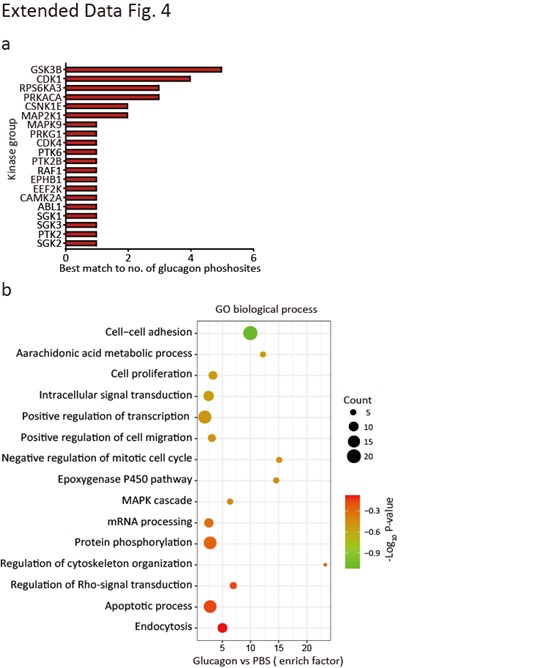
PMHs were suspended in low-glucose DMEM containing 5% FBS and seeded in 15-cm dishes at 80% confluence. The cells were washed and cultured in serum-free medium overnight 5 h later. The medium was replaced with glucose- and phenol-free DMEM supplemented with 10 mM pyruvate sodium and 10 mM sodium lactate the next day, and the cells were incubated for 1 h with PBS, rmGP73 (64 nM), or glucagon (2 μM). For cell lysate collection, the cells were washed twice with cold PBS and scraped with cold RIPA lysis buffer supplemented with protease and phosphatase inhibitors. Phosphoproteomics was performed by Oebiotech Company. Briefly, the samples were subjected to enzyme digestion and iTRAQ labeling, and the phosphopeptides were enriched and analyzed using LC-MS/MS. The raw data of this study have been deposited in the IProX database with the following accession number: PXD025381. For kinase enrichment, the Literature Based Kinase-Substrate Library with Phosphosites on the Kinase Enrichment Analysis 2 (KEA2) website was searched. The network was represented using Cytoscape ver 3.6.2 (https://cytoscape.org/). Protein-protein interactions were retrieved from STRING App (v1.51) (https://string-db.org/). Only interactions with high confidence (interaction score >0.7) from databases and experiences were kept. For KEGG and GO enrichment analysis, DAVID Bioinformatics Resources 6.8 (https://david.ncifcrf.gov/home.jsp) was used. For specific kinase substrate motif analysis, MoMo Modification Motifs 5.3.3 (http://meme-suite.org/tools/momo) was used.
In vivo imaging system (IVIS)
GP73 was labeled with Cy7 via the addition of the dye according to the manufacturer’s instructions at a pH of 8.0 and incubation of the mixture for 4 h on ice. The labeled GP73 was returned to a pH of 7.0, and the free dye was removed via overnight dialysis in PBS. The labeled GP73 was added to a Sephadex G50 size exclusion column equilibrated with PBS. Fractions of 500 µL were collected, the protein concentration was analyzed using a standard BCA protein assay kit, and fluorescence corresponding to excitation/emission of 745/800 nm was assessed.
After i.v. injections with free Cy7 and GP73-Cy7, mice were scanned using an IVIS (PerkinElmer) at the indicated time points to assess fluorescence. After whole-body imaging, the mice were sacrificed, and the major organs were imaged to assess fluorescence under the same settings as the in vivo imaging. The data were analyzed and exported using built-in Living Image Software (Version 4.5.5, PerkinElmer).
Quantitative real-time PCR (qRT-PCR)
Total mRNA was extracted from cells or various mouse tissues using NucleoZOL. cDNA was prepared from total mRNA using TransScript® One-Step gDNA Removal and cDNA Synthesis SuperMix, and the relative levels of individual mRNAs were calculated after normalization to the GAPDH mRNA level in the corresponding sample as previously described. The primer sequences are presented in Table 4.
Sandwich ELISA and Western blot analysis
For endogenous GP73 sandwich ELISA, two custom-made rat monoclonal anti-GP73 antibodies were used as the capture antibody and the detection antibody. Briefly, the plate was coated with an unlabeled capture antibody, and serially diluted standards and samples were added to the plate. After three washes, HRP-linked detection antibody was added to generate a colorimetric signal at 450 nm. For His-tagged GP73 sandwich ELISA, the same procedure was used, except a goat anti-His polyclonal antibody (Abcam) was used as the detection antibody. Increasing amounts of recombinant His-tagged GP73 were used to generate a standard curve for both assays.
For immunoblotting, cells were lysed in NP40 cell lysis buffer with fresh protease inhibitors. Whole-cell lysates were separated using SDS-PAGE after centrifugation and transferred to PVDF membranes for immunoblot analyses using the indicated primary antibodies.
Statistical analysis
The present study used GraphPad Prism 8.0 for statistical calculations and data plotting. Differences between two independent samples were evaluated using two-tailed Student’s t-tests or the Mann-Whitney test, as appropriate. Differences between multiple samples were analyzed using one-way ANOVA or two-way ANOVA followed by Bonferroni’s post hoc test, as appropriate. Correlations were analyzed using Spearman’s non-parametric test. All tests were two-tailed unless otherwise indicated. We considered a P value ≤ 0.05 as statistically significant. Significance values were set as follows: ns (not significant), P > 0.05; *, P < 0.05; **, P < 0.01; and ***, P < 0.001.