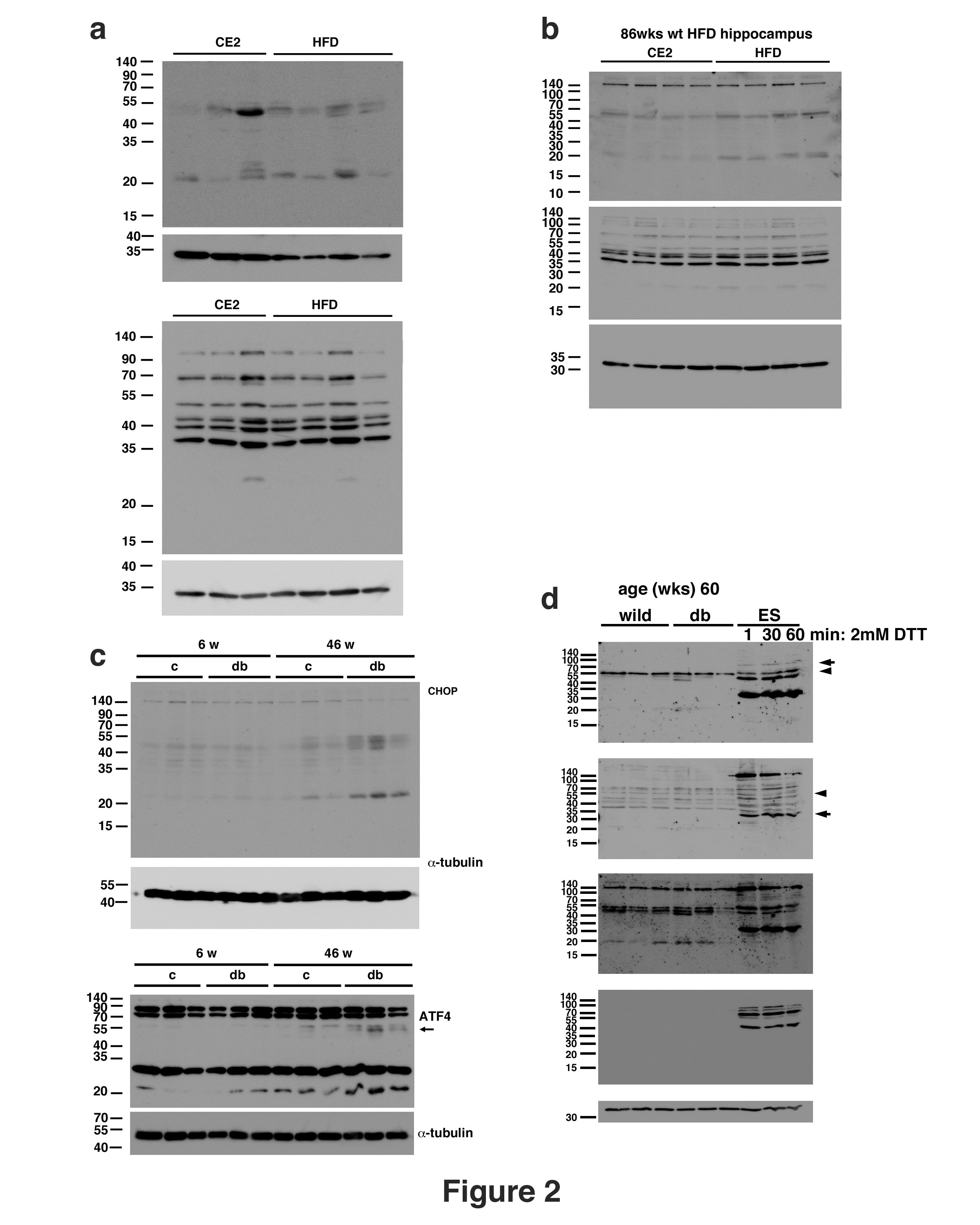
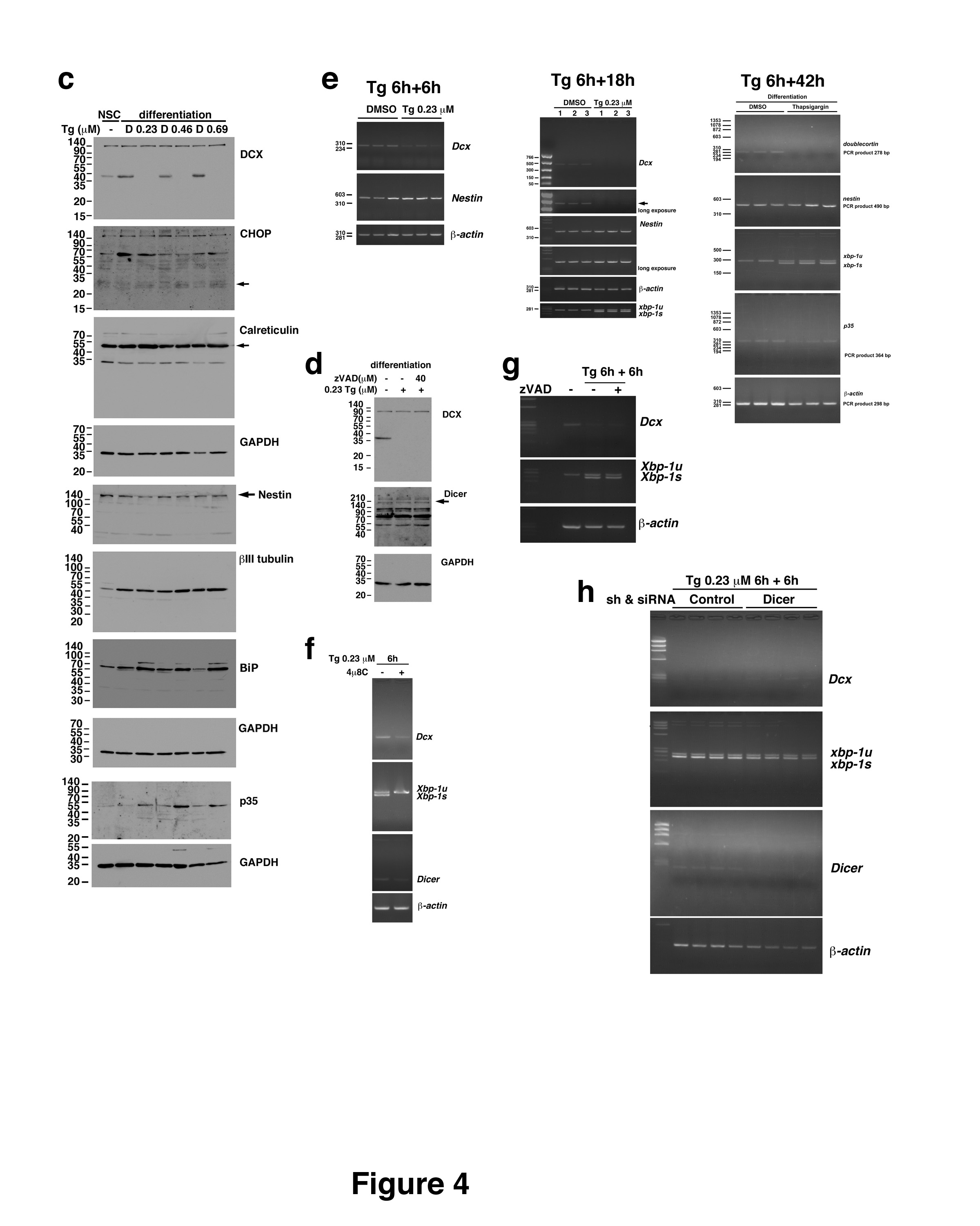
Western blot analysis and chemicals
Western blotting was performed as described previously31. Mouse tissue and cells were lysed in RIPA [50 mM Tris-HCl (pH 7.5), 150 mM NaCl, 0.1% sodium dodecyl sulfate (SDS), 0.1% sodium deoxycholate, and 1% Nonidet P-40] with Roche cOmplete™ mini tablets (Roche, Mannheim, Germany), 25 µM MG132 (EMD Chemicals, Inc; San Diego, CA), and phosphatase inhibitors, i.e., 20 mM β-glycerophosphate (Sigma-Aldrich Co., LLC; St. Louis, MO) and 20 mM sodium orthovanadate (Fujifilm, Osaka, Japan). After centrifugation at 13,100 x g, the supernatants were used for western blotting. The antibodies used for western blotting were as follows: anti-CHOP antibody (Thermo Fisher Scientific, Waltham, MA; Cell Signaling Technology, Inc; Danvers, MA); anti-ATF4 antibody (Santa Cruz Biotechnology, Inc; Santa Cruz, CA; Thermo Fisher Scientific); anti-p35 (C-19) antibody; anti-GAPDH antibody (Santa Cruz Biotechnology, Inc); anti-α-tubulin antibody (Sigma-Aldrich Co., LLC; St. Louis, MO); anti-Dcx antibody (Abcam, Cambridge, UK; Novus Biologicals, Centennial, CO; Santa Cruz Biotechnology, Inc); anti-Calreticulin antibody (StressGen Biotechnologies Corp., Victoria, Canada); anti-Nestin antibody (EMD Millipore Corp., Temecula, CA); anti-βIII tubulin antibody (Covance, Inc; Princeton, NJ); and anti-BiP (Grp78, KDEL) antibody (ENZO Life Sciences International, Inc., Plymouth Meeting, PA). HRP-conjugated anti-rat, anti-mouse, and anti-rabbit IgG (H + L) antibodies (SouthernBiotech; Birmingham, AL) and thapsigargin (Santa Cruz Biotechnology, Inc.) were used. FBS levels were measured with a FreeStyle Freedom Lite device (Abbott Diabetes Care Inc., Alameda, CA). Insulin levels were measured by a mouse insulin enzyme-linked immunosorbent assay (ELISA) Kit (RTU) (Shibayagi, Gunma, Japan). BDNF levels were measured with a Mature BDNF Rapid™ ELISA Kit (Bisensis Pty Ltd., Thebarton, Australia). All other chemicals were purchased from Wako Pure Chemical Industries, Ltd. (Osaka, Japan), Kanto Chemical Co., Inc., (Tokyo, Japan), and Sigma-Aldrich Co., LLC.
Animals and the HFD
C57BL/6J mice were purchased from Japan SLC, Inc. (Hamamatsu, Japan). APP23 mice, which express human APP751 cDNA and a Swedish double mutation on the C57BL/6 genetic background 34, were kindly provided by Dr. M. Staufenbiel (Novartis Pharma Ltd; Basel, Switzerland). Obese and diabetic db/db (Leprdb/db) mice on the C57BL/6 genetic background were purchased from The Jackson Laboratory (Bar Harbor, ME). The mice were housed in a temperature- and light-controlled room (24°C; 12-h light/dark cycle). The mice were fed on AIN93G (standard diet) or HFD-60 (Oriental Yeast Co., Ltd; Tokyo, Japan). All animal studies were approved by the Gifu University Graduate School of Medicine Animal Care and Use Committee under the guidelines for experiments on animals provided by the Ministry of Education, Culture, Sports, Science and Technology of Japan. All animal experiments were performed according to the ARRIVE guidelines.
Behavioral procedures
The NOL test was performed according to the method described by Roy et al67. Briefly, each mouse was habituated to a cage without objects for 15 min on day 1. The mice were exposed to three different objects, i.e., conical (diameter x height: 5 x 11.5 cm), cylindrical (6.5 x 10.5 cm), and reagent (5 x 13.2 cm) bottles, for 5 min three times per day at 2-min intervals. The mice were exposed to three objects on day 3 and placed in their home cages for a retention interval (two minutes). Then, one of the objects was moved to the opposite corner. The behavior of each mouse was monitored using video recording software and an automated tracking system (SMART v3.0 software; Panlab, Barcelona, Spain) and a video camera (HDC-HS350; Panasonic; Osaka, Japan). When the distance between the nose of the mouse and an object less than 2 cm or when the mouse sniffed or touched the object with its snout, the mouse was considered to be exploring the object, as previously described68. The cages and objects were cleaned with 70% ethanol and 1% acetic acid solution before each trial to eliminate dominant odors.
The MWM test was performed according to a previously described protocol69. Briefly, the apparatus was a 100-cm diameter tank containing water at a temperature of approximately 22°C water, skim milk and a submerged platform. Four acquisition trials from each of the five starting positions were performed each day. The time limit for each trial was 60 sec. Mice that did not reach the platform were guided to the platform and left on the platform for 30 sec. On day 5, the platform was removed for the probe test. Each trial was recorded and analyzed with the SMART v3.0 automated tracking system.
Immunostaining
Immunostaining was performed as described previously25 and analyzed by confocal microscopy (LSM710, Carl Zeiss; Göttingen, Germany). Briefly, mice were anesthetized and perfused with phosphate-buffered saline (PBS) followed by 4% paraformaldehyde in 0.1 M phosphate buffer (PB). The mouse brains were postfixed for 2 h in the same fixative, which was then replaced with 15% sucrose in 0.1 M PB. Sections (14 µm) were incubated in PBS containing 10% normal goat serum (Jackson ImmunoResearch Laboratories, Inc., West Grove, PA) and 0.1% Triton X-100 at room temperature for 1 h. Sections were incubated with an anti-Dcx antibody and anti-CHOP antibody or an anti-Ki67 antibody (Thermo Fisher Scientific) in PBS containing 1% normal goat serum and 0.1% Triton X-100 at 4°C for 12 h. For detection of Ki67, sections were incubated in 10 mM sodium citrate (pH 6) at 80°C for 30 min and cooled to room temperature before adding antibody. The sections were incubated with Alexa Fluor 488-conjugated anti-rabbit IgG (H + L) and Alexa Fluor 546-conjugated anti-mouse IgG (H + L) antibodies (Thermo Fisher Scientific) and Hoechst [1 µg/mL bis-benzimide (Sigma-Aldrich Co. LLC)] to detect fluorescence signals and nuclei. The fluorescence intensity profiles were analyzed with Zen software (Carl Zeiss).
Neurosphere culture, knockdown of Dicer, and thapsigargin treatment
Neurospheres were cultured and isolated from the hippocampi of 10-day-old C57BL/6J mice according to a previously described protocol37. Neurospheres were grown in neurobasal medium supplemented with B-27 without vitamin A, 20 ng/mL basic FGF, 20 ng/mL EGF, GlutaMAX, and gentamicin (Thermo Fisher Scientific). For differentiation, the cells were plated on dishes coated with natural mouse laminin (Thermo Fisher Scientific), and the medium was exchanged with medium A, which consisted of Dulbecco’s modified Eagle’s medium and Ham’s F-12 (DMEM/F12, Wako Pure Chemical Industries, Ltd.), MACS® NeuroBrew®-21 (Miltenyi Biotec, Bergisch Gladbach, Germany), 0.5 x N-2 supplement (Thermo Fisher Scientific), 20 ng/mL basic FGF, and gentamicin, the next day. On the following days, the medium A was changed in the morning replaced with medium B, which consisted of DMEM/F12, MACS® NeuroBrew®-21, 0.5 x N-2 supplement, and gentamicin, in the afternoon. Then, the medium was changed every two days. The cells were maintained at 37°C in an atmosphere containing 5% CO2. Thapsigargin was added to the medium at a final concentration of 0.23–0.69 µM, the cells were incubated for 6 h, the cells were washed with medium, and the medium was replaced with new medium, and the cells were incubated for the indicated times. Knockdown of Dicer was induced by an shRNA-expressing lentivirus (sc-4090-V, Santa Cruz) and siRNA (s101206, Thermo Fisher Scientific). Differentiating cells were infected with a lentivirus expressing shRNA targeting Dicer or control shRNA (sc-108080, Santa Cruz) with 5 µg/mL polybrene at 1 DIV and then transfected with siRNA targeting Dicer or control siRNAs at 4 DIV using TransIT-X2 (Takara Bio Inc., Shiga, Japan). The following day, the cells were treated with thapsigargin. The sequences of the siRNAs targeting Dicer were as follows: 5′-GCCGAUCUCUAAUUACGUAtt-3′ and 5′-UACGUAAUUAGAGAGAUCGGCgc-3′. For immunostaining of cells, cells were fixed with 4% paraformaldehyde in 0.1 M PB 4°C for 15 min. Cells were incubated in PBS containing 10% normal goat serum and 0.3% Triton X-100 at room temperature for 1 h. Cells were incubated with an anti-Dcx antibody and anti-CHOP antibody and an anti-GFAP antibody (Thermo Fisher Scientific) in PBS containing 1% normal goat serum and 0.1% Triton X-100 at 4°C for 12 h. Cells were incubated with Alexa Fluor 488-conjugated anti-rabbit IgG (H + L) and Alexa Fluor 546-conjugated anti-mouse IgG (H + L) and Alexa Fluor 647-conjugated anti-rat IgG (H + L) antibodies (Thermo Fisher Scientific).
Semiquantitative and quantitative RT-PCR
RNA was isolated from cells with TRIzol (Thermo Fisher Scientific) as described previously31. Reverse transcription was performed using M-MLV reverse transcriptase (Thermo Fisher Scientific) with random primers (Toyobo Co., Ltd., Osaka, Japan). RT-PCR analysis was performed using an S1000 Thermal Cycler (Bio-Rad, Hercules, CA) with TaKaRa Ex Taq (TaKaRa, Shiga, Japan). Semiquantitative polymerase chain reaction (PCR) was performed for 25 cycles. Quantitative RT-PCR was performed using a TP870 Thermal Cycler Dice (TaKaRa) with Thunderbird SYBR qPCR Mix (TOYOBO CO., LTD. Osaka, Japan). The PCR primer pairs were as follows: mouse Dcx: 5′-GCTACATTTATACCATTGACGGATCCAG-3′ and 5′-TCATCACCAAAGAAATCATGGAGACAG-3′; mouse Nestin: 5′-GAGTCAGATCGCTCAGATCC-3′ and 5′-GGAGGACACCAGTAGAACTGG-3′; mouse p35: 5′-ctgcagcccatcctcacatc-3′ and 5′-gaacacttaagtctagcggtcgttc-3′; mouse Xbp-1: 5′-GAATGCCCAAAAGGATATCAGACTC-3′ and 5′-GGCCTTGTGGTTGAGAACCAGGAG-3′; mouse Dicer: 5′-AGACCAACCTGCTCATTGCAAC-3′ and 5′-CACCATCCGCTGACTTCGAAC-3′; and mouse β-Actin: 5′-CCTAAGGCCAACCGTGAAAAG-3′ and 5′-CACGCACGATTTCCCTCTCA-3′.
Statistics
Statistical analyses were performed using SPSS Statistics 27 (IBM, Armonk, NY). Statistical significance (p < 0.05) was determined using Student’s t test (two-tailed).