human pancreatic tumor samples.
A total of 90 cases of pancreatic cancer tissues and adjacent tissues from the pancreatic surgery department of Wuhan University People's Hospital from 2009 to 2019 were collected for RNA in situ hybridization. All patients were diagnosed with pancreatic cancer according to the World Health Organization's diagnostic criteria. All samples were approved by the ethics review committee and the patient's informed consent was obtained.
Cell culture
Two human pancreatic cancer cell lines PANC-1 and MIA-PACA2 were obtained from American Type Culture Collection (ATCC, Manassas, VA, USA) and both were grown in high-glucose DMEM (Gibco, NY, USA). Both cell lines were cultured at 37 °C in a humidified 5% CO2 incubator according to ATCC protocols.
RNA in situ hybridization
The miR-489-3p was detected through an indirect labeling method using digoxin. The RNA probe labeling method was used to construct plasmids. and then synthesized by transfection. Sample processing, probe preparation, in situ hybridization, washing, blocking, and finally enzyme reaction detection. The probe was visualized under the optical microscope and images taken.
Cell viability analysis
Cell Counting Kit-8 (Beyotime, China) was used to detect the proliferation of PC cells. Following the instructions of the manufacturer, 100ul of 2x10^3 cells were seeded into a 96-well plate, and 10ul of cck8 reagent was added to each well. Absorbance values were measured every 0, 24, 48, 72, and 96 hours and recorded for statistical analysis.
Colony formation assay
Two types of pancreatic cancer cells were inoculated into six-well plates at a concentration of 500 cells per well and cultured at 37°C and 5% CO2 for two weeks. The medium was then discarded. The cultures were fixed with 4% paraformaldehyde for 20 minutes and followed by staining with 0.2% crystal violet for 30 minutes. The number of clones was observed under an optical microscope and analyzed with statistical software.
Transwell assays
Panc-1 and mia-paca2 cells of different treatment groups were seeded into the upper chamber of Transwell in a concentration of 1x10 ^ 5 per well. 200 ul of a medium containing 10% serum was added to the upper chamber, and 700 ul of serum-free medium added to the lower chamber. The transwell was then incubated for 24 hours at 37°C after which the culture medium was discarded. The transwell was washed with PBS (Hyclone, USA), and fixed with 4% paraformaldehyde(Biosharp, China) for 30 minutes. It was then dyed using 0.5% crystal violet (Solarbio, Beijing, China) solution for 30 minutes. Finally, the transwell was rinsed with pure water, observed under an optical microscope, and images taken. Finally, the software counts and analyzes.
Quantitative real-time PCR
The cells of each group were seeded in a six-well plate, and the total RNA of each group was extracted using Trizol reagent (Invitrogen, CA, USA) when the cells achieved 80% - 90% confluence. The cDNA was then synthesized using a PrimeScript RT reagent kit (Takara) according to the manufacturer's instructions. Real-time quantitative PCR was performed by Powerup SYBR Green PCR Master Mix (Life Technologies).
Luciferase reporter assay
The cells in a good state were seeded into a six-well plate. When cells in the six-well plate reached 50% confluence, they were transfected using lipofectamine reagent (Invitrogen) with miR-489-3p or TALD promoter-containing luciferin Enzyme reporter genes for 4-6 hours. The medium was replaced with High glucose DMEM and then cultured in an incubator at 37°C for 24 hours. Then disposed according to the product instructions for the double luciferase reporter gene detection kit (Beyotime Biotechnology, China). Briefly, 500 microliters of reporter gene cell lysate were discarded after discarding the medium, and the supernatant was taken for determination after sufficient lysis.
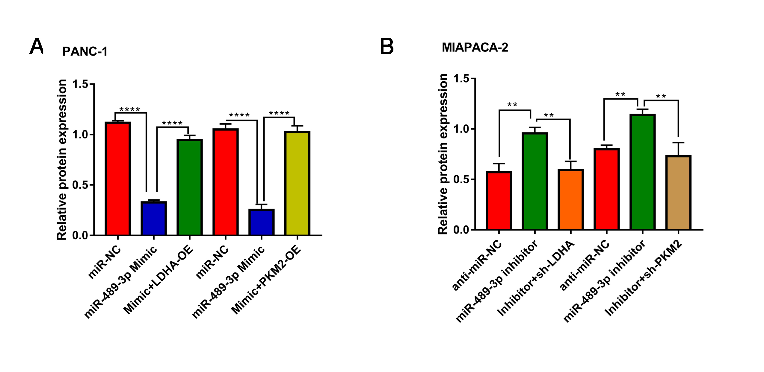
Western blot
The cells of each group were seeded in a six-well plate, and when the band fusion reached 90%, the protein was obtained by lysis with RIPA lysate (beytime, China). BCA reagent (beytime, China) was used to quantify the protein, and then the protein was diluted to the same concentration, and then boiled in a refrigerator at -20 ° C until use. Electrophoresis was performed using Solarbio reagents and 10% separation gel and 5% concentrated gel were prepared according to the manufacturer's instructions. After electrophoresis and transfer, exposure is performed.
Glucose uptake and lactate production measurements
Glucose uptake was determined using the D-2-deoxyglucose method following the manufacturer’s instructions. D-2-deoxyglucose used was purchased from Beyotime, China. Lactic acid production was assayed using a Lactic Acid detection kit (Leagene Biological, Beijing) as per the recommendations of the manufacturer.
Cellular ATP level
Experiments were performed using an ATP detection kit (Beyotime Biotechnology, China). Add 200 μl of lysate to each well of the six-well plate according to the product instructions, and centrifuge the supernatant after full lysis. Prepare ATP working solution and add it to the detection tube to measure the luminescence value.
Extracellular acidification rate (ECAR)
Hippocampal experiments were performed using Agilent equipment: Hippocampus XFe24 Micro Edition and XFe24 cartridge. Cells of each group were incubated at 500 ° C for 7.5 × 10⁴ cell seeds at 37 ° C for 1 hour at the hippocampal preparation station.
56 μl glucose (100 mm) (G8270σ), 62 μl oligomycin (10 μM) (σ) 75351 and 69 μl 2 dg (1 meter) (D6134, Sigma) were added to the cartridge wells. The ECAR values were then read.
In vivo assay
A total of 107 panc-1 cells (miR-NC group and miR-489-3p up-regulation group) were injected subcutaneously into 4 weeks old female NCr nude mice (Hua Fukang Biotechnology, Beijing) for tumor growth. The tumor size was measured with calipers every 7 days for a period of 30 days. At the end of the experiment, the mice were euthanized, and subcutaneous tumors were collected for further analyses.
IHC analysis
Immunohistochemical staining was used to detect the expression of proliferation and metabolic indicators. Briefly, the subcutaneous tumor tissue was cut into 3um sections and then dewaxed. The sections were then incubated with rabbit anti-monoclonal at 4 ° C overnight. After washing three times with PBS, each piece was incubated with goat anti-rabbit IgG for 30 minutes and then stained with 3,3 'diaminobenzidine (DAB). All antibodies used were purchased from Proteintech, USA.
Statistical analyses
The results were analyzed and presented as the mean ± standard deviation. GraphPad Prism 7.0 (San Diego, California, USA) was used for mapping and statistical analysis. Chi-square test was used to analyze the relationship between miR-489-3p expression level and clinicopathological characteristics in PC. The Kaplan-Meier curve method was used to analyze the overall survival rate. The student's t-test was used for statistical comparison between the two groups. Significance level was set at P <0.05 (*) and P <0.05 (* *).