As a member of the LIM protein subfamily, LASP1 protein, which interacts with the protruding actin on the cell membrane through the mediation of the LIM structure, can also play a role in the formation, extension and invasion of cell pseudopods through the SH3 region (Tomasetto et al., 1995). Relevant studies have confirmed that LASP1 is highly expressed in a variety of malignant tumors, and affects the development, invasion and metastasis of tumors. Although EMs are benign diseases, they have the characteristics of malignant tumor invasion and metastasis. Based on our preliminary data, we found that the expression level of LASP1 protein was significantly increased in ectopic ESCs of EMs patients. Endometrial tissue mainly contains epithelial cells and stromal cells (ESCs). The occurrence of EMS is largely dependent on ESCs. Therefore, it was speculated that LASP1 affects the migration of ESCs and promotes the development of EMs. Because AMs and EMs are both endometriotic diseases and are related, in order to fully illustrate the role of LASP1 on ESCs, this study also compared adenomyosis (AMs) ESCs with ovarian endometriosis(OEMs) ESCs. Our study is to explore the extracted primary stromal cells Q-Z1(OEMs), Q-Y19(OEMs), X-Y19 (AMs) and compared with the control ThESC. It was found that the expression level of LASP1 was relatively consistent with miR-218 in ESCs.
Many studies have confirmed that LASP1 is a downstream target gene of miRNA, and LASP1 is involved in the pathophysiological process of a variety of malignant tumors. The "eutopic endometrium determinism" of EMs, that is, the abnormal phenotype of the eutopic endometrium is determined by the specific genotype of the eutopic endometrium. Therefore, in recent years, many researches have been carried out on the specific genotype of the eutopic endometrium.. The study found that compared with the control group, the expression level of miRNA, and even the expression level of some proteins, in the endometrial tissue of EMs patients did have certain differences.
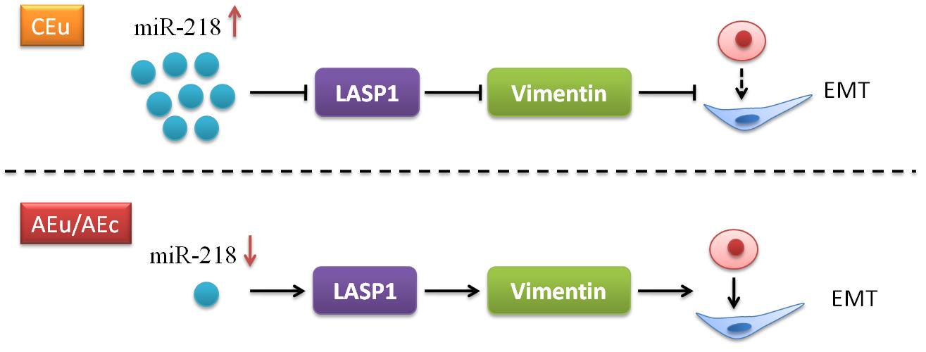
Through our qPCR and WB data, it was found that LASP1 in AMs and EMs was highly expressed in the selected original ESCs, while miR-218 was expressed at a low level. Besieds, the expression level of miR-218 and LASP1 expression were negative correlation. The results obtained verify our conjecture that LASP1 is highly expressed in EMs, and the mechanism is that miR-218 binds to the 3′UTR region of LASP1 and negatively targets and regulates LASP1, which are consistent with the results of dual luciferase reporter gene assay. After transfection according to the above transfection principles, select conditional cells with high transfection efficiency at a transfection concentration of 50nM. Through the EdU proliferation experiment, it was found that the proliferation function of control ESCs and EMs resident ESCs were are consistent with the results of Qiu Yu et al, that is, miR-218 does not affect the proliferation ability of ESCs. And in this case, low expression of miR-218 enhanced the migration ability of ESCs. Conversely, high expression of miR-218 reduced the migration ability of ESCs. This result is consistent with the results of other studies on the function of LASP1 on cell migration.
Studies have reported that miR-218 can inhibit the proliferation of cervical cancer SCC cell lines, regardless of whether its HPV expression is positive or negative; the results also show that miR-218 does have an inhibitory effect on cervical cancer(Yamamoto et al., 2013). This study found that miR-218 has no proliferation effect on ESCs. Considering that although EMs have the characteristics of malignant tumors, they are still benign diseases and may not have the characteristics of continuous proliferation of malignant tumors. And this result also ruled out that the enhanced effect of miR-218 on the migration of endometrial stromal cells may be caused by cell proliferation, making the result of cell migration function more credible.
Based on the comprehensive analysis of all experimental results in this experiment, it can be inferred that in patients with EMs, the increased expression of LASP1 protein in the resident ESCs promotes the migration of ESCs. According to the "endometrial determinism" of EMs hotspot. ESCs have a specific phenotype, and then according to the pathogenesis of EMs, "menstrual blood reflux" and many other theories, the process of EMs formation is inferred. The specific genotypes of ESCs When its migration ability is enhanced, the intimal fragments containing ESCs enter the abdominal cavity with the menstrual blood reflux, and are planted in the ectopic lesions of the abdominal cavity in a relatively adapted environment, thereby forming ectopic lesions of EMs, which promotes the occurrence and development of EMs. This also explains why most of the menstrual blood reflux did not cause EMs, and combined with the multiple existing mechanisms of EMs, this result serves as a supplementary explanation and makes the mechanism of occurrence and development of EMs more clear.
For the detection of tissue immunofluorescence, it is interesting that the differential expression of miR-218 does not appear in the endometrium (including mesenchymal and epithelial cells). However, there are obvious signals around the endometrium (Figure.3), indicating that miR-218 may be transferred to the microenvironment (the microenvironment may be also changed) through exosomes secreted from blood vessels, and further act on the endometrium, changing the physiological state of the endometrium(Phinney & Pittenger, 2017; Yu, Odenthal, & Fries, 2016). This transmission signal is a paracrine signal from secretory cell to the target cell, therefore, miR-218 is already hard to detect. It may not be differentially expressed in the endometrium by immunofluorescence experiment, but in our qPCR results (Figure.1). We need to conduct further research on the exosomes in the blood of EMs patients, explore the differential expression of microRNA among them, and carry out subsequent studies.
Besides, we also found that miR-218 play a role in EMT. To verify the above results, we first ruled out the interference of antibodies (Vimentin and E-cadherin). We used the above two antibodies for immunohistochemistry in stromal and epithelial cells, and found that E-cadherin is specifically expressed on the membrane of epithelial cells, while Vimentin is specifically expressed in stromal cells (sFig 1), indicating that the expression pattern of these two antibodies is correct. Then, in ectopic endometrial tissue, both E-cadherin and Vimentin were significantly higher in epithelial cells than in ectopic stromal cells (R3 vs. R2) (Figure.5), indicating that E-cadherin and Vimentin are specific markers for EMs epithelial cells. The above data demonstrate that ectopic endometrial epithelial cells have a tendency to transform into ectopic stromal cells when miR-218 is inhibited. In addition, E-cadherin and Vimentin expression was higher in eutopic endometrial epithelial cells than in normal human eutopic endometrium (Figure.4). These results were consistent with the results in ectopic endometrium of EMs patients (Figure.4).