4.1. Materials
NbC nanoparticles were obtained from Chaowei Company of Shanghai. The RAW264.7 macrophages and HepG2 cells lines were obtained from the Institute of Cancer Research affiliated to Harbin Medical University. The dulbecco’s modified eagle medium (DMEM, Corning), penicillin/ streptomycin (Corning), phosphate buffer saline (PBS), fetal bovine serum (FBS, Gibco) and Trypsin-EDTA solution were purchased from Harbin Shengze Biotechnology Co. Ltd. Calcein-AM, propidium iodide (PI), and 2-(2-methoxy-4-nitrophenyl)-3-(4-nitrophenyl)-5-(2, 4-disulfophenyl)-2H-tetrazolium and monosodium salt (Cell Counting Kit-8, CCK-8) were purchased from Beyotime Biotechnology Co. Ltd. 1, 3- diphenylisobenzofuran (DPBF) and 2’, 7’- dichlorodihydrofluorescein diacetate (DCFH-DA) were obtained from Sigma-Aldrich.
4.2. Characterization
The morphologies of NbC were observed by TEM (JEOL JEM-2100, Japan). The size distribution analysis was conducted on Zetasizer Nano S90 (Malvern Panalytical, UK). The crystal phase was tested by XRD (Shimadzu XD-D1). The composition and chemical valence of NbC were measured by XPS spectra (PerkinElmer PHI 5600). UV-vis-NIR absorptive spectra of samples were performed on a spectrophotometer (U-4100, Hitachi, Japan). The concentration of Nb element within cells was analyzed by inductively coupled plasma (ICP) atomic emission spectrometer (8300, PerkinElmer, USA). Two-dimensional ultrasound (2D US), Color Doppler Flow Imaging (CDFI) and contrast-enhanced ultrasound (CEUS) scans were performed using MyLab twice system (Esaote SpA, Florence, Italy) with LA523 probe. All the data of shave wave elastography (SWE) were recorded by a real-time US device (Aixplorer; SuperSonic Imagine, Aix-en-Provence, France) with 4–15 MHz liner transducer.
4.3. Photothermal Test
0.5 mL NbC aqueous dispersion of various concentrations was irradiated with 808 nm NIR laser (1.0 W/cm2) for 10 minutes. The changes of solution temperature were collected by a NIR camera (E6, FL-IR Systems, Inc, USA). The photothermal conversion efficiency was calculated according to the photoheating curve of the equivalent NbC.
4.4. Cell Lines and Cell Culture
The RAW264.7 macrophages and HepG2 cells lines were cultured in DMEM supplemented with 10 % FBS and 1% penicillin/streptomycin in a humidified atmosphere of 5% CO2 at 37 ℃. Hemocytometer (Bürker-Türk, Wertheim, Germany) was used for the total cell counting.
4.5. Preparation of the Macrophage-loaded NbC nanoparticles
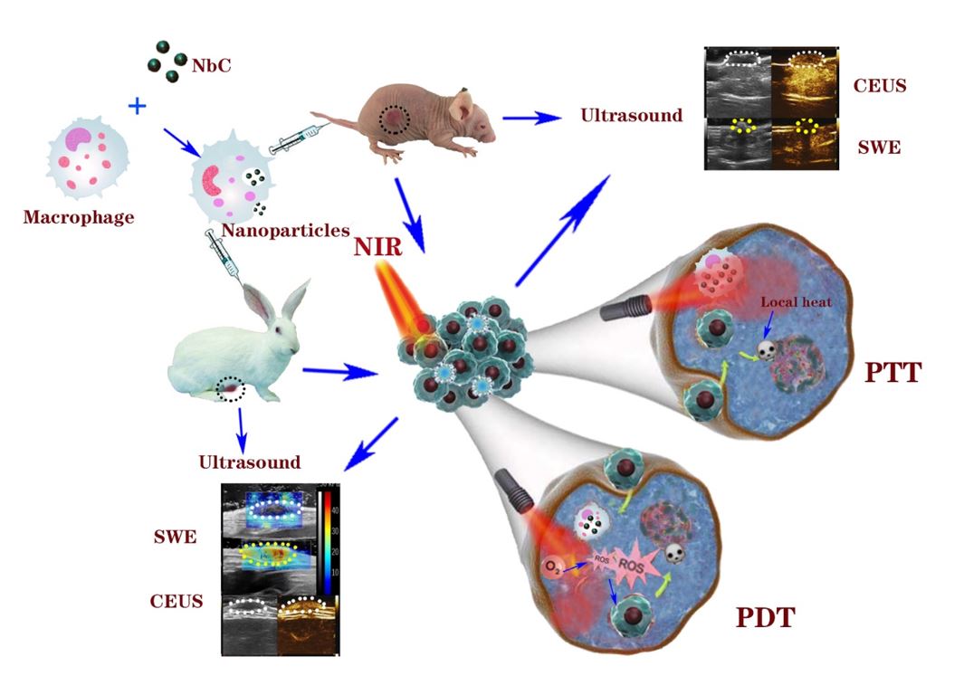
The RAW264.7 macrophages were incubated with DMEM until the cell coverage reached about 80% ~ 90%. Then, the stale DMEM was removed and the fresh DMEM supplemented with various concentrations of NbC (0.125, 0.25, 0.5, 1 and 2 mg/mL) and incubated for varied time (2, 4, 6, 12 and 24 h). After incubation, the supernatant was removed and macrophage-loaded NbC nanoparticles were washed with PBS twice. The macrophages were observed with inverted biological microscope (Olympus CKX41). ICP analysis was used to quantify the uptake amount of Nb content in the macrophage cells.
4.6. Detection of ROS.
DPBF solution with NbC or NbC@M was respectively placed in a quartz tube, and then irradiated with 808 nm NIR laser (1 W/cm2) for 10, 20, 40 and 60 min. Then, absorbance at 420 nm was determined by U-4100 spectrophotometer. In order to detect intracellular reactive oxygen species (ROS), HepG2 cells were divided into five groups, including untreated control (group 1), cells incubated with NbC@M (0.2 mg/mL) for 4 h (group 2), 808 nm NIR laser (1 W/cm2) irradiated cells (group 3), positive control with H2O2 (200 µL, 50 × 10− 3 M) at 37 oC for 1 h (group 4), NbC@M mediated phototherapy (group 5). After the treatments, the cells treated in different ways were stained with DCFH-DA (50 µL, 50 × 10− 3 M) for 1 h and then imaged on inverted biological microscope.
4.7. Cytotoxicity Assay
The standard CCK-8 assay was used to evaluate the viability of cells. HepG2 cells (8×10³ cells/well) were seeded into 96-well plates and cultured for 12 h. NbC@M solutions of different concentrations were then added into above wells and incubated for another 12 or 24 h, respectively. After washed by PBS solution for three times, 10 µL CCK-8 was added to the samples and incubated for another 1 h. The absorbance at 450 nm was then recorded on a microplate reader (BioTek, Winooski, VT, USA). The CCK-8 method was also applied for analyzing the therapeutic effect of in vitro PDT, PTT, and PDT + PTT experiments. Similarly, HepG2 cells (8×10³ cells per well) in each group were cultured in 96-well plates for 12 h. Then, 100 µL NbC@M (500 µg/mL) were added into each well and incubated for another 12 h, respectively. All groups were subjected to 808 nm NIR irradiation except control group. Additional operations are needed to handle PDT and PTT group. The process of PDT should be set in an ice bath, while sodium azide (50 µL, 1×10− 5 M) was added into the medium of PTT group before NIR irradiation.
4.8. In Vitro Phototherapeutic Effect
To evaluate the phototherapeutic effects at the cellular level, HepG2 cells (3×10⁵ cell/dish) were cultured with DMEM medium containing NbC or NbC@M (2 mL, 250 µg/mL) in 35 mm culture dishes for 6 h. Redundant NbC or NbC@M was removed by washing with PBS and then 500 µL fresh DMEM medium was added into each dish. Then, above resultant cells were irradiated with 808 nm NIR laser (1 W/cm2) for 2, 5, and 10 min. Several groups were designed as controls, including untreated cells, cells only irradiated by 808 nm NIR laser (1 W/cm2), and incubated with NbC or NbC@M. Finally, dead and live cells were labeled by Calcein-AM (1 µg/mL, 200 µL) and PI (20 µg/mL, 200 µL) for 20 min, and then washed with PBS. The fluorescence visualization images were imaged on Olympus BX53 microscope.
4.9. In Vivo Phototherapy on mice
All experimental female Balb/c nude mice were purchased from Beijing Vital River Laboratory Animal Technology Co., Ltd. HepG2 cells were digested and dispersed in DMEM to form a uniform suspension, which were then injected into the subcutaneous tissue of the upper left hind leg of nude mice to establish tumor-bearing mice model (tumor size is about 150 mm3). Then, tumor-bearing mice were randomly divided into four groups (n = 5 for each group): group 1 (100 µL PBS was injected intravenously as control), group 2 (808 nm NIR irradiation for 10 min, 1 W/cm2), group 3 (NbC@M was injected intravenously) and group 4 (intravenous injection of NbC@M plus 808 nm NIR irradiation for 10 min). NbC@M (100 µL) was injected intravenously into nude mice at a concentration of 1 mg/mL in the group 3 and 4, respectively. In the group 2 and 4, 808 nm NIR irradiation (1 W/cm2) was required to irradiate the tumor area for 10 min. Notably, the mice in the group 4 should be irradiated at 12 hours post injection of NbC@M. The change of temperature was monitored and recorded every 30 s by an FL-IR System E6 infrared camera. The data of tumor size and body weight of nude mice were recorded in detail for 14 days. The tumor volume was calculated as V = length × width2 / 2. Relative body weight was designed as dividing instant weight by initial weight (W/W0), and similarly relative tumor volume was defined as V/V0. All the animal experiments were approved by the criteria of the National Regulation of China for Care and the Ethics Research Committee of Harbin Medical University Cancer Hospital.
4.10. Rabbit VX2 breast xenograft model
All of the female New Zealand white rabbits (12 ~ 14 weeks old, 2.5 ~ 3.0 kg) were purchased from animal laboratory of the Second Affiliated Hospital of Harbin Medical University. The VX2 tumors were surgically implanted into the left second nipple areola edge for each rabbit. The VX2 tumor mass was obtained from the hind limb of a donor rabbit and minced approximately into 1 mm3 fragments, which were dispersed in normal saline to form a suspension. Extract a small amount of suspension and ensure that at least two VX2 tumor fragments were injected into the designated location. Ten days after tumor implantation, the tumors should be observed until the tumors’ volume reached up to about 300 ~ 500 mm3.
4.11. In Vivo Phototherapy on rabbits
VX2 tumor-bearing rabbits were divided into four groups randomly, including injection of PBS (1 mL) as control, receiving only NIR irradiation, receiving only injection of NbC@M (1 mL, 1 mg/mL), and receiving NIR irradiation at 8 h post-injection of NbC@M (1mL, 1 mg/mL). For the real-time thermal imaging, tumor regions were irradiated by an 808 nm laser with a power density of 2 W/cm2 for 20 min, and then monitored with an infrared imaging device (FL-IR System E6). All injections of PBS or NbC@M are performed through the posterior auricular vein.
4.12. Ultrasound on mice and rabbits
Ultrasound tests as a general term, including two-dimensional ultrasound (2D US), Color Doppler Flow Imaging (CDFI), SWE and CEUS, were completed by two radiologists with more than five years of experience. After confirming the tumor area and applying enough ultrasonic coupling gel during SWE process, the probe was kept in a stable position with no pressure for about 3 seconds and was vertical to better reduce compression artifacts. A suitable region of interest (ROI) and scale ruler were identical for the elastographic quantitative analysis (SWEmax and SWEmean). CEUS examinations were performed with intravenous vein injection of SonoVue (Bracco SpA, Milan, Italy) followed by a saline flush. 100 µL SonoVue was injected into the caudal vein for mice, while 1 mL SonoVue was injected into the posterior auricular vein for rabbits. Video clips of the examinations, including the process of enhancement and washout, were immediately recorded over a time period of 1 min after injection of SonoVue.
4.13. Histological and Blood Biochemistry Analysis
To perform histological analysis, all the mice and rabbits were sacrificed at 14th day after treatment. Hematoxylin and eosin (H&E) staining of major organs including heart, liver, spleen, lung, kidney was done for histopathologic analysis. Slices were observed by a digital microscope (magnification: ×100; DM3000; Leica, Germany). The blood (20 µL) of mice were collected and tested at 14th day after treatment by automatic blood analyzer (HF-3800).