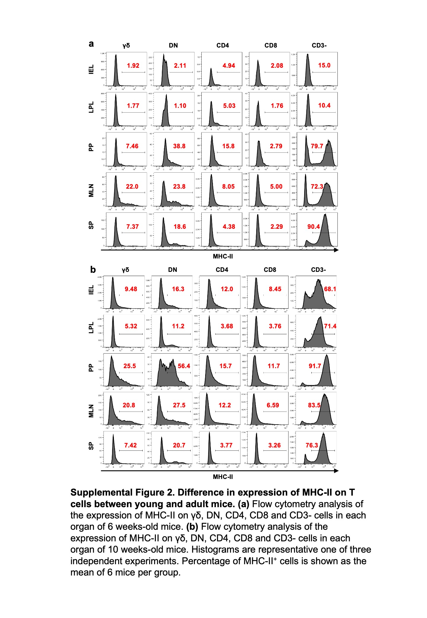
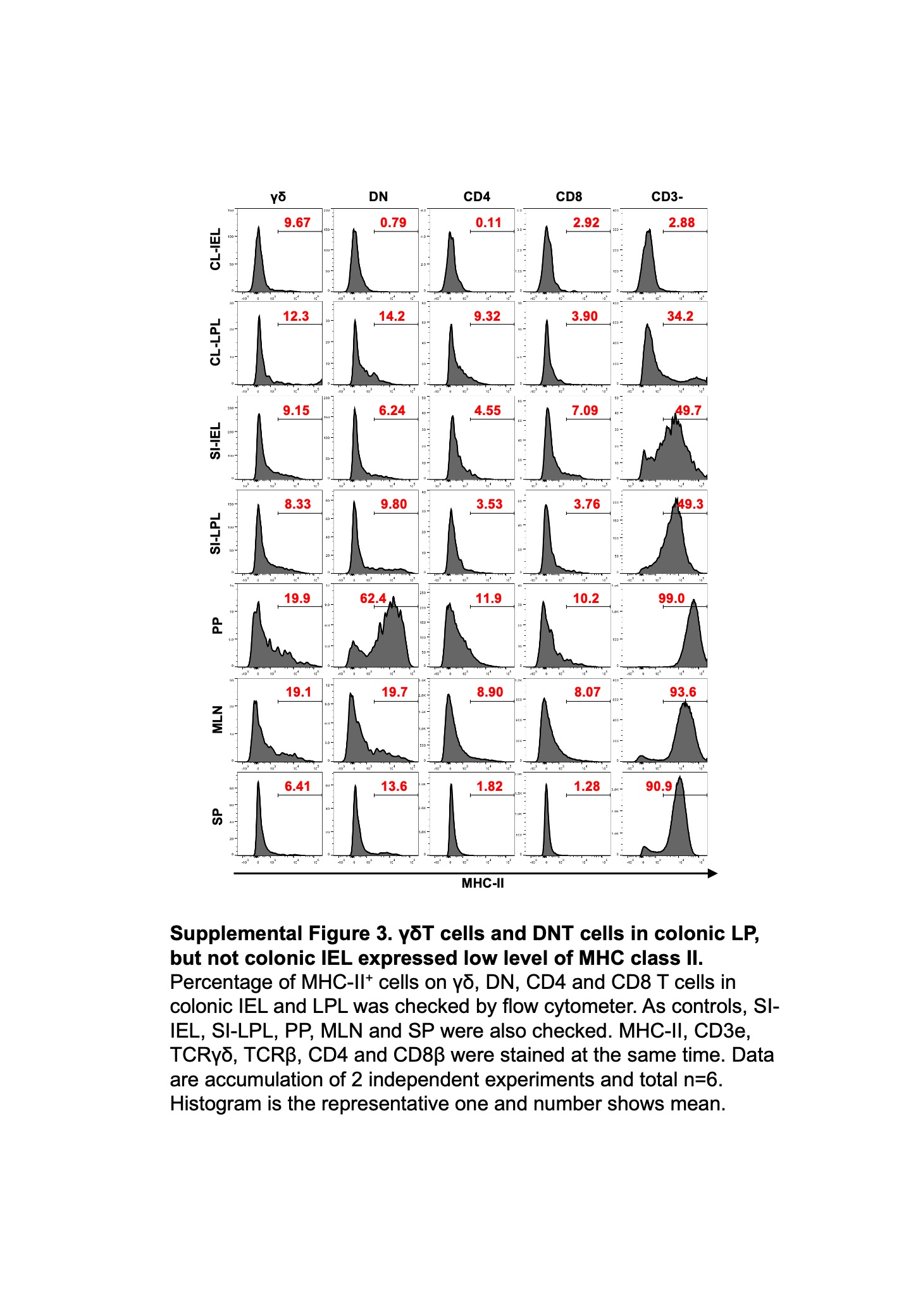
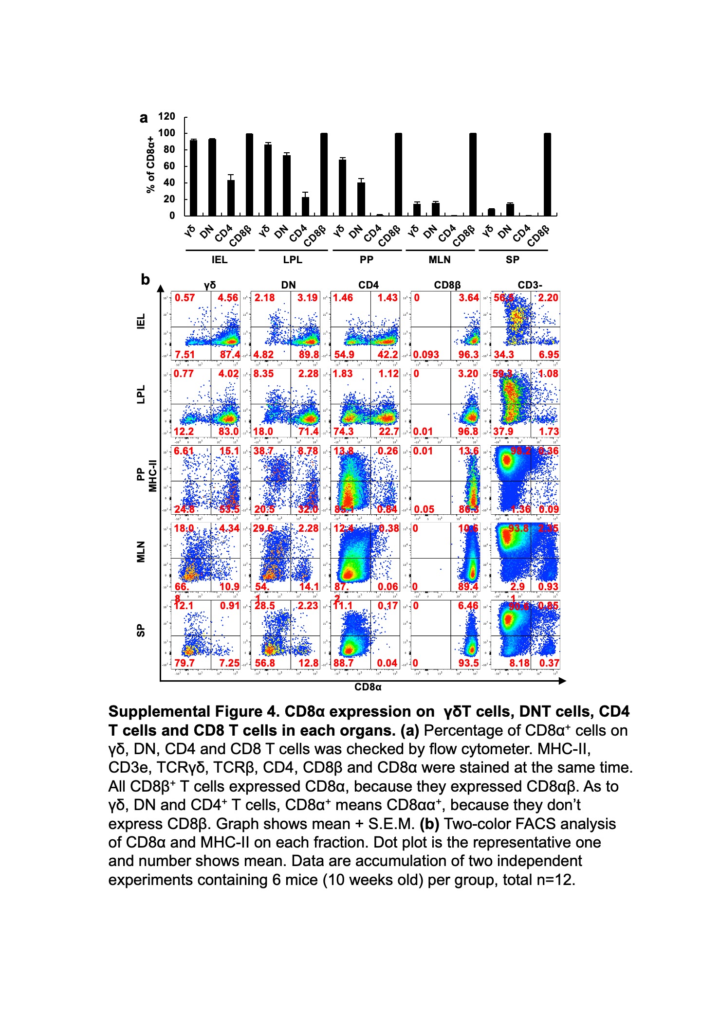
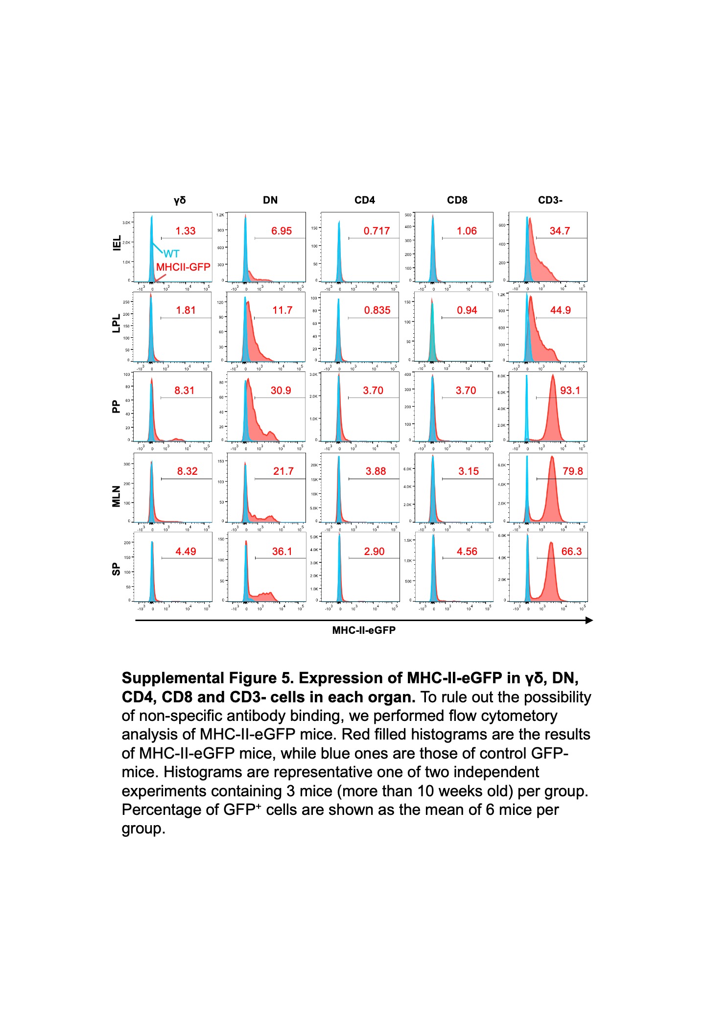
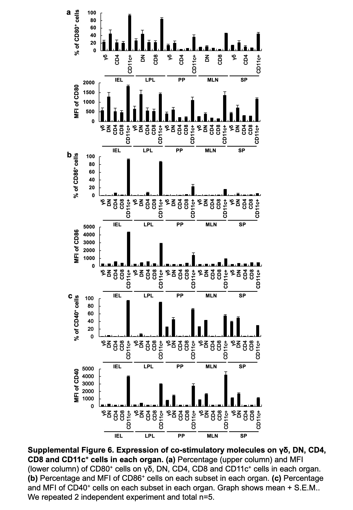
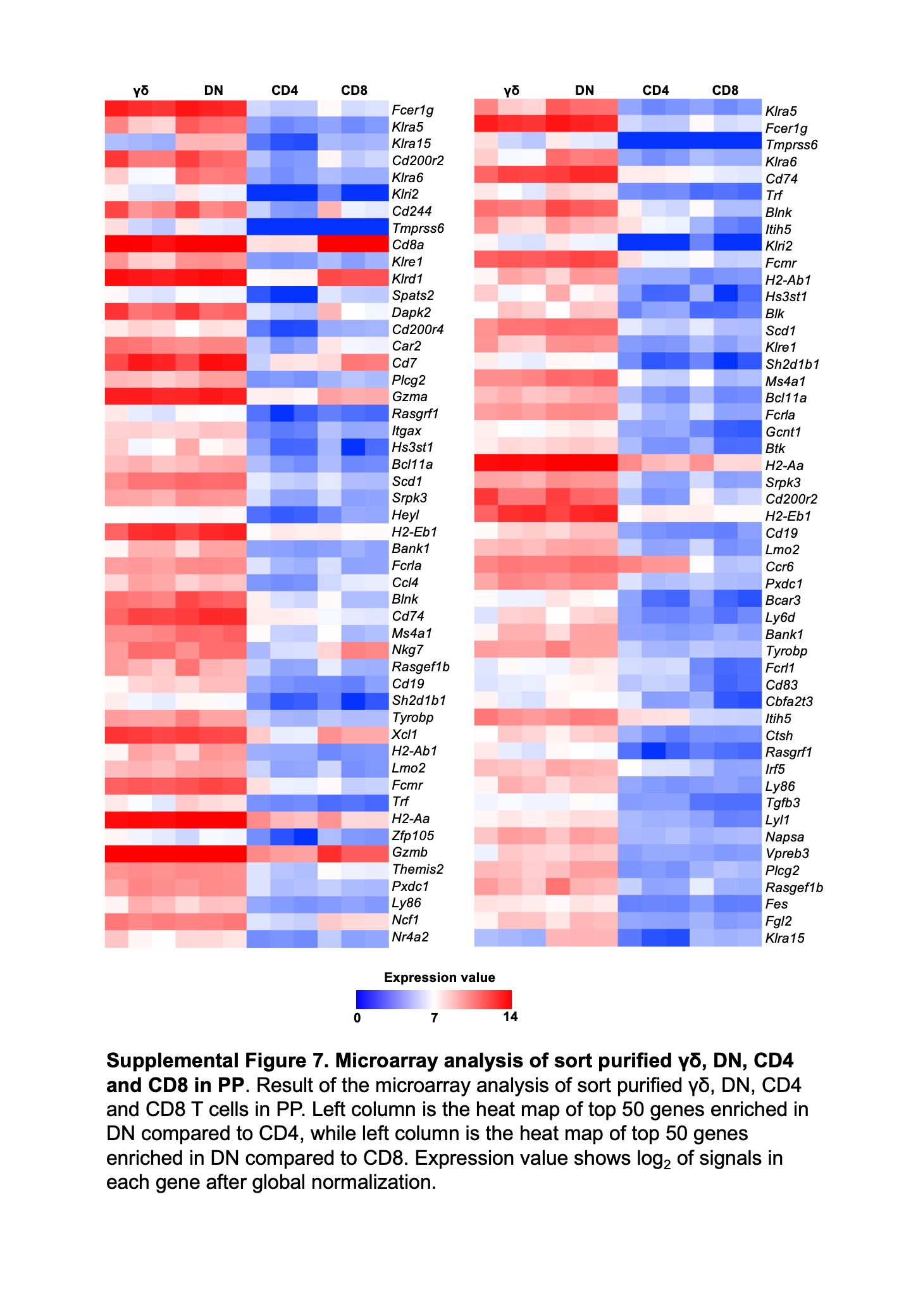
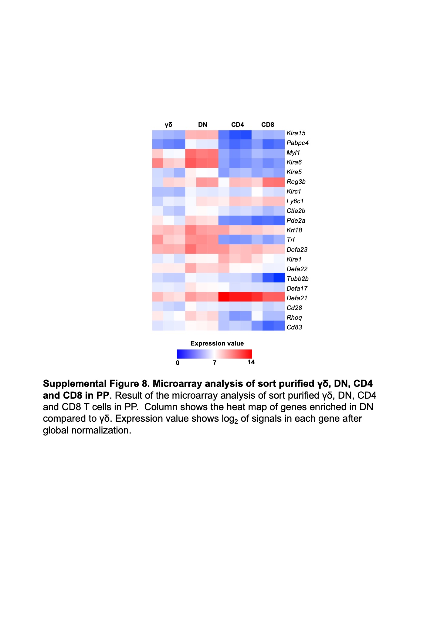
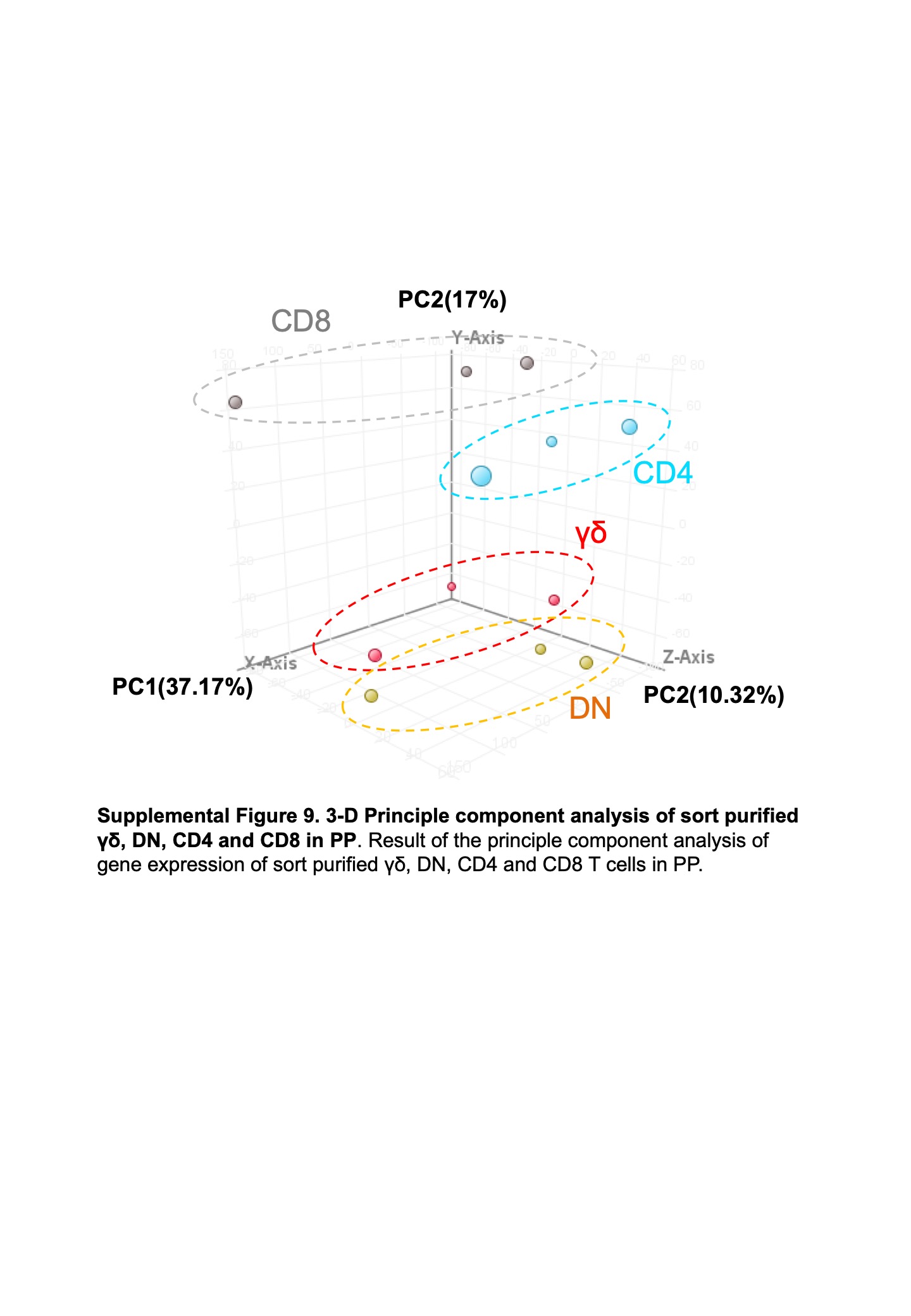
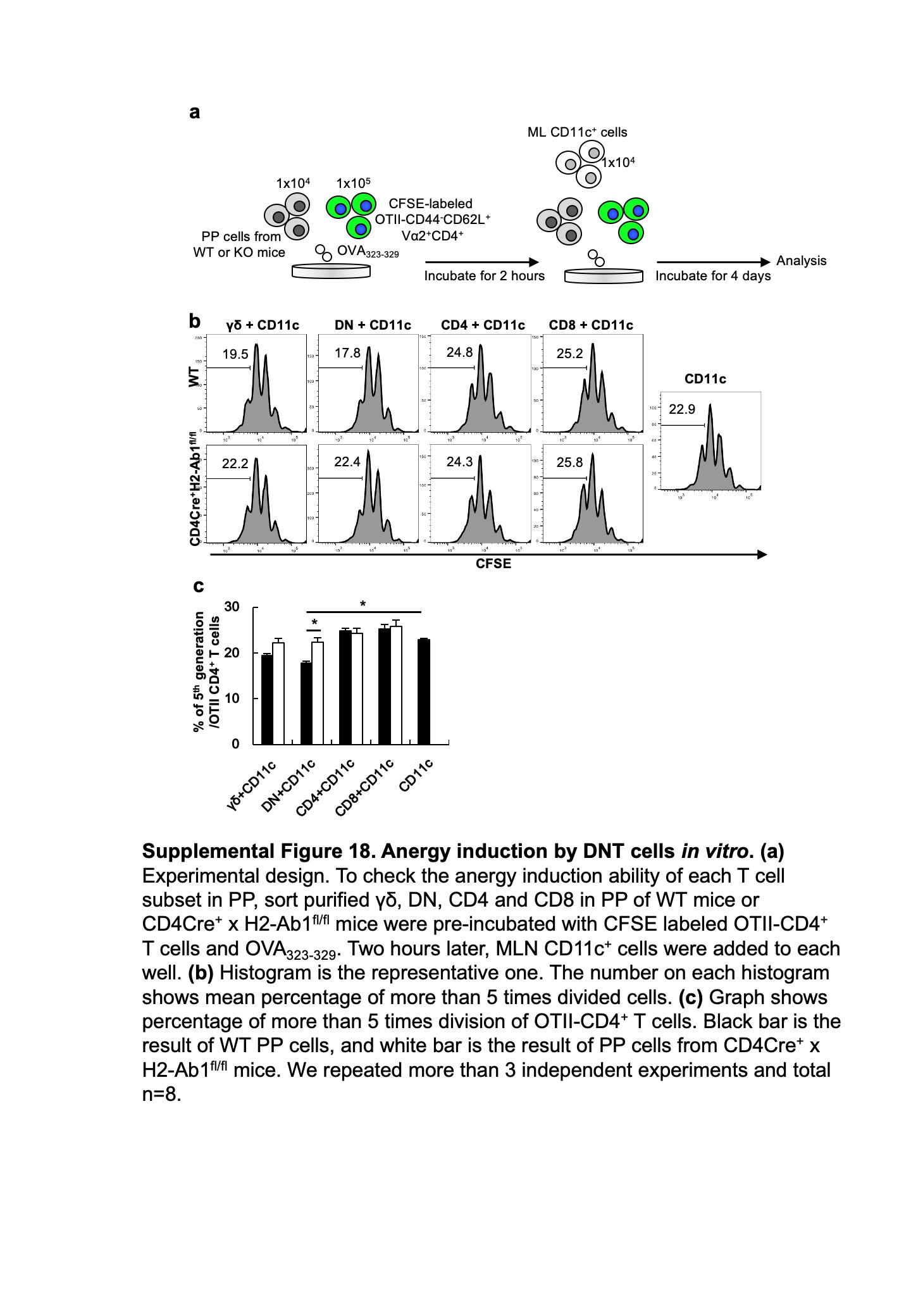
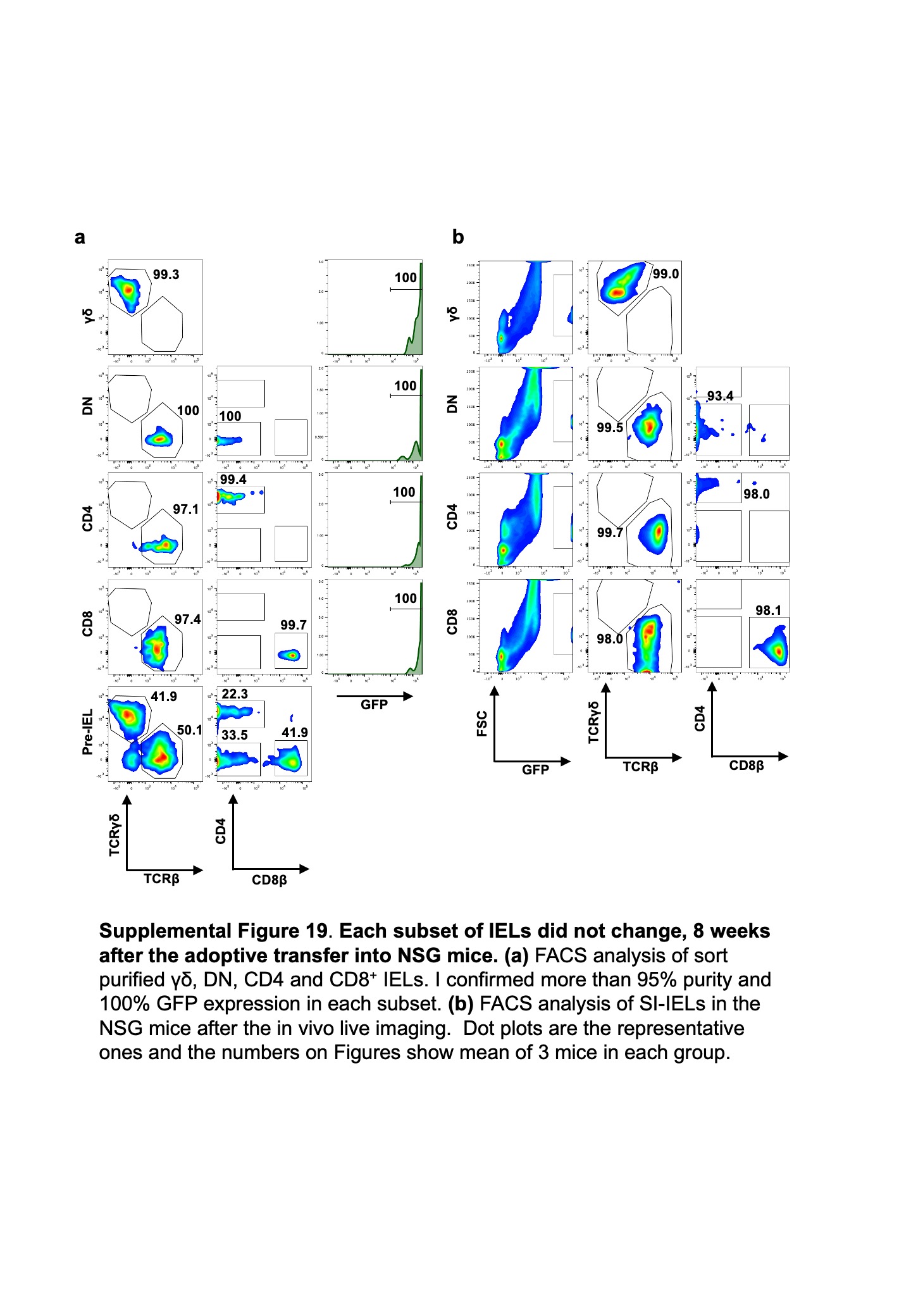
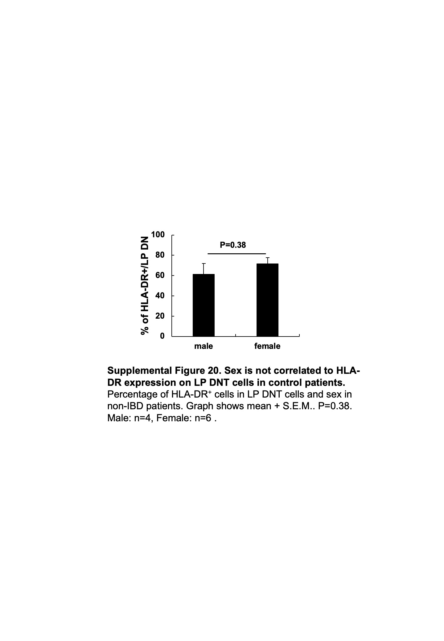
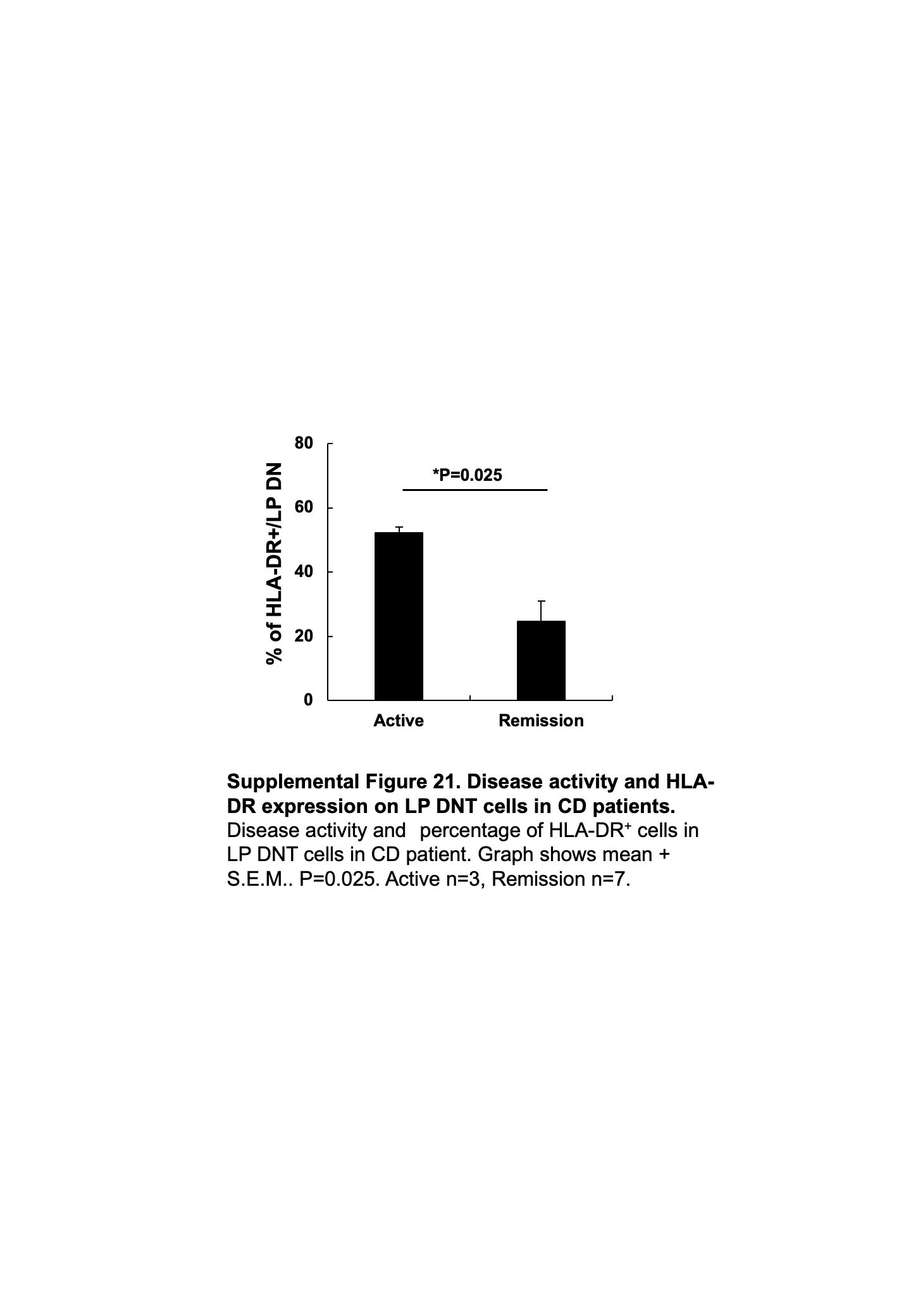
The intestinal immune system is constantly exposed to a plethora of antigens (Ags) from innocuous ingested material and the commensal flora that must be distinguished from pathogen-derived antigens. To this end, a number of anatomic, cellular and molecular mechanisms operate in the intestinal tract to acquire, process, present and interpret Ags from the intestinal lumen. The intestinal mucosa is also populated by a large number of T cells that reside within the epithelial lining (intraepithelial lymphocytes, IELs), in the underlying lamina propria (LPLs) and in gut-associated lymphoid tissues (GALT). Both IELs and LPLs are heterogeneous populations consisting of conventional CD4+ and CD8+ T cells and numerous unconventional T cells, including TCRγδ+ T cells and CD4-CD8αβ-TCRαβ+T cells (double negative; DNT cells). The latter are rarely found in non-intestinal tissues and their function is still enigmatic. Here, we show that murine DNT cells in the small intestine (SI) reach across the epithelial barrier to capture luminal Ags. A sizeable fraction of DNT cells in Peyer’s patches (PPs), mesenteric lymph nodes (MLNs) and among LPLs, but not among IELs, express MHC-II, but little or no classical co-stimulatory molecules, suggesting that DNT cells-mediated Ag presentation to naïve CD4+ T cells may trigger tolerogenic rather than effector responses. Indeed, intestinal DNT cells, particularly in PPs, acquire, process and present antigenic proteins and tolerize Ag-specific CD4+ T cells. Conditional genetic ablation of MHC-II in T cells disabled this suppressive function and rendered mutant mice hypersusceptible to DSS colitis. Moreover, similar to our findings in mice, DNT cells in human SI express HLA-DR and readily uptake exogenous Ags. Intriguingly, intestinal DNT cells in Crohn’s disease patients expressed lower levels of HLA-DR than control patients. These findings suggest that MHC-II+ DNT cells play a key role in intestinal immune homeostasis and may contribute to the pathogenesis of inflammatory bowel disease.