5.1 Materials
The backbone of the fixators consisted of PCL (molecular weight [Mn] ~ 80,000 Da), and the drug carrier was PLGA, which consisted of a 1:1 ratio of lactide and glycolide and an Mn of 33,000 Da. The drugs used included commercial grade vancomycin hydrochloride, ceftazidime hydrate, and lidocaine hydrochloride. Recombinant human bone morphogenetic protein-2 (BMP-2) was used as the growth factor. The solvents used included dichloromethane (DCM) and 1,1,1,3,3,3-hexafluoro-2-propanol (HFIP). All these chemicals were purchased from Sigma-Aldrich (St. Louis, MO, USA).
5.2 Design and fabrication of biodegradable fixators
Three types of self-locking biodegradable fixators were designed (H2, H3, and X), as shown in Fig. 7A. The fixators were fabricated on a lab-developed solution-extrusion 3D printer [39], which included a solution-extrusion feeder, driving stepper motors and related components, power source, syringe controlled by a user interface, accumulation platform, and dispensing tip with an outlet inner diameter of 0.18 mm (Fig. 7B). An open Cura interface (Ultimaker B.V., Geldermalsen, The Netherlands) was used to manage the entire 3D printing procedure (Fig. 7C).
To print the fixators, 2.5 g of PCL was mixed with 3.5 mL of DCM on a magnetic stirrer for 6 h. The mixed solution was then fed into the feeding system of the printer, which consisted of a syringe and plastic dispensing tip. The dispensing tip was moved using a microprocessor-controlled servo motor during the 3D printing process, and the PCL solution was pushed out of the tip and deposited on the collection platform. Upon solvent evaporation, biodegradable fixators of different geometrical designs and thicknesses (0.2 and 0.4 mm) were obtained.
5.3 Mechanical properties of the biodegradable fixators
A pilot in vitro study was initially conducted to assess the mechanical properties of the biodegradable fixators prior to in vivo animal studies. Four 6-month-old New Zealand white rabbits weighing 2800–3200 g were used in the biomechanical tests. Each rabbit was placed in an enclosed chamber, and inhalation euthanasia was carried out using carbon dioxide (2 L/min for 3 min). Carbon dioxide was pumped for an additional 2 min to ensure a complete lack of respiration and faded eye color. All animal procedures received institutional approval from the Institutional Animal Care and Use Committee at the Animal Center of Chang Gung University (CGU 14–102). Additionally, each rabbit was cared for in accordance with the regulations of the National Institutes of Health of Taiwan.
In total, 21 ribs were harvested (bilateral 5th to 7th ribs in each rabbit) and examined. The ribs were prepared by removing the attached muscles and periosteum after euthanasia. Each fixator was fixed to the ribs by passing belts through matching holes and wrapping around the fracture site; the time required for fixing was recorded. Subsequently, the control ribs and fixator–rib assemblies were analyzed using 3-point bending on a mechanical testing device (LRX, Lloyd Instruments, Bognor Regis, UK). The specimens were placed on two supporting pins 25 mm apart from each other, and the loading pin was moved at a speed of 1 mm/s in a downward direction until the specimen fractured. The applied force and strain in the ribs (control and fixator) during the loading process were recorded.
5.4 Fabrication of biomolecule-loaded nanofibers
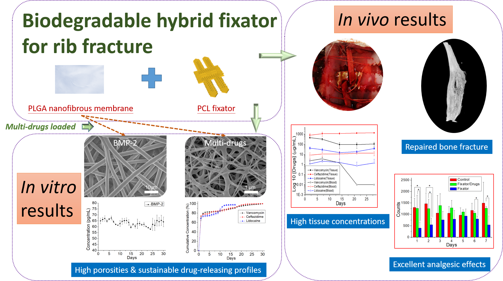
Bilayered biomolecule-loaded nanofibers, i.e., a regular nanofibrous layer and a sheath-core structured layer, were prepared using electrospinning and co-electrospinning. An electrospinning setup, consisting of a high-voltage supply generating positive direct current voltage (maximal power: 35 kV) and current (4.16 mA/125 W), aluminum sheet, ground electrode, and syringe with a needle (0.4 mm internal diameter), was used to fabricate the regular nanofibers. To fabricate these nanofibers, PLGA (1120 mg), vancomycin hydrochloride (93.3 mg), ceftazidime hydrate (93.3 mg), and lidocaine hydrochloride (93.3 mg) were mixed with 5 mL of HFIP and used to fill the syringe pump. This solution was extruded from the syringe at a rate of 0.8 mL/h and electrospun on the aluminum sheet, which acted as the collector. The distance from the needle tip to ground electrode was 15 cm, and an 18 kV positive voltage was applied to the polymer solution.
Meanwhile, to fabricate the sheath-core structured nanofibers, a special coaxial device that concurrently deposited two solutions on the aluminum sheet was used [5]. PLGA (840 mg) was dissolved in 1 mL of HFIP and applied as the sheath solution, whereas the core solution consisted of 20 µg of BMP-2 and 1 µL of bovine serum albumin in 1 mL of phosphate-buffered saline (PBS). For coaxial electrospinning, the PLGA and core liquids were placed in two separate feeding tubes equipped with needles. During co-electrospinning, the liquids were conveyed and spun onto the aluminum sheet at a rate of 0.9 and 0.3 mL/h for the shell PLGA and core BMP-2 solutions, respectively, using two independently controlled pumps. A 17 kV positive voltage was applied to the solutions, and the distance between the needle tip and ground electrode was maintained at 15 cm.
All electrospinning experiments were carried out at room temperature (25°C), and the manufactured nanofibrous membranes were heated in a vacuum oven at 40°C for 72 h to vaporize any remaining solvents.
5.5 SEM
The morphology of the electrospun nanofibers was evaluated using SEM (JSM7500F, Jeol, Tokyo, Japan). To enhance sample conductivity, before testing, the membrane samples were initially coated with a thin layer of gold. Fiber diameters were analyzed from 100 randomly selected fibers (n = 3) using ImageJ software (National Institutes of Health, Bethesda, MD, USA).
5.6 TEM
The structure of the sheath-core nanofibers was analyzed using TEM (JEM-2000EXII, Jeol); samples for TEM analysis were prepared by directly depositing the fibers onto copper grids.
5.7 Water contact angle
The hydrophilicity of the electrospun nanofibers was examined in terms of their water contact angles (First Ten Angstroms, Newark, CA, USA). Nanofibrous membrane samples (10 × 10 mm2) were prepared, and distilled water was slowly dropped onto their surfaces; the contact angles between water droplets and the membranes (n = 3) were evaluated using a video monitor. The water contact angle of PCL fixators was also measured for comparison.
5.8 Fourier-transform infrared (FTIR) spectroscopy
To analyze the functional groups present in the synthesized samples, the FTIR spectra of pure PLGA and drug-embedded PLGA nanofibers (in the form of pressed KBr disks) were recorded on a Nicolet iS5 spectrometer (Thermo Fisher Scientific, Waltham, MA, USA) at a resolution of 4 cm− 1 over 32 scans in the wavenumber range of 400–4000 cm− 1.
5.9 In vitro release of pharmaceuticals and BMP-2
An in vitro elution method was employed to characterize the release patterns of vancomycin, ceftazidime, lidocaine, and BMP-2 from biomolecule-loaded PLGA nanofibers. Nanofiber samples (2 × 3 cm2, 216–220 mg) were cut from the electrospun membranes and incubated in 1 mL of a PBS dissolution medium (0.15 mol/L, pH 7.4) at 37°C for 24 h. Subsequently, the dissolution medium was isolated and analyzed at 24 h intervals. PBS (1 mL) was replaced every 24 h until the sample was fully dissolved. Drug concentrations in the eluents were evaluated using a Hitachi L-2200R multisolvent delivery system (Tokyo, Japan), whereas the BMP-2 levels were analyzed using enzyme-linked immunosorbent assay (R&D Systems, Inc., Minneapolis, MN, USA).
5.10 Rib-fracture model
Twelve 6-month-old New Zealand male rabbits weighing 3.0 ± 0.2 kg were used for in vivo experiments. The right 6th rib was selected as the target rib. Each rabbit was anesthetized by inhalation of isoflurane after pre-oxygenation for 5 min, and the anesthetic was circulated throughout the experimental process. After administering the anesthetic, each rabbit was placed in the decubitus position with the surgical field upward. The skin was prepared and disinfected according to standard antiseptic procedures. A 3 cm incision was made along the rib, and the target rib was reached after dissecting the soft tissue layer and leaving the periosteum undamaged; a short oblique osteotomy was performed on each rib. These rabbits were then randomly divided into control, fixator, and fixator/drug groups (n = 4 per group). No osteosynthesis was performed in the control group, whereas in the remaining two groups, the osteotomized ribs were fixed either with the fixator alone or with the selected fixator accompanied by biomolecule-loaded PLGA nanofibers, which were circumferentially wrapped around the target rib (Fig. 8). Following the surgical procedure, the wound was repaired in layers with absorbable sutures. Post-operatively, the surgical wound was smeared with an antibiotic ointment, and the rabbits were returned to their cages after complete recovery from anesthesia and surgery.
5.11 In vivo release of drugs and BMP-2 from the electrospun nanofibers
To evaluate the local release and systemic diffusion of antimicrobial agents, analgesic, and growth factors embedded in PLGA nanofibers, the blood and muscle tissue surrounding the implanted PLGA nanofibers were obtained at regular intervals (post-operative days 2, 7, 14, and 28). After anesthetizing the rabbits, as described in Sect. 5.10, 1 mL of blood was extracted from the central auricular artery in their ears. Meanwhile, muscle tissue was obtained through the same skin incision on the operated chest wall. Both samples were preserved in 10% formalin solution and later analyzed using high-performance liquid chromatography (Hitachi L-2200 Multisolvent Delivery System).
5.12 Animal bioactivity
We assembled an ABC (30 × 30 × 30 cm3) and divided it into nine lattice zones (numbered 1–9). Rabbit movement in these lattice zones was spontaneously detected using electric sensors placed above each lattice and connected to a recording computer. Food and water were supplied in lattice 1 and refilled every 24 h. The rabbits were sent to this designed cage after recovering from anesthesia and surgery for a 7-day observation and pain evaluation period.
During the evaluation process, each ABC was placed in an isolated room without people walking around, and constant temperature (21 °C ~ 24 °C), pressure (1 atmospheric pressure), and humidity (45%~70%) were maintained. Evaluation was completed after a 7-day bioactivity-evaluation course, after which the rabbits were sent to regular cages for routine care.
5.13 Microcomputed tomography (micro CT) and histology evaluation
Micro CT images (nanoScan SPECT/CT, Mediso, Hungary) were acquired to monitor the healing of fractured ribs in the fixator/drugs group. The osteotomized rib specimens were excised from rabbits euthanized 28 days after osteosynthesis. The specimens were sent for histological examination after hematoxylin and eosin staining.
5.14 Statistical analyses
All statistical data were processed using SPSS software (V17.0 for Windows; SPSS Inc, Chicago, IL, USA). Descriptive statistics are displayed as mean ± standard deviation. One-way analysis of variance (ANOVA) was conducted for data analysis and calculating statistical differences. The post-hoc Bonferroni method was used to compare pairs of groups to identify significant differences. The observed differences were considered statistically significant at p < 0.05.