mPEG-PLA-PAE was obtained from Xi’an Ruixi Biotechnology Co., Ltd. DSPE-PDP was purchased from Shanghai Langxu Biotechnology Co., Ltd. mPEG-DSPE was provided by Shanghai Fansuo Biotechnology Co. Ltd. Arsenic trioxide was kindly provided by the Chinese Academy of Sciences. RIPA lysis buffer was purchased from Beyotime Institute of Biotechnology. Monoclonal antibodies against GSDME, GPX4, and Cyt-c were obtained from Abcam. Monoclonal antibodies against GAPDH and Caspase3 were purchased from Cell Signaling Technology. β-actin-HRP and Cyt-c were obtained from Proteintech.
Preparation of As/miR-34a-mPEG-PLA-PAE nanoparticles
mPEG-PLA-PAE NPs were prepared as previously described. As/miR-34aNPs (As/miR-34a-mPEG-PLA-PAE nanoparticles) were prepared by a double emulsion method as follows. miR34a-SH is conjugated to DSPE-PDP as previously reported. Briefly, a 0.1 ml aqueous solution of As2O3 (1 mg/ml) and a 0.2 ml DSPE-miR34a (20 mg/ml) were emulsified in 0.5 ml of dichloromethane containing 20 mg mPEG-PLA-PAE and 4 mg mPEG-DSPE copolymer using an ultrasonic processor at 400 W for 2 min. Next, the water/oil (w/o) emulsion was added to 1.7 mL of pluronic F68 water solution (1 mg/mL) and the mixture was sonicated at 400 W for 2 min. Subsequently, the resulting w/o/w emulsion was stirred at room temperature to evaporate the organic phase.
Characterization of nanoparticles
Nanoparticle morphology at pH 7.4 and pH 6.5 was observed by transmission electron microscopy (TEM). The potential of nanoparticles under different acidities (pH=7.4, 6.5, 5.5) was determined by a Nano-ZS analyser. As/miR-34aNPs were incubated with GSH at pH 7.4 or pH 6.5, and then the DNA gel block method was used to determine miRNA stability after serum incubation. The film dialysis method was used to measure the amount of the drug released at different time points to evaluate the drug release properties as described in a previous report. Inductively coupled plasma mass spectrometry (ICP-MS) was used to determine the encapsulation efficiency and drug loading of As2O3 (As) according to a previous procedure.
Sublethal heat treatment HCC cell lines as an insufficient microwave ablation model
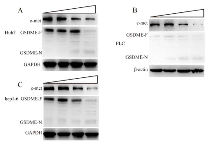
Huh7, PLC/PRF/5, and Hep1-6 cells at 5x10⁵ cells/cm3 were inoculated into DMEM containing 10% foetal bovine serum (FBS) and cultured (37°C) overnight. Then, they were treated at 47°C for 10 minutes. After heating, DMEM was replaced three times a day until the 3rd day to remove debris and dead cells. Then, the cell number was adjusted to 1x10⁶ on the 5th day after heating, the surviving HCC cells were subcultured into new culture dishes, and the cell lines were named H-Huh7, H-PLC, and H-Hep1-6. This procedure was performed three times. c-Met expression was detected by western blot. H-Huh7, H-PLC, and H-Hep1-6 cells at 5x10³ cells/cm3 in 96-well plates were cultured overnight according to a procedure similar to that for studying viability using the MTT method. As2O3 and miR34a (according to the agent procedure) were applied using the same methods for determining cell viability and c-met expression. Different concentrations of blank nanoparticles were added to LO2 human liver cells to determine viability using the MTT method.
As/miR-34aNPs inhibit the growth of HCC cell lines subjected to sublethal heat treatment
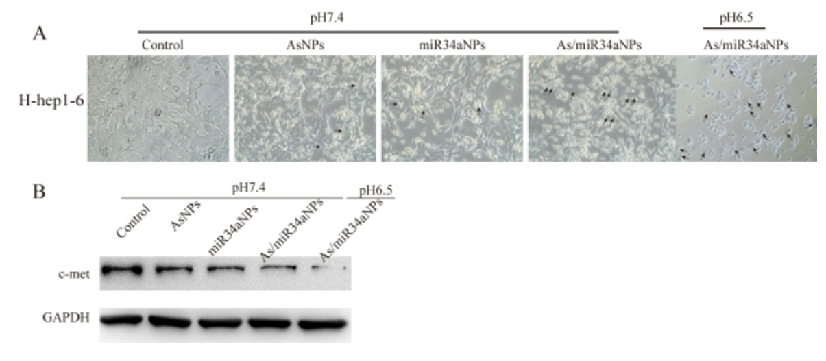
H-Huh7, H-PLC, and H-Hep1-6 cells were inoculated into 96-well plates at a fusion rate of approximately 50%. PBS, miR-34aNPs and AsNPs (As2O3NPs) were added to the culture at pH 7.4 for 48 h. As/miR-34aNPs were added at pH 6.5 and pH 7.4. Finally, MTT was added, and the cells were cultured for 4 h.
H-huh7 insensitivity to ferroptosis after treatment with As2O3
H-Huh7 cells were inoculated into 6-well plates at a fusion rate of approximately 50%. RSL3 and RSL3+Vit. E were added to the culture for 48 h and then observed by microscopy. H-Huh7 cells were treated with RSL3, RSL3+Vit. E, RSL3+DFOM, As, As+Vit. E, As+DFOM, RSL3, RSL3+As, and RSL3+As for 48 h. Then, the MTT method was used to detect cell viability as described above. GPX4 was detected after treatment with As2O3 and RSL3.
Cell uptake of nanoparticles in H-Huh7 and H-PLC
Cy3-labelled nanoparticles were prepared according to the procedure. Cy3NPs and free cy3 were inoculated into H-Huh7 and H-PLC cells in a normal physiological environment. Cy3NPs were inoculated into H-Huh7 and H-PLC at pH 6.5, further verifying that the acid-sensitive charge reversal mechanism was beneficial to cell uptake. Cellular uptake was observed by confocal microscopy.
Antitumour mechanisms of As/miR-34aNPs in vitro
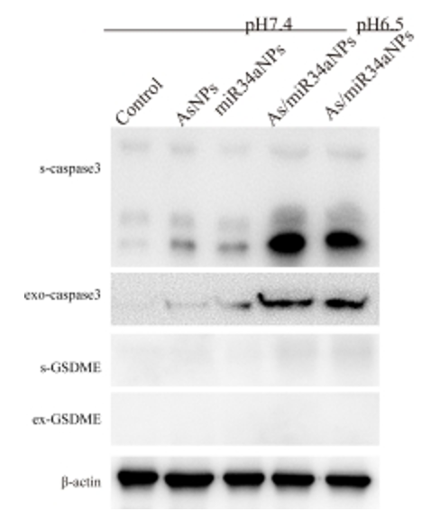
H-Huh7, H-PLC, and H-Hep1-6 cells were inoculated into 6-well plates at a fusion rate of approximately 50%. PBS, miR-34aNPs and AsNPs (As2O3NPs) were added to the culture at pH 7.2 for 48 h. As/miR-34a@NPs were added at pH 6.5 and pH 7.2. The expression of C-met, caspase3, GSDME, Cyt-c, and cell medium Cyt-c was detected by western blot.
As/miR34aNPs trigger Cyt-c release
HEK293T cells were transiently transfected with GSN-GFP or GSC-GFP and then observed by fluorescence microscopy after treatment with As2O3 for 24 h. GSN-mCherry and GSC-mCherry were transiently transfected into HEK293T cells. Then, mitochondria and mCherry were observed by confocal microscopy, MitoTracker (Green) was used to stain mitochondria, and Hoechst 33342 was used to mark nuclei. H-Huh7 cells were treated for 48 h with PBS, AsNPs and miR34aNPs at pH 7.4 and As/miR34aNPs at pH 7.4 and pH 6.5. The expression levels of Cyt c and Cyt c in the culture media (s-Cyt c) were detected by western blot.
Cell uptake of nanoparticles in vivo
Six-week-old BALB/c female nude mice were obtained from Slack Laboratory Animal Co., Ltd. (Shanghai, China). All animal experimental procedures were carried out according to protocols approved by the Animal Care and Use Committee of the Shanghai Institute of Biochemistry and Cell, Chinese Academy of Sciences. An insufficient microwave ablation HCC model was established by subcutaneous inoculation of the sublethal heat-treated HCC cell line H-Huh7 into nude mice. When the tumour grew to a size of 200 mm3, Cy3NPs were administered to mice via the tail vein and then the mice were sacrificed after 48 h. Tumour sections were prepared to observe the fluorescence distribution by confocal microscopy.
Antitumour therapy in an insufficient microwave ablation model
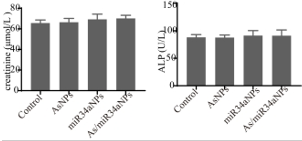
H-Huh7 suspension was inoculated on the right back of mice to establish a subcutaneous transplanted tumour model. When the tumours grew to 200 mm3, the mice were randomly divided into four groups with six mice per group. All miR-34a formulations were equivalent to 1 mg/kg body weight, and As formulations were equivalent to 0.5 mg/kg body. All formulations were administered intravenously once a week. Tumour growth was monitored and recorded by callipers eight times over three weeks. The tumour volume (V) was calculated based on the following formula: V=[major axis(minor axis)2]/2. Then, the mice were sacrificed and the tumours were excised and photographed. To evaluate the potential cytotoxicity of formulations in vivo, at the end of the treatment, serum biochemical analysis was carried out. Liver function was estimated with the serum levels of ALT, AST, and ALP. Kidney function was verified based on the serum levels of BUN and CRE.
Statistical analysis
All values were presented as the mean ± S.D. Statistical significance was determined by one-way analysis of variance using SPSS software (version 17.0, IBM Inc, Chicago, IL, USA). The differences were considered significant at p < 0.05 and highly significant at p < 0.01.