Materials
2, 6-diaminopyridine, FeCl3•6H2O and Sodium hyaluronate were commercially provided by Shanghai Aladdin Reagent CO, Ltd. (China). N-hydroxysuccinimide (NHS) and 1-(3-Dimethylaminopropyl)-3-ethylcarbodiimide hydrochloride (EDC·HCl) were brought from Sigma-Aldrich Trading Co., Ltd. (China). [Ru(dpp)3]Cl2 (RDPP), 1, 3-diphenylisobenzofuran (DPBF) and 2′, 7′-dichlorofluorescein diacetate (DCFH-DA)were purchased from Shanghai Medpep Co., Ltd (China). Roswell Park Memorial Institute (RPMI) 1640, fetal bovine serum (FBS), penicillin–streptomycin, and trypsin were supplied by GIBCO Invitrogen Corp. (USA). Cell counting kit-8 (CCK-8) and 4′,6-Diamidino-2-phenylindole (DAPI) were provided by Shanghai Yeasen Biotech Co., Ltd. (China). All other reagents were recieved and used without further purification. Deionized (DI) water was obtained from experimental water purification system .
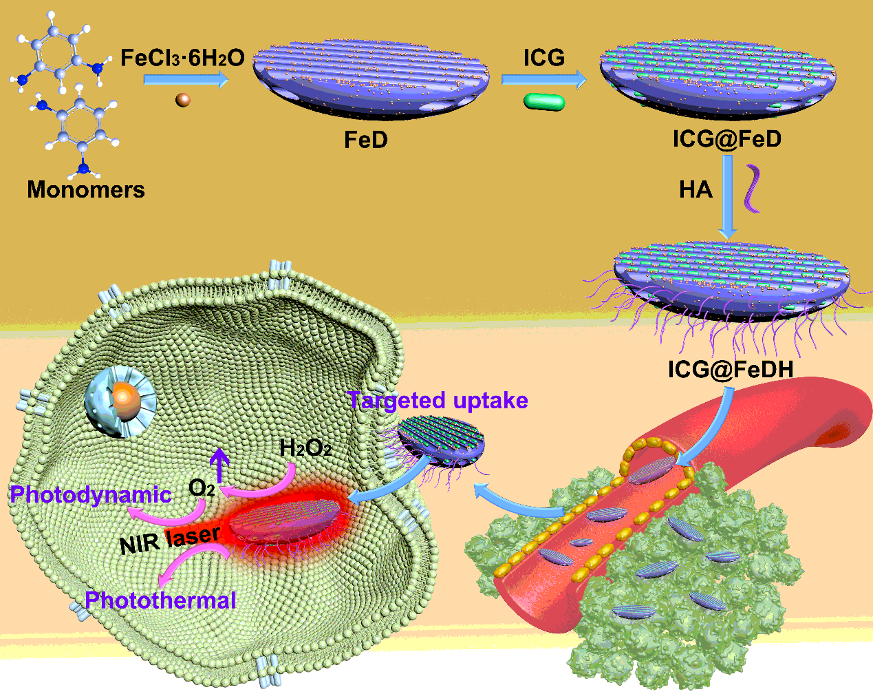
Synthesis of FeD nanoparticles
In a 50 mL round-bottom flask, 8.0 mmol of FeCl3•6H2O was stirred in 20 mL of deionized water for 0.5 h. Then, 2.0 mmol of 2, 6-diaminopyridine was added and the mixture was heated at 40 °C for 24 h. The obtained FeD nanoparticles were collected by dialysis.
Preparation of FeDH
First, 0.1 g of HA, 36.2 mg of EDC and 27.9 mg of NHS were stirred in 10 mL of deionized water for 4 h. Then, 10 mL of FeD solutions (5 mg/mL) was added and reacted with activated HA for another 20 h. The HA modified FeD (denoted as FeDH) were obtained by high-speed centrifugation. To ICG-loaded FeDH (ICG@FeDH), 5 mg of ICG was added in FeD solutions and the modification is proceeded with the same steps. The samples were isolated and purified by centrifugation.
In vitro photothermal effect of ICG@FeDH
To evaluate the photothermal performance of ICG@FeDH, 0.5 mL of ICG@FeDH solution at different concentrations (100, 50, 25 μg/mL) was added into a Eppendorf tube (0.5 mL) and irradiated by the 808 nm laser (1 W/cm2, 5 min). The temperature changes were timely measured by a infrared thermal imaging camera. As controls, the temperature of pure water or FeDH solution with concentration of 100 μg/mL were also recorded under the same conditions.
Assessment of oxygen generation ability
5 mL of H2O2 (2 mM) was mixed with 5 mL of FeDH dispersion with a dissolved oxygen meter inserted in the solution. The pure water and H2O2 solutions were also tested under the same conditions for comparison.
To evaluate the intracellular oxygen-evolving ability of FeDH, an oxygen-sensitive probe molecule RDPP was used to monitor the intracellular oxygen level. In brief, cells were seeded in culture dishes and then installed with RDPP (10 µM) at 37 °C for 2 h. Subsequently, the cells were treated with FeDH at 200 µg/mL for another 2 h. Finally, the human prostatic cancer cells (PC-3 cells) were rinsed with PBS for several times and directly imaged by confocal laser scanning microscope under an excitation of 488 nm.
Reactive oxygen species detection
Typically, DPBF was used as a probe molecule to detect the singlet oxygen generation of different samples [35]. 1 mL of ICG@FeDH aqueous solution at concentration of 100 μg/mL was mixed with 50 μL of DPBF dissolved in dimethyl sulfoxide (10 mM) and irradiated by a 808 nm laser (1 mW/cm2). The absorbance at 419 nm was measured by a UV-vis spectrophotometer at different time intervals.
To investigate the intracellular ROS generation, PC-3 cells were seeded in culture dishes and incubated with ICG@FeDH (50 μg/mL) for 4 h. Then the cells were incubated with DCFH-DA (2 μM) for 0.5 h. After removing the excess probe, the cells were further illuminated by 808 nm laser for 10 min, and imaged by CLSM under the excitation of 808 nm. The cells treated with complete medium alone, laser irradiation alone, and ICG@FeDH alone were also stained with DCFH-DA for control. Moreover, the cells pre-treated with HA were also set as the receptor-inhibition group, in which the same protocol was performed except that the PC-3 cells were pre-treated with free HA (5 mg/mL) for 2 h before the incubation and irradiation.
Targeted ability of ICG@FeDH
Targeted ability of ICG@FeDH was also investigated by CLSM. Typically, PC-3 cells were seeded in culture dish at density of 2 × 10⁵ cells/dish for 24 h. Subsequently, the cells were cultured with ICG@FeD and ICG@FeDH for 2 h. After removing the medium, the cells were washed, fixed by 4% paraformaldehyde and imaged by CLSM under oil lens. Moreover, the cells pre-treated with free HA were also incubated with ICG@FeDH and observed by CLSM. The cells without treatments were set as control.
In vitro cytotoxicity and cell killing effect
The cytotoxicity of free FeDH was assessed by standard CCK-8 assay. In a typical process, PC-3 cells were seeded into 96-well plates at a density of 1×10⁴ cells/well for 24 h. Then the medium was replaced with different concentrations of FeDH, and the cells were further incubated for another 24 h. After that, the cells were rinsed with PBS. After 4 h incubation, the medium was replaced with CCK-8 work solution and incubated at 37 °C for 2 h. The absorbance at 450 nm of each well was measured by a microplate reader. The cell viability was calculated by the value of the control group divided by the values of the samples [36]. Four parallel experiments were set for each sample.
To evaluate the cancer cell killing effect, PC-3 cells were seeded into 96-well plates at a density of 1×10⁴ cells/well for 24 h, and further incubated with ICG@FeDH (100 μg/mL) for 4 h. Then the cells were irradiated by 808 nm laser for 10 min, followed by incubated at 37 °C for another 20 h. For comparison, the cells treated with complete medium, NIR laser alone, ICG@FeDH alone, and ICG@FeDH + NIR laser with the pre-treatment of free HA were set as different groups with corresponding procedures. Last, the cell viability was evaluated by CCK-8 assay as described above.
In vivo antitumor effect
All animal experiments were approved by the Animal Care and Use Committee of Southern Medical University, Guangzhou, China. 4-6 weeks-old male nude Balb/c mice were provided by Experimental Animal Center of Southern Medical University. The tumor model was established by subcutaneously injecting PC-3 cells (2×10⁶ cells) into right back of the mice. After the tumor size reached ~50 mm3, the tumor-bearing mice were randomly divided into four groups (n = 4): PBS (control group), NIR laser alone (irradiation group), ICG@FeD with laser irradiation (non-targeted group) and ICG@FeDH with laser irradiation (targeted group). Correspondingly, the experimental mice were intravenously injected with different samples (4 mg/mL, 0.2 mL). For irradiation groups, the tumor site of mice was exposed to 808 nm laser for 10 min. The treatments were performed day 1 and day 3. After that, the tumor size and body weight of experimental mice were monitored every two days. Moreover, the tumors of each group were extracted for hematoxylin&eosin and immunofluorescent staining.
Statistical analysis
The results were expressed as mean ± standard deviation. The significance was analyzed by one-way analysis of variance (ANOVA) statistical method and Scheffe's post hoc test. The criteria was set as *p < 0.05 and **p < 0.01 for statistical significance.