1. Knip M. Diabetes: Loss of beta-cell mass - an acute event before T1DM presentation? Nat Rev Endocrinol 2017; 13: 253-4. doi: 10.1038/nrendo.2017.33.
2. Olehnik SK, Fowler JL, Avramovich G, Hara M. Quantitative analysis of intra- and inter-individual variability of human beta-cell mass. Sci Rep 2017; 7: 16398. doi: 10.1038/s41598-017-16300-w.
3. Wang H, Bender A, Wang P, Karakose E, Inabnet WB, Libutti SK, et al. Insights into beta cell regeneration for diabetes via integration of molecular landscapes in human insulinomas. Nat Commun 2017; 8: 767. doi: 10.1038/s41467-017-00992-9.
4. Kahn SE, Carr DB, Faulenbach MV, Utzschneider KM. An examination of beta-cell function measures and their potential use for estimating beta-cell mass. Diabetes Obes Metab 2008; 10 Suppl 4: 63-76. doi: 10.1111/j.1463-1326.2008.00945.x.
5. Chintinne M, Stange G, Denys B, In 't Veld P, Hellemans K, Pipeleers-Marichal M, et al. Contribution of postnatally formed small beta cell aggregates to functional beta cell mass in adult rat pancreas. Diabetologia 2010; 53: 2380-8. doi: 10.1007/s00125-010-1851-4.
6. Golson ML, Bush WS, Brissova M. Automated quantification of pancreatic beta-cell mass. Am J Physiol Endocrinol Metab 2014; 306: E1460-7. doi: 10.1152/ajpendo.00591.2013.
7. Coppens V, Leuckx G, Heremans Y, Staels W, Verdonck Y, Baeyens L, et al. Semi-automated digital measurement as the method of choice for beta cell mass analysis. PLoS One 2018; 13: e0191249. doi: 10.1371/journal.pone.0191249.
8. Hara M, Wang X, Kawamura T, Bindokas VP, Dizon RF, Alcoser SY, et al. Transgenic mice with green fluorescent protein-labeled pancreatic beta -cells. Am J Physiol Endocrinol Metab 2003; 284: E177-83. doi: 10.1152/ajpendo.00321.2002.
9. McGirr R, Hu S, Yee SP, Kovacs MS, Lee TY, Dhanvantari S. Towards PET imaging of intact pancreatic beta cell mass: a transgenic strategy. Mol Imaging Biol 2011; 13: 962-72. doi: 10.1007/s11307-010-0435-5.
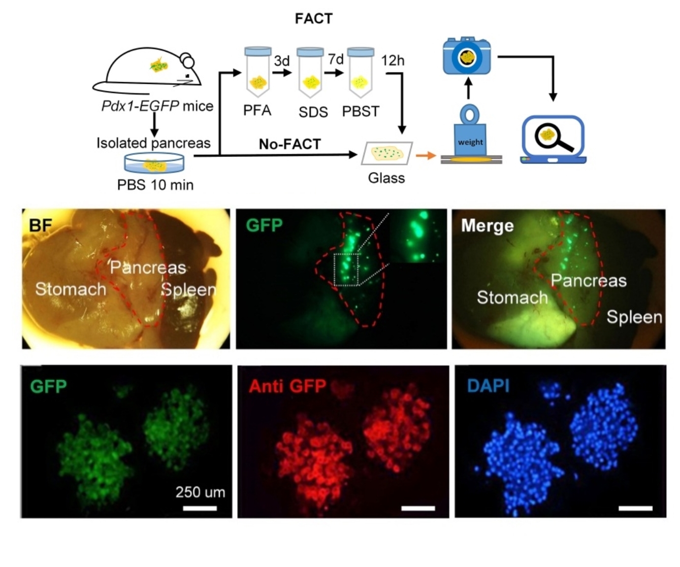
10. Xu N, Tamadon A, Liu Y, Ma T, Leak RK, Chen J, et al. Fast free-of-acrylamide clearing tissue (FACT)-an optimized new protocol for rapid, high-resolution imaging of three-dimensional brain tissue. Sci Rep 2017; 7: 9895. doi: 10.1038/s41598-017-10204-5.
11. Khoradmehr A, Mazaheri F, Anvari M, Tamadon A. A Simple Technique for Three-Dimensional Imaging and Segmentation of Brain Vasculature U sing Fast Free-of-Acrylamide Clearing Tissue in Murine. Cell J 2019; 21: 49-56. doi: 10.22074/cellj.2019.5684.
12. Mohammad Rezazadeh F, Saedi S, Rahmanifar F, Namavar MR, Dianatpour M, Tanideh N, et al. Fast free of acrylamide clearing tissue (FACT) for clearing, immunolabelling and three-dimensional imaging of partridge tissues. Microsc Res Tech 2018; 81: 1374-82. doi: 10.1002/jemt.23078.
13. Basiri M, Behmanesh M, Tahamtani Y, Khalooghi K, Moradmand A, Baharvand H. The Convenience of Single Homology Arm Donor DNA and CRISPR/Cas9-Nickase for Targeted Insertion of Long DNA Fragment. Cell J 2017; 18: 532-9. doi: 10.22074/cellj.2016.4719.
14. Sylvestersen KB, Herrera PL, Serup P, Rescan C. Fgf9 signalling stimulates Spred and Sprouty expression in embryonic mouse pancreas mesenchyme. Gene Expr Patterns 2011; 11: 105-11. doi: 10.1016/j.gep.2010.10.001.
15. Brouwers B, de Faudeur G, Osipovich AB, Goyvaerts L, Lemaire K, Boesmans L, et al. Impaired islet function in commonly used transgenic mouse lines due to human growth hormone minigene expression. Cell Metab 2014; 20: 979-90. doi: 10.1016/j.cmet.2014.11.004.
16. Elsner M, Tiedge M, Guldbakke B, Munday R, Lenzen S. Importance of the GLUT2 glucose transporter for pancreatic beta cell toxicity of alloxan. Diabetologia 2002; 45: 1542-9. doi: 10.1007/s00125-002-0955-x.
17. Jorns A, Munday R, Tiedge M, Lenzen S. Comparative toxicity of alloxan, N-alkylalloxans and ninhydrin to isolated pancreatic islets in vitro. J Endocrinol 1997; 155: 283-93. doi: 10.1677/joe.0.1550283.
18. Malaisse WJ, Malaisse-Lagae F, Sener A, Pipeleers DG. Determinants of the selective toxicity of alloxan to the pancreatic B cell. Proc Natl Acad Sci U S A 1982; 79: 927-30. doi: 10.1073/pnas.79.3.927.
19. Wang H, Khoradmehr A, Tamadon A. FACT or PACT: A Comparison between Free-Acrylamide and Acrylamide-Based Passive Sodium Dodecyl Sulfate Tissue Clearing for whole Tissue Imaging. Cell J 2019; 21: 103-14. doi: 10.22074/cellj.2019.5989.
20. Ighodaro OM, Adeosun AM, Akinloye OA. Alloxan-induced diabetes, a common model for evaluating the glycemic-control potential of therapeutic compounds and plants extracts in experimental studies. Medicina (Kaunas) 2017; 53: 365-74. doi: 10.1016/j.medici.2018.02.001.