Materials and reagents
All oligonucleotides used in this work were synthesized and purified by Sangon Biotechnology Co., Ltd. (Shanghai, China), and their sequences are listed in Additional files 1: Table S1. Fetal bovine serum (FBS) and Dulbecco’s modified Eagle’s (DMEM) medium were obtained from Biological Industries (Israel) and HyClone (USA), respectively. Lipofectamine 3000 was ordered from ThermoFisher scientific (USA). Annexin V-FITC apoptosis detection kit was purchased from CoWin Biosciences (Beijing, China). Hoechst 33342 solution, 4S Red Plus Nucleic Acid Stain and N, N, N', N'-tetramethyl ethylenediamine (TEMED) were ordered from Sangon Biotechnology Co., Ltd. (Shanghai, China). All the reagents were of analytical grade and used without further purification.
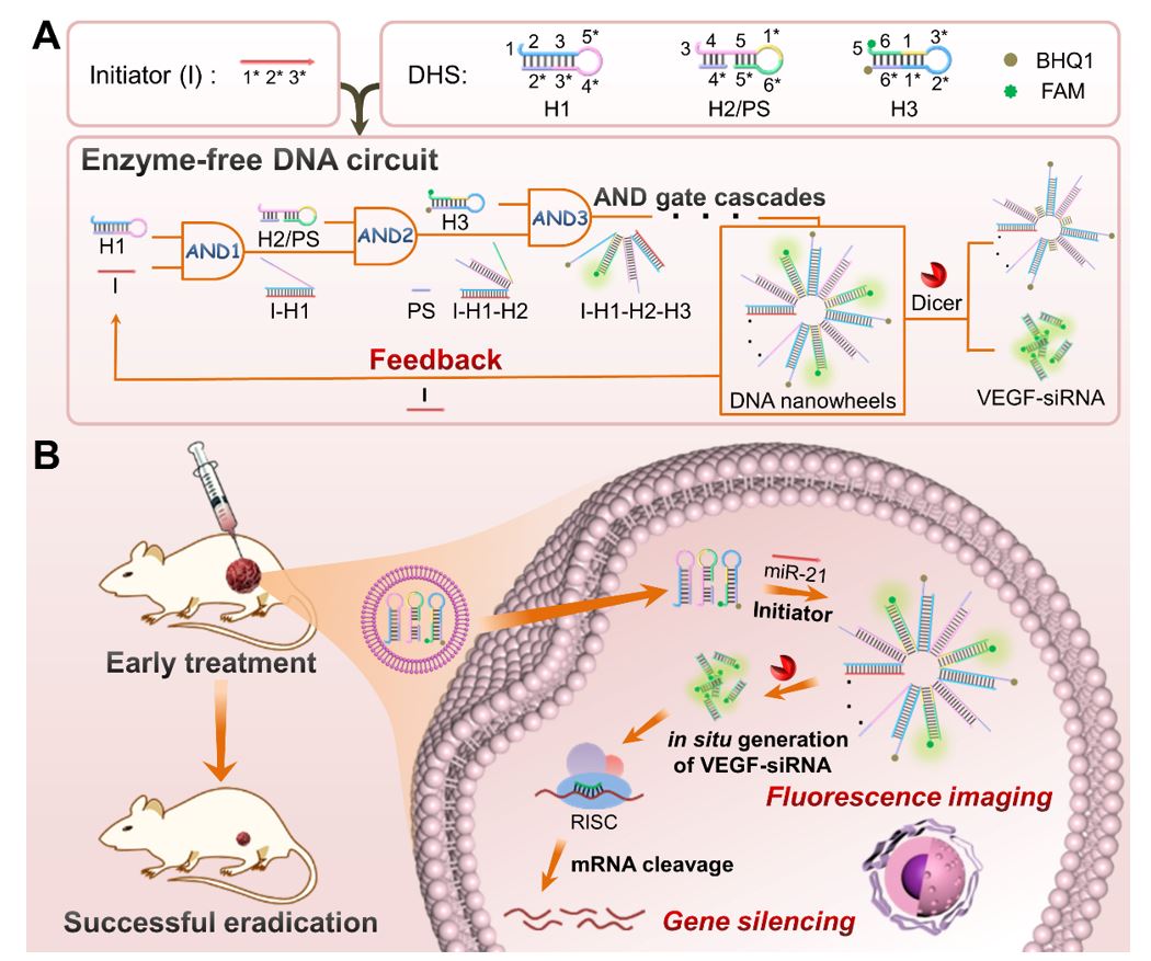
Preparation of DNA hairpins
The stock solutions of oligonucleotides (100 μM) were prepared using deionized ultrapure water, which were further diluted to 10 μM using TE buffer (10 mM Tris-HCl, 1 mM EDTA-2Na, 12.5 mM MgCl2, pH 7.4). To obtain the desirable secondary structures, the DNA hairpin strands (H1, H2 and H3) were respectively annealed in TE buffer by heating to 95 oC for 10 min, followed by cooling to 25 oC with a rate of 0.1 oC/s and standing at 25 oC for 4 h before each use. Further, H2 was incubated with equal amount of protect strand (PS) at 37 oC for 2 h to form the H2/PS hybrid.
Native polyacrylamide gel electrophoresis
The 8% polyacrylamide gel (PAGE) was prepared according to the previous study [32], followed by adding 10 µL of each sample and 2 μL of 10× loading buffer. Then, the native PAGE was running at 170 V for 5 min and 110 V for 35 min in 1× TAE buffer (10 mM Tris, 1 M glacial acetic acid, 1 mM EDTA-2Na, 12.5 mM MgCl2, pH 8). Finally, the resulting gel was stained with 4S Red Plus solution for about 30 min and recorded by Tanon 2500R gel imaging system (Shanghai, China).
Atomic force microscopy imaging
A 10 µL of each sample was deposited onto the freshly cleaved mica for 30 min. Then, the mica surfaces were washed using deionized ultrapure water for three times, followed by drying at room temperature. Finally, the samples were scanned by Being Nano-Instruments CSPM-4000 system (Guangzhou, China) under the tapping mode. The obtained images were analyzed using CSPM Console software.
Fluorescence measurements
A 2 μL of miR-21 with different concentrations was added to the mixture of H1, H2/PS and H3 (10 μL and 200 nM for each species), respectively. The real-time fluorescence was monitored on a LineGene 9600 Real-Time detection system (Hangzhou, China) at an interval of 30 s (λex = 470 nm and λem = 525 nm) for 4 h. The reaction temperature was set at 37 oC. The corresponding fluorescence spectra at 4 h were recorded on a F-7000 fluorescence spectrophotometer (Hitachi, Japan).
Stability assays
The naked VEGF siRNA (100 nM) and VEGF siRNA formed in DNWs (100 nM) was incubated in 100 μL of HeLa cell lysate solution for 1 h, 2 h, 3 h and 4 h, respectively. Subsequently, 15% PAGE was performed to study the stability [36].
Cell culture
The HeLa cells (human cervix carcinoma cell), HepG2 cells (human liver cancer line) and L-02 cells (human normal liver cell line) were purchased from Shanghai Institutes for Biological Sciences (SIBS) (Shanghai, China) and cultured in DMEM supplemented with 10% FBS, streptomycin (100 μg/mL) and penicillin-streptomycin (100 μg/mL) at 37 oC under 5% CO2 humid atmosphere. The cells were counted using a cell counting chamber (ThermoFisher scienticfic, USA) before each assay.
Confocal laser scanning microscopy imaging
The HeLa cells, HepG2 cells and L-02 cells were seeded in 48-well plate (3 × 10⁴ cells/well, 500 μL) and cultured in DMEM medium for 12 h, respectively. According to the manufacturer instructions, HeLa cells were transfected with DNA circuit, C-circuit and R-circuit, respectively, in which the DNA circuit contains H1, H2/PS and H3 to generate VEGF siRNA triggered by miR-21, C-circuit contains H1-C, H2-C and H3-C to generate negative control siRNA triggered by miR-21, and R-circuit contains random DNA (H1-R, H2-R and H3-R) that is not responsive to miR-21. In addition, the antisense miR-21 pretreated HeLa cells were transfected with DNA circuit. The final concentration of each DNA hairpin was 200 nM. After incubation for 4 h at 37 oC, the cells were washed with PBS for three times, and the fluorescence images were recorded on a Nikon Confocal Microscope A1 (Nikon, Japan) (λex = 488 nm and λem = 520 ± 20 nm). For nuclear localization imaging, the HeLa cells were incubated with DNA circuit (200 nM for each DNA hairpin) for 4 h, followed by staining with Hoechst 33342 solution (5 μg/mL) for 30 min. After washing with 1× PBS for three times, the fluorescence images were recorded with an excitation wavelength of 405 nm and an emission wavelength of (475 ± 25) nm using a Nikon Confocal Microscope A1
Flow cytometry
The HeLa cells, HepG2 cells and L-02 cells were seeded in 6-well plates (5 × 10⁵ cells /well) for 12 h and then transfected with DNA circuit for 4 h, respectively. Then, the cells were washed three times with 1× PBS and detached from the plate by trypsin. Finally, the flow cytometry assays were performed on a CytoFLEX system (Beckman Coulter, US) under the excitation of 488 nm.
Quantitative real-time PCR
The HeLa cells and HepG2 cells were seeded in 6-well plates (5 × 10⁵ cells /well) for 12 h and then transfected with different samples (naked siRNA, DNA circuit, C-circuit, R-circuit and Lipo for HeLa cells; C-circuit, R-circuit and DNA circuit for HepG2 cells) for 48 h at 37 oC, respectively. The final concentration of each DNA hairpin was 200 nM. The total RNAs were extracted from the transfected cells using Trizol reagent (Invitrogen, USA). Then, the cDNA was generated using PrimeScriptRT reagent kit (Takara, Japan). Finally, the qRT-PCR analysis was performed using TB GreenR Premix Ex TaqTM II (TaKaRa, Japan) according to the manufacturer instructions. The PCR primer sequences are listed in Additional files 1: Table S1. The operation conditions of PCR were as follows: an initial step (95 oC for 30 s), followed by 40 cycles (95 oC for 5 s and 60 oC for 30 s). The data were analyzed by normalizing to the expression of GAPDH and using the 2-ΔΔCT method. The process of RNA extraction was performed on ice.
Western blot assay
The protein expression of cells was examined using western blot. In brief, the HeLa cells and HepG2 cells were seeded in 6-well plates (5 × 10⁵ cells/well) for 12 h and then transfected with different samples (PBS, naked siRNA, DNA circuit, C-circuit and R-circuit for HeLa cells; PBS, naked siRNA and DNA circuit for HepG2 cells) for 48 h at 37 oC, respectively. The final concentration of each DNA hairpin was 200 nM. Then, the cells were lysed in RIPA (Radio Immunoprecipitation Assay) lysis buffer for 30 min on ice and then scraped immediately. The extracted solution was transferred to an EP tube and centrifuged at 4 oC for 15 min (12000 r/min). The concentration of the total protein was measured using a BCA protein assay kit (CoWin Biosciences, Beijing, China). A 25 μg total protein of each sample were loaded on 10% sodium dodecyl sulfate-polyacrylamide gel electrophoresis (SDS-PAGE) and electro-transferred to polyvinylidene fluoride (PVDF) membrane. After being blocked with PBS buffer containing 5% nonfat dry milk for 2 h, the membranes were incubated with the rabbit anti-VEGFA polyclonal antibody (1:1000 dilution) (Absin Bioscience Inc, Shanghai, China) and β-actin (Santa Cruz Biotechnology, USA) overnight at 4 oC, followed by incubation with goat anti-rabbit IgG-HRP secondary antibody (Absin Bioscience Inc, Shanghai, China) (1:5000 dilution) for another 1 h at room temperature. The protein bands were visualized with a Sparkjade ECL super (Sparkjade Biotechnology, Jinan, China) using Vilber Fusion FX7 Spectra (France).
Cell apoptosis experiments
The HeLa cells and HepG2 cells were seeded in 6-well plates (5 × 10⁵ cells/well) for 12 h and then transfected with different samples (naked siRNA, DNA circuit, C-circuit, R-circuit, and Lipo for HeLa cells; C-circuit, R-circuit and DNA circuit for HepG2 cells) for 48 h at 37 oC, respectively. The final concentration of each DNA hairpin was 200 nM. After washing three times with 1× PBS, the collected cells were stained using Annexin V-FITC/PI Apoptosis Detection Kit according to the manufacturer instructions. Finally, the fluorescence signals of cells were analyzed using Cytomics FC 500 (Beckman, USA).
CCK-8 assay
The HeLa Cells and HepG2 cells were seeded in 96-well plates (5× 10³ cells/well) for 12 h and then transfected with different concentrations of DNA hairpins (50 nM, 100 nM, 150 nM and 200 nM) for 48 h, respectively. After washing twice with PBS, 100 μL of 10% CCK-8 solution (10 μL of CCK-8 reagent and 90 μL of DMEM without FBS) was added into each well and incubated with cells for 30 min. Then, the optical density (OD) at 450 nm was measured using a microplate reader (TECAN Safire 2, Switzerland). The relative cell viability (%) was calculated by (Atest - Ablank)/(Acontrol - Ablank) × 100%, in which Atest, Ablank and Acontrol represent the OD450 of experimental group, blank group and control group, respectively. In addition, to demonstrate the therapeutic efficiency of in situ generated VEGF siRNA, the HeLa cells and HepG2 cells were transfected with different samples (naked siRNA, DNA circuit, C-circuit, R-circuit and Lipo for HeLa cells; C-circuit, R-circuit and DNA circuit for HepG2 cells), respectively, followed by determining the relative cell viability using the above methods.
In Vivo antitumor efficacy
All animal experiments were carried out in agreement with the Institutional Animal Care and Use Committee. The female BALB/c nude mice (5-6 weeks) were acquired from Beijing Vital River Laboratory Animal Technology Co., Ltd (Beijing, China) and randomly divided into five groups with four mice per group. The tumor xenograft models were developed by subcutaneous inoculation with 6 × 10⁶ HeLa cells suspended in 100 μL of PBS. When the tumor volumes grew to 75 mm3, the groups were intratumorally injected with 50 μL of PBS, naked siRNA, DNA circuit, C-circuit and R-circuit every two days for eight treatments, respectively (the equivalent siRNA dose for each mouse is 0.25 mg/kg). Meanwhile, the bodyweight and tumor growth were monitored during therapy. The tumor volumes were calculated using the formula of V = (L × W2)/2, where L and W represent the length and width of tumor, respectively. At the end of the experiment, the mice were sacrificed. In addition, the pathological changes of tumors were analyzed using hematoxylin and eosin (H&E) staining assay and terminal deoxynucleotidyl transferase-mediated dUTP nick-end labeling (TUNEL) staining. To further study the biocompatibility of the proposed DNA circuit in vivo, the H&E assay was performed to analyze the pathological changes of major organs including heart, liver, spleen, kidney and lung from mice in different groups. The images were obtained with Pannoramic MIDI (3D HISTECH Ltd., Hungary).
Statistical analysis
The data was expressed as mean ± standard deviations. We used one-way analysis of variance (ANOVA) to analyze the statistical difference (* means P ˂ 0.05, ** means P ˂ 0.01, and *** means P ˂ 0.001).