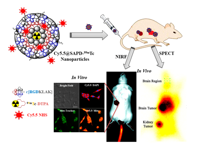
This study describes the development of self-assembled peptide nanoparticles followed by modifications using near-infrared fluorescent dye (Cy5.5 NHS) and bifunctional chelating agent radiolabeled with 99mTc for multimodal imaging and enhanced therapeutic efficacy against brain tumor glioblastoma multiforme.
The development of malignant cells in the circulatory system is progressively assisted by the growth of new blood vessels by utilizing oxygen and nutrients ultimately raising tumor angiogenesis. Self-regulation of angiogenesis causes the progression of diseases like the proliferation of cancer cells, glioblastoma multiforme (GBM), myocardial infarction, and atherosclerosis [1]. For the treatment of cancer, anti-angiogenic therapy in combination with anticancer therapy has been emerging as a novel strategy to combat tumor cell growth by discontinuing their nutrient oxygen supply. Multidomain-Targeted drug delivery serves as a guided missile to effectively and efficiently target the cancer cells vasculature. These targeted drugs have been developed using peptides/ proteins, integrin-receptor ligands, antibodies, and aptamers possessed recognition domains and effector domains [2]. There are many cognate receptors and pro-angiogenic factors involve to promote vessel formation in tumors such as fibroblast growth factor-2 (FGF-2), vascular endothelial growth factor (VEGF), and platelet-derived growth factor (PDGF) [3]. These growth factors up-regulate the expression of integrins including α1β1, α2β1, α4β1, α5β1, α9β1, and αvβ3-integrins on blood and lymphatic vessels. Several investigations implicate integrins as key regulators of tumor angiogenesis which are also regulating endothelial cell survival, migration and mediate cell-cell adhesion and cell–ECM events [4, 5]. Basic clinical studies reveal that angiogenesis can be blocked by inhibiting the angiogenic signaling pathways, ultimately resulting in tumor dormancy and metastasis [6, 7].
Integrin-mediated signaling pathways play an important role in tissue development and homeostasis, while its deregulation causes multiple brain diseases [8]. Interestingly, integrins are crucial glycoproteins essential for many physiological processes such as proliferation, cell migration, wound healing, hemostasis, bone remodeling, and oncogenic transformation [9, 10]. They are also important for cell-cell and cell-extracellular matrix interactions and comprised of nineteen α- and eight β-subunits [11, 12]. Among them, αvβ3-, αvβ5-, αvβ8-, α5β1-, and αIIbβ3-integrins have been studied extensively for their active role as an excellent candidate for cancer theranostic [13]. More specifically αvβ3-integrin serves as a receptor for extracellular matrix proteins such as fibronectin, vitronectin, fibrinogen, collagen, laminin, and osteopontin with exposed arginine-glycine-aspartic acid (RGD) sequence [14]. It is expressed on epithelial and mature epithelial cells in low levels, while, highly expressed on the surface of many tumors including carcinomas of breast and lungs, melanomas, osteosarcomas, and glioblastoma [15-17]. Hence, αvβ3-integrin is considered as a molecular target of interest for the early diagnosis of cancer and selective attachment and internalization of RGD-containing peptides and peptidomimetics for cancer therapy [18]. The cyclic monomeric (RGDfV) and multimeric (Galacto-RGD) peptides have emerged in phase-III clinical trials for diagnosing glioblastoma and in phase-II trials for many other tumors [19]. It is the first anti-angiogenic small molecule drug that specifically targets the αvβ3-, αvβ5-, and α5β1-integrins and is the first potent and superactive αvβ3-integrin receptor antagonist.
Over the last two decades, both linear and cyclic RGD peptide analogs have been discovered, radiolabeled (99mTc, 111In, 68Ga, and 18F), and evaluated as radiotracers for tumor diagnosis using single-photon emission computed tomography (SPECT) or positron emission tomography (PET) imaging [20]. Linear RGD peptides showed a lack of αvβ3-integrin specificity, low binding affinity, and high enzymatic degradation of the aspartic acid residue due to free rotation around a single bond. On contrary, cyclic RGD peptides were observed to be highly stable towards proteases and increased affinity to αvβ3-integrin receptors and have reduced structural flexibility [21]. Therefore, many researchers have studied the effect of hybrid peptides by incorporating RGD tripeptide with other anticancer peptides analogs including RGD-111In-DTPA-octreotate [22, 23], 18F-bombesin-RGD [24], 99mTc-RGD-Bombesin [25], 64Cu-RGD2-PG12-bombesin heterodimer [26], 5FU-loaded SF-cRGDfK-Ce6 [27], and RG301-MTX peptide [28] for targeted drug delivery and improved diagnostic as well as therapeutic efficacy.
In the light of above knowledge, we hypothesized that by designing the short head-to-tail cyclic peptide sequence using RGD homing motif (αvβ3-integrin receptor binding) along with KLAK (mitochondria-targeting) motif without addition of unnecessary long chains of linking amino acids which can improve integrin receptor targeting and mitochondrial damaging potentials for therapeutic applications [29]. Secondly, the modification of this cyclic peptide by incorporation of Near-Infrared fluorescent (NIRF) dye for optical imaging as well as bifunctional chelating agent for radiolabeling with β/γ-emitting radionuclides for SPECT imaging can introduce novel dual-imaging agent for SPECT/ NIRF diagnosis. To the best of our knowledge, we are the first who have designed novel head-to-tail cyclic RGD-KLAK heptapeptide sequence [2]. Further, the effectiveness and efficacy of this newly developed novel NIRF-dye conjugated self-assembled peptide nanoparticles radiolabeled with 99mTc (to give Cy5.5@SAPD-99mTc) was investigated for diagnosis of glioblastoma as well as therapeutic potentials as dual-imaging and dual-targeting probe. The in vitro cell studies as well as in vivo diagnostic and therapeutic studies were finally carried out to assess the capabilities of Cy5.5-SAPDN-99mTc as SPECT/ NIRF imaging agent for brain tumor glioblastoma multiforme (GBM).
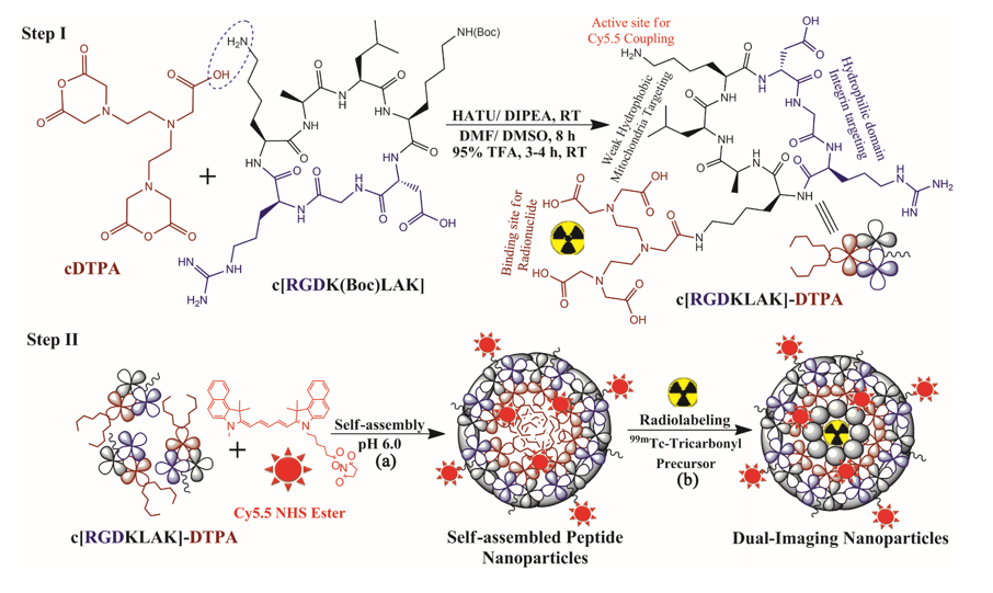
To achieve this goal, firstly, we modified the ɛ-amino group of terminal lysine residue attached to this peptide sequence by coupling with deprotected free β-carboxylic group of diethylenetriamine pentaacetic acid (DTPA) via covalent linkage as bifunctional chelating agent (BFC). This BFC is useful tool for selective radiolabeling of γ-emitting radionuclide Technetium-99m (Half-life 6 h; Er = 140 keV) as SPECT imaging probe. The reaction was successfully accomplished by HATU/DIPEA-chemistry with a satisfactory yield of ~ 78% as presented by Step-I in Scheme 1. The Boc protecting group at the amino group of central lysine residue was removed using 95% TFA to obtain corresponding free amine preferably available for conjugation with a NIR-fluorescent probe [30]. Secondly, this Peptide-DTPA complex was intrinsically converted to self-assembled nanoparticles via co-assembly of NIRF-probe (Cy5.5 NHS), which covalently conjugated with free amino group of intermediate lysine residue to introduce optical imaging features. A simple and facile synthesis approach was used to design novel dual-targeting self-assembled cyclic peptide-DTPA (SAPD) nanoparticles with high chemical yield and efficacy.
The purity of the cyclic peptide-DTPA (cPD) complex was confirmed by HPLC analysis indicating a single peak with ≥ 98% purity at retention time Rt = 3.780 min (Figure S1), HR-MS analysis shows molecular mass peaks for calculated for C47H81N15O18 with m/z found [M+2H+] = 1144.01 a.m.u (Figure S2). Furthermore, FTIR-ATR analysis shows superimposed spectrum of cDTPA (black line), cyclic peptide (red line), and cPD complex (blue line) having peaks at 3265.1 cm-1 and 1640.2 cm-1 assigned for stretching vibrations of –NH2 and –NC=O group; respectively and peaks at 2109.7 cm-1 designated to bend vibrations of –COOH group (Figure S3).
To rationalize the generality of self-assembly strategy for cPD complex and optical imaging, we choose Cyanine 5.5 NHS ester as near-infrared fluorescent (NIRF) dye. The cPD complex was successfully self-assembled with Cy5.5 NHS via covalent interactions to form uniform nanoparticles with well-defined spherical shape and “Always ON” NIR-fluorescence property as presented in Step-II (a) of Scheme 1 by using the pH-sensitive method [31, 32]. This will facilitates in enhancing the pharmacokinetics and improving the diagnostic as well as therapeutic efficiencies of dual-targeting dual-imaging peptide nanoparticles, whilst maintained intrinsic biocompatibility and biodegradability [33]. The effect of self-assembly on change in fluorescence intensity was assessed by fluorescence spectrophotometer, spectrogram presented in Figure 1a shows peaks at an excitation wavelength of 650 nm and emission wavelength of 702 nm in aggregation state, which is nearly consistent with the parent NIRF dye (Cy5.5 NHS; Ex/ Em = 650/ 700 nm) with a slight increase in emission wavelength indicating successful co-assembly of Cy5.5 with cPD complex [34]. The freshly synthesized Cy5.5@SAPD nanoparticles were further radiolabeled with Technetium-99m by using fac-[99mTc-(CO)3(H2O)3]+ core complex for SPECT/ CT imaging.
The radiosynthon introduced by Alberto et al. has been used widely for preferential labeling of peptides/ proteins to achieve relatively high specificity keeping retained biological property of bioactive molecules [35]. The radiosynthon was successfully prepared with high radiochemical purity of ≥ 97% showing a single high-intensity peak at retention time Rt = 4.727 min as indicated by Radio-HPLC analysis (Figure S4). The inset figure shows TLC-SG results depicted that fac-[99mTc(CO)3(H2O)3]+ moves with the solvent front at Rf = 0.90, while free 99mTc remain at the point of spotting (Rf = 0.00). The electron donor nitrogen and oxygen groups of DTPA provide an easy platform for radiolabeling with gamma-emitting radionuclides in the presence of suitable reducing agent and optimum pH value as shown in Step-II (b) of Scheme 1. Consequently, the radiolabelled nanoparticles were observed to remain stable at room temperature over 4 h incubation period and no change in radiolabeling efficiency was seen by increasing the concentration of Cy5.5@SAPD-99mTc nanoparticles with a percent radiochemical purity of > 96% (Figure 1b), obtained using ultracentrifugation technique. The same results were calculated from TLC-SG/ Methanol technique, as inset images presented in Figure S4 shows ~ 97% yield at Rf = 0.65, while < 3% impurities were found in saline [36].
Additionally, Transmission electron microscope (TEM) images presented in Figure 1 (c, d) shows clear spherical morphology of Cy5.5@SAPD and Cy5.5@SAPD-99mTc nanoparticles; respectively with uniform dispersion in an aqueous medium. The size of Cy5.5@SAPD nanoparticles was observed to be in between 30 – 40 nm as indicated in inset Figure 1c′ acquired by High resolution-TEM as well as for Cy5.5@SAPD-99mTc nanoparticles the size reduced to 20 – 25 nm as shown by inset Figure 1d. The clear consecutive bight and dark lattice fringes with interplanar lattice fringe distance of 0.294 nm as presented in Figure 1d′ indicate a tight interface among all three-ingredient which could facilitate balancing the charge on the surface of nanoparticles as well as demonstrates the successful self-assembly of cPD complex with Cy5.5 NHS having crystallinity in novel designed nanoparticles [37]. The dynamic light scattering (DLS) measurements reveal the hydrodynamic size of Cy5.5@SAPD nanoparticles with a diameter of less than 100 ± 28 nm as depicted in Figure S5 enabling the capabilities of novel designed nanoparticles in vitro and in vivo [38].
Upon successful modification of dual-targeting cyclic peptide to design dual imaging cyclic peptide nanoparticles, in vitro cancer cell studies were carried out to evaluate the effectiveness, specificity, and efficacy. The dual-targeting capabilities of SAPD nanoparticles were assessed by using αvβ3-integrin positive cancer cell line (U87MG), and αvβ3-integrin negative cells (HEK-293). Both cancer cells were separately treated with Cy5.5@SAPD and Cy5.5@SAPD-99mTc nanoparticles to estimate the cytotoxicity potential. The results presented in Figure 2a showed that the cytotoxicity effect against U87MG cells was slightly higher for Cy5.5@SAPD-99mTc nanoparticles as compared to Cy5.5@SAPD nanoparticles, with EC50 values of 20 µM and 25 µM; respectively. On contrary, the MTT assay showed that both nanoprobes were weakly cytotoxic towards HEK-293 cancer cells as ≥ 80% viable cells were found in 96-well plates as presented in bar graph (Figure 2b). The significantly higher cytotoxicity of Cy5.5@SAPD-99mTc nanoparticles might be due to the attachment of γ-emitting radionuclide compared with Cy5.5@SAPD nanoparticles [39]. So, further cell studies were performed using Cy5.5@SAPD nanoparticles to avoid the effect of diagnostic radionuclides.
The CLSM images acquired after treatment with Cy5.5@SAPD nanoparticles showed very less red fluorescence intensity in HEK-293 cells (Figure 2c), alternatively showed bright red fluorescence intensity in U87MG cells (Figure 2d). The co-localization of DAPI staining in the nuclear region shows blue fluorescence, while the merged image showed localization of red fluorescence in the nuclear periphery region. These results highlight the specificity and efficacy of newly designed nanoparticles for αvβ3-integrin positive cancer cells due to RGD tripeptide [11].
Further, we investigated the potential of apoptosis induction in U87MG cancer cells upon treatment with Cy5.5@SAPD nanoparticles. The results presented in Figure 2e showed 95.6% live cells as control and figure 2f showed 17.2% live cells as well as 26.8% early apoptotic and 52.7% late apoptotic cells, while only 1.3% necrotic cells. This study confirms our hypothesis that our proposed nanoparticles have the potential to kill cancer cells by inducing apoptosis in glioblastoma cancer cells due to the presence of the KLA motif [40]. Moreover, to investigate the appropriate cell apoptosis-inducing pathway, we also performed CLSM imaging study by treating the U87MG cancer cells with Cy5.5@SAPD nanoparticles and stained the cells with Mito-Tracker Green as well as Caspase-3 dyes. The CLSM images acquired with Cy5.5 filter after 30 min incubation time showed that Cy5.5@SAPD nanoparticles internalized into cells via pinocytosis and enters into the mitochondria by disrupting the mitochondrial membrane as indicated in Figure 2g, merged images showed well-overlapped fluorescence intensities to give yellowish-green color, scale bar 20 µm was set for all images. This ultimately produces reactive oxygen species (ROS) that causes mtDNA damage, whilst, it releases cytochrome c to promotes the formation of apoptosome, causing activation of Caspase-3 enzyme as showed in Figure 2h [41]. The bright green fluorescence is due to activated Caspase-3 which is well-overlapped with red fluorescence as presented in the merged image to give bright yellow fluorescence intensity in nuclear periphery region [42, 43]. The merged CLSM images with blue colored holes show stained nuclear region (Figure c, d) while blank holes showed unstained nuclear region (Figure g, h) indicates improved specificity of our novel SAPD nanoparticles to selectively target mitochondria and induces cell apoptosis.
Additionally, we also performed a Bio-TEM imaging study using Cy5.5@SAPD nanoparticles treated U87MG cancer cells, the TEM images showed pinocytic internalization of nanoparticles as shown in Figure 2i. The zoom image shows the accumulation of nanoparticles in the inner-layers of the cell membrane (Figure 2j) and targets the mitochondria to induce apoptosis by damaging the mitochondrial membrane as illustrated by HR-TEM image presented in Figure 2k. Bio-TEM images are in agreement with the CLSM study evidenced the induction of cancer cell apoptosis by damaging the mitochondria with high specificity and improved efficacy [38, 44, 45].
Additionally, we also investigated the dual-imaging potential of Cy5.5@SAPD-99mTc nanoparticles in brain tumor glioblastoma (using U87MG cells) and human embryonic kidney (HEK-293) tumor-induced (5×10⁷ cells/ mice subcutaneously) female Balb/c mice models. Firstly, the nanoparticles with a concentration of 20 µg/ 200 µL (~ 74 MBq) saline were injected via tail vein, and images were acquired using a dynamic SPECT/CT camera (NM/ CT 670 Pro Discovery) as well as fluorescence imaging camera (IN VIVO FX Pro Carestream). Images presented in figure 3 (a-c) indicates CT, SPECT, and SPECT/ CT images; respectively acquired after 30 min post-injection (p.i) with coronal, sagittal, and transaxial directions show the occurrence of brain tumor glioblastoma and kidney tumor with excellent accumulation of proposed nanoparticles as tumor-to-background contrast showed at the left side of Balb/c mice [46]. The results presented in Figure 3 (d-g) depicted planar SPCET images acquired at 30 min p.i (Figure 3d left), and 2 h p.i (right) before therapeutic dose treatment, showed high internalization of radiotracer in brain tumor glioblastoma with 4.7 ± 0.8 % ID/ g as compared to kidney tumor (2.1 ± 0.9 % ID/ g). The results of the planar SPECT imaging study are comparable with that biodistribution study.
After therapeutic dose (30 mg/ kg b.w) treatment within a very short period of nearly one week, Figure 3f clearly shows very less or negligible accumulation of radiotracer at the site of a brain tumor while prominent uptake can be seen at the kidney tumor site, this is because of the binding potential of RGD motif to αvβ1-integrin overexpressed on HEK-293 cells [9]. The sharp decrease in brain tumor size indicates the insightful excellent therapeutic potential of Cy5.5@SAPD-99mTc nanoparticles for GBM. Moreover, Figure 3e indicates an ex vivo image presenting pharmacokinetic of Cy5.5@SAPD-99mTc nanoparticles before and (figure 3g) after therapeutic dose treatment. The main accumulation of radiotracer was found in the brain, liver, lungs, and kidneys and nonspecific uptake was observed in the heart, stomach, and spleen. Furthermore, the brain tumor-bearing female Balb/c animal models were subjected to fluorescence camera imaging, and results presented in Figure 3 h shows in vivo as well as Figure 3 (i, j) showed ex vivo images after 30 min and 2 h p.i; respectively. The in vivo live imaging study shows the accumulation of Cy5.5@SAPD nanoparticles at the site of brain glioma tumors. The tumor uptake was observed to be highly prominent than normal brain tissues and other body organs pointed out the efficacy, specificity, and effectiveness of our newly designed novel Cy5.5@SAPD nanoparticles as compared to previously reported nanoparticles [47-50].
In the light of the above-mentioned results, we firstly designed a dual-targeting peptide sequence consisting of RGD motif for targeting αvβ3-integrin and KLAK pro-apoptotic motif for targeting mitochondria and induce cancer cell apoptosis by activation of the Caspase-3 enzyme. This dual-targeting peptide probe was further modified with diethylenetriamine pentaacetic acid (DTPA) for radiolabeling with γ-emitting radionuclide and serves as SPECT/ CT imaging agent. Further, the peptide-DTPA complex was self-assembled along with NIR-fluorescence dye Cy5.5 NHS to form uniform spherical shaped nanoparticles for molecular optical imaging study as novel dual-imaging probe. Consequently, these novel dual-imaging and dual-targeting self-assembled cyclic peptide nanoparticles were successfully designed to possess improved diagnostic and therapeutic capabilities with enhanced specifically and efficiently for GBM. The in vitro cytotoxicity assay, apoptosis assay, and CLSM imaging studies illustrated that these newly synthesized Cy5.5@SAPD nanoparticles has potential to internalize specifically and efficiently into U87MG brain tumor cells as compared to HEK-293 kidney tumor cells for early diagnosis of GBM. SPECT/ NIRF theranostic studies in tumor-bearing female Balb/c mice models shows the excellent potential of our novel designed Cy5.5@SAPD-99mTc nanoparticles to diagnose brain tumor more prominently as well as showed remarkable therapeutic effectiveness within one-week treatment. These outcomes suggested that our novel theranostic nanoparticles (Cy5.5@SAPD-99mTc) may serve efficiently, and specifically as potential SPECT/ NIRF nanoprobe for future pre-clinical and clinical studies against brain tumor glioblastoma multiform.