Structural analysis of green synthesized IONPs
XRD patterns of iron oxide nanoparticles (IONPs) synthesized by using spinach leaves and (black coffee) BC extract, subjected to various microwave powers i.e., 100 W to 1000 W (Fig. 2). XRD results demonstrated the presence of phase pure spinal structured magnetite nanoparticles for all of the microwave powers. Peaks exhibiting at 2θ angle of 30.02o, 35.64o, 43.44o, 49.79o, 54.1o, 57.35o, 62.72o and 64.18o corresponds to diffraction planes of (202), (311), (400), (313), (422), (511), (404) and (531). The peaks of Fe2O3 nanoparticles were index with JCPDS card no. 19-629. In present study, BC was utilized as reducing agent. Major constituents of BC are caffeine and tannin. Tannins are composed of polyphenolic compounds (non-toxic) which are reducing and stabilizing agents for the synthesis of IONPs. The phenolic-OH as well as ortho-dihydroxyphenyl groups present in the chemical structure of tannins are responsible for complex formation with iron. These groups also participate in redox reactions [35]. Therefore, production of Fe3O4 nanoparticles is mainly governed by the tannins present in BC extract. Also, the use of microwave radiation makes it possible to synthesize pure phase Fe3O4 nanoparticles without any further heat treatment. Microwave energy is converted into heat energy which is highly depends upon the nature of utilizing solvent. The parameter which determines the capability of material for this conversion is termed as “tangent loss” (tan δ). Usually for microwave radiations having typical frequency of 2.45 GHz, solvents with higher value of tan δ are preferable choice as they can absorb excellent amount of radiations thus convert it into heat energy [36]. Due to higher tangent loss of polyphenols (i.e., constituents of BC, tannin), high heat will be produced in microwave oven thus leading towards the phase pure Fe3O4 nanoparticles even without further heat treatment.
Crystallite size (t) and dislocation density (δ) were plotted (Figure S1) and calculated using Eqs. (4-5), respectively [37].


Where, k is shape factor taken as 0.9, λ is the wavelength of Cu ka source, β is full width at half maximum (FWHM), θ is the diffraction angle of highest intensity peak. Variation in crystallite size was observed with the variation in various extracts used during microwave powers. Black coffee along with microwave heating played a critical role not only in achieving Fe3O4 phase of iron oxide (Fig. 2) but also in achieving a small value of crystallite size (Figure S1). XRD analysis also revealed that synthesized nanoparticles using 100 W - 200 W power possess amorphous behavior while for higher microwave powers crystalline nature of iron oxide nanoparticles along with phase stability was observed (Fig. 2). Major factors influencing crystallite size are lowest surface energy, grain boundary energy and diffusion of surface atoms. Microwave energy was observed to be effective to tune these parameters thus leading to formation of stabilized and pure phased Fe3O4 nanoparticles. Highest crystallite size i.e., ~15 nm along with lowest dislocation value was observed after using microwave power of 1000 W (Figure S1).
Dielectric analysis of green synthesized IONPs
Short ranged electrical conduction of material is dependent on the dielectric properties which it exhibits. Charged particles in the material experience a displacement as a result of applied electric field and gets pile up at the interfaces and hence creation of dipoles takes place. Frequency dependent dielectric constant of iron oxide nanoparticles (IONPs) was obtained by impedance analyzer using Eq. (6), whereas the tangent loss was calculated using Eq. (7) [38].
ε = (Cd)/εoA (6)
tan δ = 1/(2πfεεoρ) (7)
Where, ‘C’ represents capacitance, ‘d’ is for specimen’s thickness, ‘A’ is for area, ‘ρ’ is for resistivity and permittivity of free space is represented by ‘εo’.
Variation in dielectric constant and tangent loss as function of frequency at room temperature was shown in Fig. 3A, B. Different types of polarization i.e., ionic, electronic, orientational and dipole are accompanied with dielectric constant under the effect of an alternating electric field. At low frequency space charge polarization is dominant whereas, at high frequency ionic and electronic polarizations dominate in polycrystalline material. When an external electric field is applied dispersion occurs in space charges and these space charges require some time to align themselves in the direction of applied field. At low frequency space charges get enough time for their arrangement as per applied field. However, at high frequency values charges do not get enough time to orient themselves in the direction of the applied field, thus, resulting in low dielectric values (Fig. 3A). Dielectric constant almost remained same for samples synthesized with low microwave powers i.e., 100W–200W. Small values of dielectric constant at low microwave power is due to amorphous behaviour of iron oxide nanoparticles as observed in XRD patterns (Fig. 2). Whereas increase in dielectric constant was observed with the increase in microwave power from 300W–1000W. High value of dielectric constant is attributed to the presence of both Fe3+ and Fe2+ cations in Fe3O4 phase of iron oxide. Heterogeneity in Fe3O4 structure arises because of the existence of Fe2+ cations that gives high polarization, leading to higher value of dielectric constant [39,40]. High dielectric constant can be employed in agricultural field i.e., plant pathology for investigating activities of pathogenic microbes including fungi.
Tangent loss for as synthesized nanoparticles is shown in Fig. 3B. The plot of tangent loss shows normal dispersion behaviour due to space charge polarization. Relatively, higher values of tangent loss are observed for nanoparticles prepared at low microwave power i.e., 100 W – 200 W whereas, low values of tangent loss are observed for high microwave powers. Comparison of dielectric constant as well as tangent loss with respect to varying microwave powers at log f =5 and log f =1.3 (Figure S2).
Conductivity of as synthesized nanoparticles of IONPs are calculated by Eq. (8) [38].

Conductivity is categorized in two regions, i.e., low frequency region and high frequency region. At low frequency region conductivity is known as dc-like conductivity because of free charge carriers. Whereas, at high frequency it is known as ac conductivity because of bound charge carriers. Figure S3A shows variation in conductivity as a function of frequency for all of the samples prepared using various microwave powers i.e., from 100 W-1000 W. Small values of conductivity, because of the hopping mechanism of charge carriers, are observed even at high frequency values. Comparison of conductivity values at log f =5 and log f =7.3 by using various microwave powers is exhibited in Fig. S3B.
Impedance is a property that offered an opposition to the flow of electric current. Conduction in nanomaterials can be best comprehended on the base of complex impedance spectroscopy which is an effective technique to differentiate the resistive and conducting elements in the circuit. Complex impedance (Z*) is presented in Eq. (9) [38] while real (Z') and imaginary (Z'') impedance are given in Eq. (10) and (11) respectively [38].
𝑍* = 𝑍 ' ‒ 𝑗𝑍'' (9)
𝑍' = 𝑍𝑐𝑜𝑠𝜃 (10)
𝑍'' = 𝑍𝑠𝑖𝑛𝜃 (11)
The variation of real and imaginary impedance in iron oxide nanoparticles with respect to frequency is depicted in Fig. 4A,B. Two regions are observed in Z' plots (Fig. 4A). First is the region of low frequencies in which there is a slight decrease in Z'. Second is the region in which Z' decreases monotonically and becomes constant at higher frequencies. This behavior is connected with conductivity of charge carriers observed in Fig. 4A where conductivity increases at high frequencies [41,42]. Z'' (Fig. 4B) shows different relaxation peaks at different values of frequency for changes in microwave powers. Information regarding grains and interface effects in poly crystalline materials can be realized on the basis of these relaxation peaks which demonstrate different relaxation mechanism happening in nanoparticles [42]. Relaxation peaks at different microwave powers spread in different zones of frequency explains the distinguished participation of grains and grain boundaries. To obtain the exact contribution of grains and grain boundaries towards the conduction process Cole-Cole plots i.e., plots between Z'' and Z' were studied (Fig. 4C). Generally, Cole-Cole plot contain of three semi circles. First semi-circle in high frequency regime represents the effect of grains resistance, second semicircle in the middle range of frequency correspond to grain boundary resistance and the third one in low frequency range represents the resistance offered by grain-to-grain boundary interface. These plots provide information about electrical characteristics of material (grains and grain boundaries) [43].
FTIR analysis of green synthesized IONPs
FTIR spectra of green synthesized IONPs using BC extract and dried using various microwave powers is depicted in Fig. 5A,B. Peak appeared at ~ 560 cm-1 was assigned to characteristic band of Fe-O that also in well agreement with previously reported literature [44]. The band at 1068 cm-1 was appeared due to C-N stretching. While, peaks appearing at 1561 cm-1 and 1650 cm-1 were ascribed to C=C stretching vibrational bands due to presence of aromatic rings / phenolic groups present in BC extract. Presence of phenolic feature indicates the capping effect on the surface of IONPs. Absorption band appeared at 2352 cm-1 corresponds to atmospheric CO2 [45].
Magnetic response of green synthesized IONPs
Superparamagnetic response of green synthesized IONPs at various microwave powers i.e., 100 W -1000 W, was detected through M-H loops (Fig. 6). However, high saturation magnetization (~21.72 emu/g) was observed for IONPs synthesized at 1000 W (Fig. 6J). Superparamagnetic behavior arises when size of single domain becomes so small that thermal energy can easily overcome anisotropy energy barrier. As the particle size is decreased number of surface spins contributing to magnetization increases [46]. Such behavior of IONPs makes it potential candidate for agricultural applications in terms of targeted delivery of nutrients and controlled release of pesticides in plant. Variation in saturation magnetization (Ms) was observed by using various microwave powers (Figure S4).
X-ray photoelectron spectroscopic (XPS) analysis of green synthesized IONPs
XPS analysis of IONPs was performed by using spinach as a precursor along with black coffee (BC) extract at various microwave powers (100 W-1000 W) with interval of 100 W (Fig. 7 A, B). It can be observed that binding energy peaks of Fe 2p3/2 and Fe 2p1/2 at 710.9 eV and 724.5 eV are due to magnetite (Fe3O4), respectively (Fig. 7A) [47]. Splitted spin-orbit peak of Fe 2p are wide due to less chemical shift between Fe2+ & Fe3+[48]. Figure. 7b represents the spectra of O1s core level. Peak present at 529.9 eV is due to existence of O-2 species and 531.7 eV is due to existence of OH- species present on iron oxide surface. Other peak at 532.9 eV is associated with adsorbed H2O molecules [49].
Surface morphology and size distribution of green synthesized IONPs
The benefits of using microwave-assisted approach in contrast to chemical-methods includes in-core heating of materials in a rapid way and its specific chemical bonds gives discerning absorbance, which results as nano size particles, having uniform size and shape, depicted from microscopic images (SEM and TEM). The morphology and particle size distribution of green synthesized iron oxide nanoparticles (IONPs) at 1000 W power were revealed by SEM and TEM analysis (Figure S5). Microscopic images indicate uniform, spherical, polydisperse, less-aggregated crystalline nature of IONPs (Figure S5 A, C) which endorses the purity of the sample without any other phases. Figure S5B showed the particles size distribution of IONPs by SEM, was in the range of 3-23 nm with an average diameter of 4.99 ± 0.17 nm. Correspondingly, the mean particle size of 4.08 ± 0.19 nm was determined by TEM, varying from 1-15 nm, conferring to Gaussian fit of particle size distribution (Figure S5D). These results display the narrow size distribution of green synthesized IONPs, which are somehow coherent with previous studies [50-53].
IONPs as inducer of antifungal activity-inhibiting fungal growth and spore germination
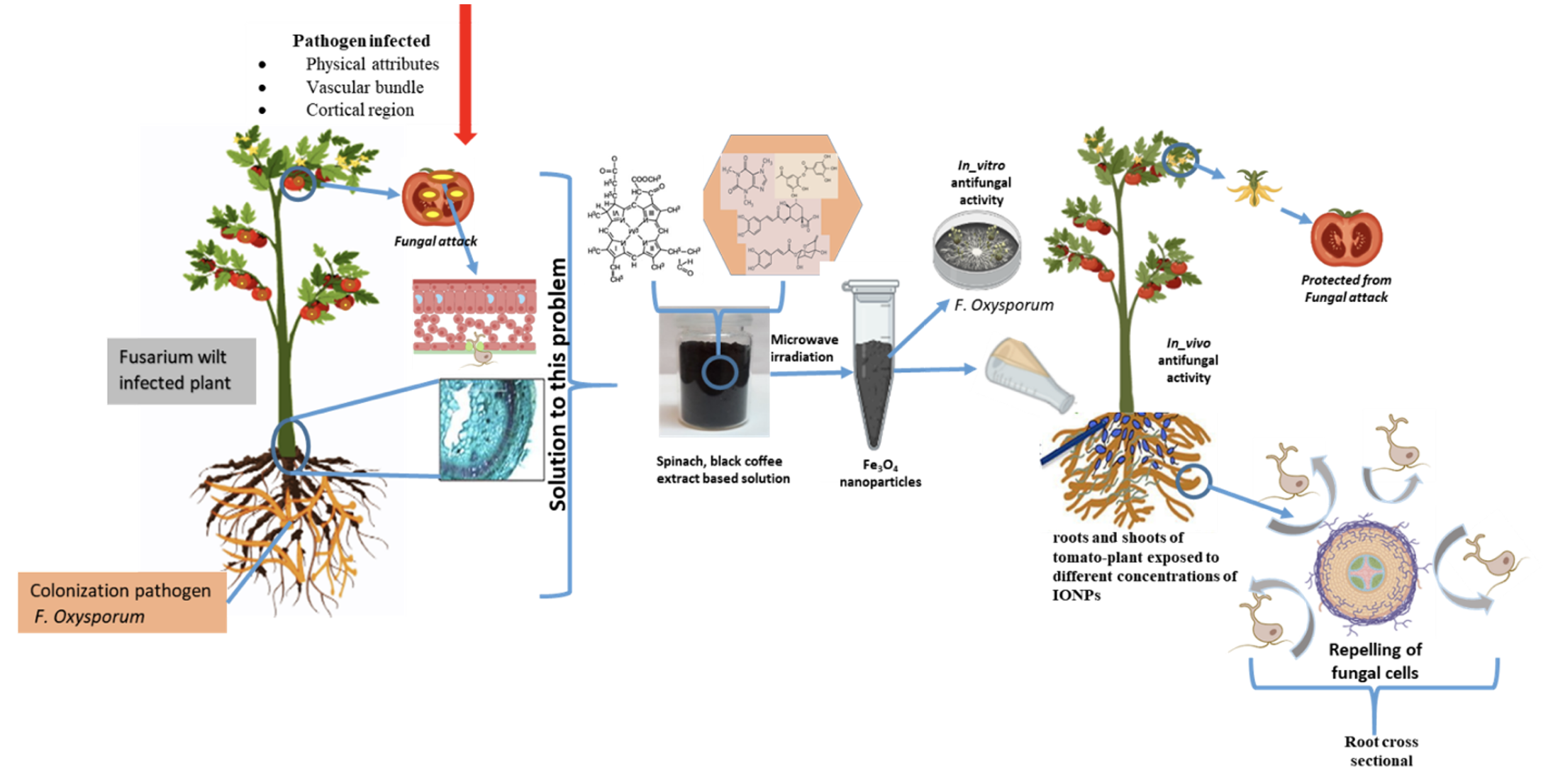
We investigated the in-vitro antifungal activity of biosynthesized iron oxide nanoparticles (IONPs) synthesized at various microwave powers (100 W-1000 W) as shown in Table S1. After initial screening, the IONPs at 1000W was selected and used for next experiments which also justify the phenomena of highest magnetic saturation at this microwave power. The emergence of resistance in plant fungal pathogens against agrochemicals leads towards development of more efficient antifungal agents which must be eco-friendly. Therefore, antifungal potential of nanoparticles is beneficial in agriculture sector as they emerged as “innovative-generation fungicides” [54]. Figure 8 shows the effect of INOPs on fungal mycelial growth and spore germination at various tested concentrations (0.01 µg/mL -15 µg/mL) against F. oxysporum, the causal agent of tomato wilt.
IONPs exhibited strong inhibitory effect on radial growth of fungal mycelia on PDA medium as compared with control treatment (Fig. 8A). It is evident from the results that antifungal activity of iron oxide nanoparticles significantly increases in a dosage-dependent form. After seven days of post inoculation, 15 µg/mL of IONPs strikingly minimized F. oxysporum growth by 90.84 ± 0.56%, in contrast to the control treatment. For lower concentrations, ranges from 0.01- 1.5 µg/mL, displayed minimum growth inhibition i.e., below 50% whereas, fungicide exposure yielded 89.77 1.24%, relative to the growth rate in control group (Fig. 8B). Former studies indicated the antimicrobial activity of IONPs, and their finding suggests that this activity increases gradually from lower to higher concentration [55, 22-23].
The sporicidal activity of IONPs on spore’s germination of F. oxysporum was illustrated in Fig. 8C,D. Disruption of fungal membrane indicates the inhibiting action of nanoparticles as observed in this study is owing to the biocidal action of nanoparticles, furthermore, smaller size NPs retain large surface area gets readily attached and absorbed to disassemble the microbial cell membrane, leads to deterioration of intracellular organelles, eventually results in death of microorganism [56,57]. After six hours of incubation with various concentrations of iron oxide nanoparticles, the microscopic images revealed that spore suspension of F. oxysporum displayed a sharp decline in spore germination rate in contrast to the untreated control samples (Fig. 8D). In fungal life cycle, spore germination and maturation are vital phases for successful plant colonization, but once the germination is subdued after treated with graphene oxide, spore cannot develop into mature mycelium to initiate the infection cycle [35]. It can be clearly indicated from the results that germination rate of spores was gradually reduced in response to the increasing concentrations. A statistically significant reduction in germination rate was observed at concentrations ranges from 1.5 -15 µg/mL, while minimum germination rate was up to 5.38 ± 1.38% at 15 µg/mL in comparison with the control group (88.58 ± 0.69%). Analogously, in case of fungicide, significant inhibition to spore germination was determined to be 18.56 ± 0.86% respectively (Fig. 8C). Devi et al. worked on two fungal species (A. niger and M. piriformis) proposed that greater surface interaction among iron oxide nanoparticles and fungal membranes played a significant role in antifungal activity [58]. Analogously, Saleem et al. also exhibited antifungal potency of green synthesized iron-oxide nanoparticles against A. flavus and F. oxysporum, their finding suggests that IONPs have potential to be used for biological applications [59].
IONPs induce changes in cell-wall morphology, viability and ROS production in F. oxysporum
The SEM micrographs revealed malformed mycelium after treatment with iron oxide nanoparticles can be attributed to distortion of chitin synthesis and cell envelop; that shields the leakage of cellular component into extracellular environment [19]. SEM visualization indicates the detrimental effects of iron-oxide NPs on F. oxysporum. The IONPs treated mycelia revealed some eccentric morphological characteristics as compared to the control (Fig. 9A). In control, cylindrical shaped mycelium has healthy smooth turgid surface with clear conidiation (Fig. 9A-a). Whereas, after contact with IONPs, the remarkable structural changes were induced in fungal hyphae as observed in Fig. 9A-b,c. At 10 µg/mL, hypha became deformed showing irregular shrinkage with minute granules on surface (Fig. 9A-b). The impairment was intensified at 15 µg/mL, the hyphae become recessed, slender and stacked together including rifts or blebs (Fig. 9A-c). Earlier investigations suggested that consecutive interactions occurred due to magnetic nanoparticles which stimulated microbial toxicity, such as discharging of metal-ions, affecting protein synchronization and cellular-homeostasis; lipid-peroxidation and nucleic acid impairment by accumulation of reactive oxygen species (ROS) and mutilation of cell integrity by membrane-depolarization [60].
The antifungal property of IONPs was further verified by using fluorescent dyes i.e., Propidium iodide (PI) and 2’,7’-dichloro-dihydro-fluorescein-diacetate (H2DCFDA). The fluorescence intensity of fungal hyphae was significantly enhanced in dose-dependent manner on the treatment of IONPs in contrast to the untreated control. Alike outcome was perceived for IONPs bound with amphotericin B, by a reaction between aldehyde and amine groups against candida strains [61]. Propidium iodide is a DNA-fluorescent-probe that invades disrupted plasma membrane of cell, emitting red color fluorescence from stained nucleus [62]. The effect of IONPs on membrane integrity of F. oxysporum in control and treated fungal mycelial samples were depicted in Fig. 9B. As pragmatic from the RFP (red fluorescence protein) images a slight red-fluorescence was detected in the control hyphae whereas, stronger fluorescence was observed in treated ones. After 15 mins of exposure, the PI fluorescence intensity in treated groups (5,10 and 15 µg/mL) were 2.21, 3.09 and 3.69 folds, significantly higher than the control group (Fig. 9D). The GFP (Green fluorescence protein) micrographs of control and IONPs treated fungal mycelia of ROS accumulation were illustrated in Fig. 9C. Treatment of F. oxysporum with 5, 10 and 15 µg/mL of IONPs significantly increased H2DCFDA fluorescence intensity by 2.07, 2.73 and 3.05 times than the control (Fig. 9E). Cell genotoxicity and cytotoxicity affected by the surface charge of IONPs, furthermore, NPs with positive charge acts to be more lethal, endures adsorptive endo-cytosis and nonspecific-interactions with the negatively charged cell-membrane, hence distressing the membrane permeability by enhancing intracellular-accumulation [63]. Thus far, it is documented that IONPs reveals antimicrobial affects by inducing production of reactive oxygen species (ROS), following disruption of microbe’s electron-transport, NADH oxidation and cellular- homeostasis thus contributing towards anti-fungal effects.64 No doubt, magnetic oxide nanoparticles retain the ability to aggravates oxidative stress by disturbing the redox potential of cells additionally, augmenting the host antimicrobial resistance by targeting the infection sites with direct approach to eradicate microbial pathogens [61, 65].
Our results indicate that IONPs inactivates the oxidation reduction balance by generating ROS, maybe associated with action mechanism of magnetic NPs, outcomes as pore-formation in cell membrane and stipulates to transports nanoparticles into Fusarium cells. Earlier reports unveiled that cell after uptake of IONPs produces ROS via either pathway: Iron ions contributes in the Haber-Weiss cycle by releasing ions into cytosol where chelation by adenosine-phosphate or citrate occurs or the surface of IONPs catalyzed Fenton reaction or Haber-Weiss cycle that could have a detrimental effect on fungal cells while highly reactive-hydroxyl radical is formed as a result of both pathways [66]. By comparing the fluorescence intensities as shown above specifies that the fungal-nano interactions were relatively stronger after treatment with IONPs that eventually increases the variation in free-energy content, resulting more ROS generation. In line up with current investigation, Arakha et al. found that chitosan-coated IONPs in culture media has ability to enhance ROS production by altering the interaction pattern among bio-nano interface, plays a critical role for antimicrobial affinity of iron oxide NPs [27].
IONPs as inducer of DNA-fragmentation in F. oxysporum
DNA-fragmentation is another biochemical key feature of apoptosis (programmed-cell-death) [67]. The inter-nucleosomal cleavage of genomic DNA was studied in F. oxysporum subjected to various concentrations of IONPs. DNA was isolated from fungal cells and analyzed by agarose gel electrophoresis. The electro-phoretogram is presented in Figure S6. As it is clear from the gel image that typical DNA ladder were formed in control group however “DNA-laddering” of non-chronological DNA fragments were found in IONPs treated group. Moreover, it can be observed that DNA-cleavage increases in IONPs treated samples in concentration-dependent manner. Gel containing DNAs of F. oxysporum also indicates single high molecular-DNA-bands in lanes 1, 2 and 3, treated with 0.01, 0.5 and 1.5 µg/mL IONPs. However, the intensities of these DNA-bands were less than the control group (lane 10). Additionally, the smeared-DNA in all lanes is less than 1Kb and apparently appears weaker than the control. Smeared-DNA almost disappears in lane 8 and 9, implying that IONPs completely impaired the fungal DNA. The results indicated that antifungal effect of IONPs on fungal cells was triggered through the initiation of cell-apoptosis. Alarifi et al. observed breakage of DNA double helix strand by iron-oxide nanoparticles in time and dose-dependent manner [68]. ROS is reported to be implicate in DNA mutilation by IONPs effecting DNA bases such as pyrimidine and purine, contributing to reduce the biofilm formation in bacterial cell [29].
Impact of IONPs on tomato growth-parameters
All concentrations of Iron oxide nanoparticles (0.01-15 µg/mL) significantly enhanced the growth parameters (length (root and shoot), biomass (fresh and dry weight) and plant height of tomato plants infected with Fusarium wilt under pot bioassay (Fig. 10: Figure S7). Root-shoot length and germination-rate are the chief indicators to investigate the impact of nanoparticles in different plant species [69]. The concentration at 12.5 µg/mL revealed the best results by depicting a substantial increase in growth attributes in comparison to the control and other treatments. A gradual increase in plant height was observed for all treatments however, plants treated with 10, 12.5 and 15 µg/mL of IONPs showed the maximum plant height of 45.1, 48.9 and 43 cm (Figure S7A), exceeding the control treatment by 64%, 77.8% and 56.3% respectively. Similarly, consistent result was observed for fungicide with an increase of 51.6%. Correspondingly, root and shoot length of plants were substantially improved after the treatment of IONPs (Figure S7B). Furthermore, the average root and shoot length under 5-15 µg/mL were greatly analogous to the control treatment, predominantly for 12.5 µg/mL showed an increase of 76.3% (roots) and 79.05% (shoots) respectively. Moreover, statistically significant difference was found for plant biomass (fresh and dry weight) in contrast to the control treatment (Figure S7C). The fresh and dry weight was superior with 12 µg/mL treatment surpassing the control by 60.9% and 67.1%. While fungicide treatments followed the same trend for fresh and dry weight by showing an increase of 43.8% and 47.7%, with respect to the control. Earlier studies reported increase in seed germination, plant-biomass, seedling growth/vigor and yield after application of iron oxide nanoparticles [70-74]. Treatment of tomato seeds with Fe3O4 nanoparticles had no side effects on plant growth and development [75]. Similarly, our results verified the previous research by observing the enhancement in growth parameters of tomato plants treated with various concentrations of IONPs.
Impact of IONPs on disease attributes of tomato wilt
Virulence of many pathogens relies on iron procurement so; occasionally microbial infections can be avoided by using iron-chelating products that inhibit pathogen’s ability to approach iron [76]. Pot bioassays were performed to evaluate whether IONPs, considered as an agricultural antifungal agent, have potential to control Fusarium wilt disease (Fig. 10D,E). As shown in Figure S8A,B treating infected tomato seedlings with IONPs reduced the severity and incidence of Fusarium wilt caused by F. oxysporum. Furthermore, a clear positive correlation exists between the concentrations of NPs and disease-index. The disease in control plants were specifically severe after 25 days of post inoculation, and the disease severity came up to 96.67% , however the disease severity of tomato seedlings exposed to IONPs at concentrations of 10, 12.5 and 15 µg/mL were reduced to 47.78%, 43.33% and 45% respectively. Though, with fungicide treatment a decline of 55% in disease severity was achieved (Figure S8A). The corresponding disease incidence in IONPs treated plants was 46.6%, 33.3% and 46.7% in contrast to the 100% control (Figure. S8B). Plants activate a toxic oxidative-burst by increasing iron levels to minimize pathogen virulence; roots mutualistic interactions also encounter phyto-diseases via iron uptake as well as competition for iron acquisition induces a systemic resistance that signal components in roots for iron-uptake [77]. There has no work been done so far on the application of iron oxide nanoparticles to combat plant diseases under field conditions, so recent work is considered as novel, and the results clearly indicates that these NPs has potential to become a part of disease management.
Impact of IONPs on photosynthetic pigments
The impacts of IONPs on the photosynthetic pigments were also accessed in this study by comparing diseased plants with treated ones (Figure S9A,B). Plant stress can also be indicated by changes in photosynthetic pigments [78]. Results indicated that various treatments of IONPs induces an increase in photosynthetic pigments. After exposure to 12.5 µg/mL IONPs, total chlorophyll and carotenoid contents in treated plants were significantly increased by 75.6% and 70.3% respectively in comparison to the control however, lower doses (0.01 and 0.5 µg/mL) showed decline up to 27.7% and 1.94% for chlorophyll content and 19.05 and 8.54% for carotenoid content respectively. Askary et al. reported the similar results with application of nano-iron fertilizer [9]. Compared to the control, increased pigment production was detected for fungicide treatment, with an elevation of 56.1% and 55.1% (p< 0.001) respectively. Iron plays a vital role in synthesis of chlorophyll; iron chlorosis reduces the level of photosynthetic pigments in plants effected the process of photosynthesis, while Fe-chlorosis is more frequent in photosystem-II analogous to photosystem-I [9]. The results demonstrated that higher doses of IONPs enhanced the synthesis of photosynthetic pigments in diseased plants by reducing chlorosis.
Impact of IONPs on phenolic content and antioxidant enzymatic activities
Plants endures stress environment by generating higher quantities of antioxidant enzymes, which enhances tolerance against oxidative burst [13]. Figure 11, 12: Figure S10 shows the phenolic content and activities of antioxidant enzymes (SOD, CAT, APX, GPX and POD) in the roots and shoots of diseased tomato plants in the presence of 0.01-15 µg/mL IONPs. Defensive response in plant induced as outcome of biotic-stress commonly involved phenolic-compounds [79]. The total phenolic content in roots and shoots of tomato plants significantly increased with increasing concentration of IONPs (Fig. 11A). In comparison to the control, total phenolic content in plants treated with 12.5 µg/mL of IONPs, showed an upsurge of 3.04 and 2.96-folds in roots and shoots respectively. In fungicide treatment the fold in roots and shoots increased up to1.96 and 1.89 respectively. Avio et al. reported increased phenolic content in lettuce, infected with Rhizoglomus irregulare positively associated with activities of antioxidant enzymes [80]. Increased phenolic compounds were detected in Molvadian balm under salinity stress after application of iron oxide nanoparticles [81]. Nourozi et al. demonstrated that iron oxide nanoparticles act as abiotic elicitor in Dracocephalum kotschyi enhancing accumulation of phenolic compounds [82].
SOD, a metalloenzyme, plays a key role in plant defence system by mobilizing the disproportionation of free-hydroxyl-radicals (O2•–) to H2O2 to mediate ROS-toxicity [83]. SOD activity significantly enhanced in roots and shoots of infected tomato plants exposed to IONPs in parallel to the control treatment. The maximum activity, both in roots and shoots was recorded at 12.5 µg/mL, which showed 3.11 and 2.79-folds higher than the control treatment, whereas its activity dropped 0.39 and 0.42 times at 15 µg/mL compared with the control (Fig. 11B). Rui et al. observed increased SOD activity in peanut plant after treatment with iron-oxide nanoparticles in comparison to control and Fe-EDTA-treatment [84]. Interaction of iron-oxide nanoparticles and stress environment (salinity) enhances the activity of SOD enzyme; as iron is responsible for higher production of SOD; leads towards oxidative stress [82], therefore scavenging activity of some other antioxidant-enzymes such as POD, APX and CAT reduced the level of H2O2 in order to improve plants immunity against ROS burst [85]. Hence, lesser SOD activity at highest concentration of IONPs may induce reduction in ROS scavenging that ultimately increased mutilation in plants [86].
In plants, cellular destruction is being prevented by CAT involves in regulating H2O2 status in tissues by causing disintegration into oxygen and water molecules [87]. The CAT activity presented a dose-dependent effect, higher concentration of IONPs indicates greater enzymatic activity both in roots and shoots (Fig. 11C). The CAT activity in tomato plants at various concentrations of IONPs revealed a fold increase of 1-3.21 in roots and 1.2-2.7 in shoots respectively as compared to the control (p<0.05). Additionally, it can be assumed that higher activities of CAT interrelated with greater production of H2O2 and SOD activities [13]. APX consumes ascorbate and GPX employs glutathione as an electron-donor to downgrade ROS levels [88]. The activities of APX and GPX were significantly enhanced after application of IONPs both in roots and shoots. In plants treated with 12.5 µg/mL of IONPs, both root and shoot showed APX activity up to 1.98 and 2.03 times higher than the control (Fig. 12A). Similar trend was observed for GPX activity that gradually increased with doses of IONPs, surpassing the control by 71.8% and 73.7% at 12.5 µg/mL in root and shoots respectively (Figure. 12B). Both APX and GPX activities showed decline at 15µg/mL Peroxidases is another defence related enzyme utilizes pyrogallol and guaiacol for detoxification of H2O2, involved in activating plant resistance against invading pathogen and wound healing [87]. A significant elevation was observed for POD activity in roots and shoots after exposure to IONPs. In comparison to the control the peroxidase activity, enhanced by 2.09, 2.13 and 2.05-folds in roots and 2, 2.02 and 1.9-fold in shoots at 10, 12.5 and 15 µg/mL respectively (Fig. 12C). However, in all enzymatic activities fungicide treatment also indicates significant increase in comparison to the control treatment. Enzymes such as CAT, APX and POD containing iron-group participating in plant-metabolism by neutralizing hydrogen peroxide [89]. In agreement with the current work, earlier studies also reported the increase in secondary-metabolites and antioxidant enzymatic activities after treatment with iron-oxide nanoparticles in Citrus-maxima plant [90], Hyoscyamus reticulatus [91], Dracocephalum kotschyi [82], Oenothera biennis [92] and Dracocephalum moldavica [81] respectively.
Impact of IONPs on growth and non-enzymatic compounds of tomato fruit
Fruit quality is imperative for merchandise, pulpy fruits are putrescible, certain biotic and abiotic agents are intricating in deteriorating the quality of product [93]. Table 1 indicates substantial improvement in fruit variables (average weight and number) and non-enzymatic compounds of tomato fruit exposed to various concentrations of IONPs (Fig. 10F,G). The average fruit weight was significantly increased by 48.8% in presence of 12.5 µg/mL IONPs in comparison to the control. Similarly, the same trend was noted for fungicide treatment surpassing the control by 46.2% respectively. Likewise, under the same concentration (12.5 µg/mL) highest number of fruits, 32.6 per plant was counted with an increase of 51.6% respectively. Kumar et al. reported increases in fruit mass and fruit number per plant in strawberry after combined application of iron oxide and zinc oxide nanoparticles [94]. Furthermore, Hernandez-Hernandez et al. observed 25% increase in tomato fruit weight after application of selenium and copper NPs [95].
Table 1: Average weight, number, antioxidant compounds and protein content in tomato fruits treated with IONPs
|
Treatments
|
Average Fruit Weight
(g)
|
Fruits
Number
|
Lycopene
(mg 100 g-1 FW)
|
Flavonoids
(mg 100 g-1 FW)
|
Vitamin C
(mg 100 g-1 FW)
|
Protein
(Umin-1 mg-1)
|
|
Control
|
59.61
|
21.53
|
2.16
|
14.12
|
15.82
|
5.26
|
|
Fungicide
|
87.19***
|
30.12***
|
3.42**
|
20.42***
|
17.42*
|
8.92***
|
|
0.01µg/mL-IONPs
|
37.71***
|
16.89***
|
1.84
|
13.92
|
14.29*
|
4.75
|
|
0.5 µg/mL-IONPs
|
42.29***
|
18.67***
|
1.98
|
14.19
|
14.75
|
4.86
|
|
1.5 µg/mL-IONPs
|
57.81
|
20.53
|
2.06
|
15.25
|
15.37
|
5.12
|
|
2.5µg/mL-IONPs
|
69.63***
|
21.84
|
2.49
|
16.52***
|
16.21
|
5.67
|
|
5µg/mL-IONPs
|
74.64***
|
23.74***
|
2.87
|
18.83***
|
16.64
|
6.74**
|
|
7.5µg/mL-IONPs
|
79.93***
|
25.52***
|
3.15
|
19.42***
|
17.07
|
8.49***
|
|
10µg/mL-IONPs
|
83.59***
|
28.59***
|
3.66***
|
20.76***
|
17.86***
|
8.74***
|
|
12.5µg/mL-IONPs
|
88.75***
|
32.63***
|
3.95***
|
21.37***
|
18.12***
|
9.15***
|
|
15µg/mL-IONPs
|
84.26***
|
31.09***
|
3.78***
|
21.03***
|
18.02***
|
8.96***
|
Significant-difference (* P< 0.05; **P < 0.01; *** P < 0.001) between different concentrations of IONPs and control group performed by one-way-ANOVA at P < 0.05 and Tukey-multiple-comparisons analysis.
Tomato fruit has carotenoids such as lycopene, is one of the potent antioxidants, counteracts the ROS; another faction of lycopene in plants is associated with chemo- and photo protection [28, 96-97]. The lycopene content of tomato fruit significantly increased at 10, 12.5 and 15 µg/mL of IONPs, with an increase of 69.4%, 82.8% and 75% respectively in contrast to the control. Lower treatment (0.01-1.5 µg/mL) in comparison to the control showed non-significant decrease of 14.8%, 8.33% and 4.62% respectively. Some former studies specified that soil and foliar treatment of TiO2, ZnO and CuO nanoparticles enhanced lycopene content in tomato plants [98-99]. Foliar application of various treatments of copper nanoparticles significantly increased the lycopene content from 56.8% - 105.3% in comparison to the control in tomato [100].
Flavonoids are the naturally available phyto-chemicals present in vegetables and fruits; having anti cancerous properties, acts as antioxidants to regulate ROS homeostasis [101]. The flavonoid-content in tomato fruit presented statistically substantial difference among treatments. The maximum flavonoid-content was observed at 12.5 µg/mL generated a surge of 51.3% in relative to the control. Similarly, application of fungicide exceeding the control by 44.6%. Nanoparticles induced the generation and accumulation of antioxidants like flavonoids, Vitamin C, carotenoids in plants as natural response against plant pathogens [102]. Regarding the vitamin C content, there was 9.67%- 2.84% decrease when lower treatment (0.01-1.5µg/mL) of IONPs were applied. While three highest doses of IONPs (10-15 µg/mL) including fungicide significantly increased the vitamin C content in tomato fruit by 12.9%, 14.5%, 13.9% and 10.1% with respect to the control. Vitamin C is most vital component of tomato fruit, signifies a key role by avoiding oxidative-damage [96]. Quiterio-Gutiérrez et al. reported increase in flavonoid and Vitamin C content in tomato fruit after application of selenium and copper nanoparticles [102]. Proteins has a key role in fruit growth and quality, tomato ripening is associated with function of regulatory proteins involved in initiation of ethylene- biosynthesis [103-104]. Total protein content significantly increased at the highest concentrations of IONPs in comparison to the control, reaching up to 28.1%, 61.4%, 66.2%, 73.9% and 70.3 % with 5-15 µg/mL of IONPs respectively, the value also increased by 69.6% for fungicide treatment. However, the rest of the treatments didn’t show significant difference from the control. Zaho et al. noticed increased protein content in cucumber fruit after application of nanoparticles [105].