HCC is characteristic of high malignancy and high mortality because of the heterogeneity of tumor microenvironment. Recently, although advances in treatment of HCC, the prognosis of patients with advanced HCC remains unsatisfactory. Indeed, the response of each HCC patients to drug treatments is quite different, due to molecular heterogeneity. Numerous studies have focused on ferroptosis, for it influences therapeutic vulnerabilities and may predict clinical outcomes [37]. In addition, it has been confirmed that ferroptosis plays a key role in HCC migration, invasion, proliferation, which provides a new angle for the therapy as well as prognosis for HCC [10]. Evidences have demonstrated that lncRNAs play a crucial regulatory role in cancer-associated ferroptosis process.
For instance, LncRNA MT1DP could aggravate oxidative stress in hepatocytes through interplay with miR-365, and as the intermediate to repress NRF2‐mediated antioxidant signaling [38]. LINC01554 closely related to tumor invasion, tumor size, tumor staging and shorter survival of HCC, exerted its tumor-suppressive function
through regulating PKM2 and Akt/mTOR signaling pathway [39]. Although a few studies have indicated that ferroptosis-associated genes and lncRNAs might regulate drug-induced ferroptosis in HCC [22], their correlation with the prognosis of HCC patients remains undefined. In this study, we constructed 12 FRLS to predict OS of HCC patient. Meanwhile, we explored the relationship between the FRLS and immune microenvironment, for it can effectively predict the prognosis of patients. Therefore, this study proposes the potential biomarkers and therapeutic targets for HCC.
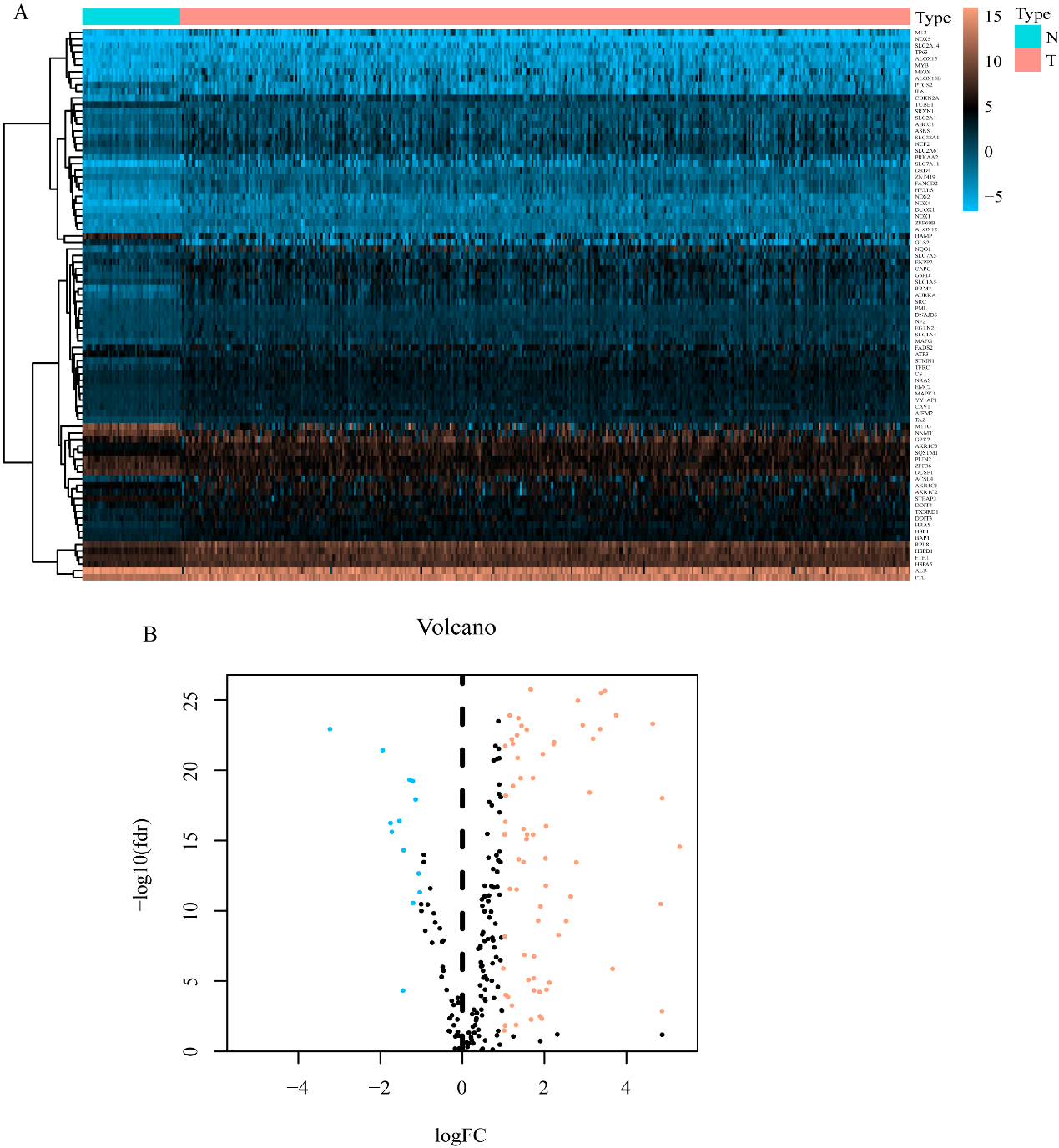
Firstly, 84 ferroptosis-related DEGs and 784 ferroptosis-related lncRNAs were obtained. KEGG analyses revealed the genes mainly participated in synthesis of microRNAs in cancer cells, central carbon metabolism in cancer cells, VEGF signaling pathway, HIF-1 signaling pathway. A recent study found that exosomal miR-522 derived from cancer-associated fibroblasts suppressed ferroptosis, and the knockdown of miR-522 could enhance sensitivity to drug [40]. iASPP (inhibitor of apoptosis-stimulating protein of p53) was found to inhibit ferroptosis through the Nrf2/HIF-1 signaling pathway, thus played protective roles in acute lung injury [41]. In this study, ferroptosis-related lncRNAs (LUCAT1, LINC01224, THUMPD3-AS1, AC116025.2, LINC00942, SNHG10, AC131009.1, POLH-AS1, MKLN1-AS, LINC01138, LNCSRLR, AL031985.3) were selected as independent prognostic factors for HCC. LUCAT1 over-expressed in HCC and other malignant tumors, was found to associated with shorter OS [42] and as well regulate breast cancer stemness by activation of Wnt/β-catenin pathway [43]. LINC01224 enhanced the malignant phenotype of HCC cells by elevating expression of CHEK1 [44]. The high expression level of THUMPD3-AS1, significantly related to the progression and recurrence of NSCLC, regulated cancer cells self-renewal via miR-543 and ONECUT2 [45].LINC00942 (LNC942) elicited pro-tumorigenic roles
by promoting METTL14-mediated m6A methylation and subsequently enhancing the stability and expression of CXCR4 and CYP1B1 [46]. SNHG10, associated with poor OS of HCC, could facilitate the carcinogenesis and metastasis of HCC cells through modulating the expression of SCARNA13 [47]. MKLN1-AS highly expressed in HCC tissues, was reported to be associated with shorter OS, the knockdown of MKLN1‑AS tremendously suppressed HCC cells proliferation, invasion, and migration [48].LINC01138 played a nonnegligible role in tumorigenicity and metastasis of HCC by activating PRMT5 [49]. LNCSRLR, involved in predicting the prognosis of patients of renal cell carcinoma, exerted its carcinogenic role by directly binding to NF-κB, thus leading to the development of intrinsic sorafenib tolerance [50]. However, to date, there is no study on the role of AC116025.2, AC131009.1, POLH-AS1, AL031985.3 in tumors, thus requires subsequent experiments.
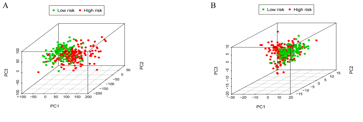
The risk score of each HCC patient was calculated on the basis of the expression of the 12 prognostic ferroptosis-related lncRNAs and patients were grouped into low- or high-risk groups according to the median value of the risk score. The Kaplan-Meier curves showed that FRLS could distinguish the patients with good prognosis from those with poor prognosis (P < 0.001). The AUC predictive value of the FRLS corresponding to 1 year, 3 years, and 5 years of survival were 0.817, 0.760 and 0.749. Besides, the Cox regression analysis concluded that the signature is an independent factor of HCC. Combining the risk model and clinical value, such as age, gender, clinical stage, and TNM stage, we found that the FRLS was closely related to the malignant clinical-pathological characteristics of the samples. To further analyze these 12 lncRNAs of the signature, we established a nomogram and validated their predictive value. All above results indicated that this FRLS had superior prognostic value to clinicopathological factors. The results of PCA and t-SNE suggested that the significant difference in OS between the two groups might originate from different ferroptosis status induced by FRLS. GSEA analysis indicated that ferroptosis-related and tumor-related pathways, such as sphingolipid-metabolism, notch signaling pathway, mTOR signaling pathway, homologous recombination, endocytosis, cell cycle, non-small cell lung cancer, bladder cancer were enriched in the high-risk groups. Previous studies have shown that cell proliferation and cancer-related pathways have long been known to participate in ferroptosis regulation and progression of tumors [15, 51], thus affecting survival in patients with HCC. Furthermore, mTOR pathway has been confirmed to be involved in mediating ferroptosis in cancers cells, and inhibition of the PI3K-AKT-mTOR signaling pathway sensitizes cancer cells to ferroptosis induction [52]. On the other hand, the metabolism-related pathway belonged to the low-risk groups. Indole-3-pyruvate (I3P) was highly protective against ferroptosis by activation of anti-oxidative stress pathways, recently, interleukin-4-induced-1 (IL4i1) was reported to suppress ferroptosis by generating I3P from tryptophan [53].Besides, elongation of very long-chain fatty acid protein 5 (ELOVL5) and fatty acid desaturase 1 (FADS1) participated in regulating polyunsaturated fatty acid biosynthesis, lack of these enzymes rendered cells resistant to ferroptosis [54].The results suggested that ferroptosis-associated lncRNAs strongly related to the biological characteristics of HCC progression, which may benefit future precision targeted therapy.
Recently, immunotherapy as a new paradigm of cancer treatment has attracted more attention. Considering immune microenvironment strongly associated with the process of hepatocarcinogenesis, proliferation, metastasis, and thus significantly influence cancer progression, immunotherapy response and patient prognosis. Researchers also have witnessed ferroptosis and lncRNAs play a crucial role in cancer progression and therapeutic effect via diverse biological ways. A few studies have evaluated the relation between immune checkpoint inhibitors and ferroptosis. Wang et al. [16] reported that CD8 + T cells activated by checkpoint inhibitors could regulate cancer cells ferroptosis through releasing IFN-γ and subsequently reducing the expression of SLC3A2 and SLC7A11. Lang et al. [55] also confirmed that immunotherapy based on immune checkpoint inhibitors can enhance antitumor activity by induction of ferroptosis. However, the potential molecular mechanisms linking the ferroptosis-related lncRNAs to cancer immunity remain to be further investigated. Herein, we utilized the lncRNAs-mRNA co-expression network to explore the function of related lncRNAs, which is of great significance to innovation of immunotherapy strategies. Besides, we further assessed the relative immune cell infiltration of each sample by using multiple algorithms. Results indicated that patients in the two risk groups exhibited different immune status, immune-related functions including cytolytic activity, type II IFN response and MHC class I responses were different significantly between the two groups (P < 0.05). Significant differences in the expression of immune checkpoints between high- and low-risk groups indicated the differences in the sensitivity to immunotherapies. which illustrated that targeting tumor-specific ferroptosis pathways in combination with immune checkpoint inhibitors is a promising regimen in the future, especially for the types with immune checkpoint inhibitors-resistant [56]. These results indicated that the prognostic signature was related to the immune microenvironment of HCC, should be taken into account in clinical treatment.
N6-methyladenosine (m6A), recognized as one of the most frequent chemical modifications that occurs in mRNAs and lncRNAs in many eukaryotic species, exerts important effects on RNA metabolism including translation, splicing, export, degradation and microRNA processing [57, 58]. It has been reported that m6A was closely associated with various biological pathways, including ferroptosis-related pathways and tumor-related pathways [59], thus played a nonnegligible role in the heterogeneity and complexity of the TME [60]. In the study, we found that the expression of m6A (YTHDF1/2, YTHDC1/2, FTO, HNRNPC, RBM15, WTAP, METTL3/14) was significantly different between the two risk groups, which may aid in development of personalized immunotherapy strategies.
Nowadays, limited issues have confirmed that some drugs and immunotherapies could induce ferroptosis, which serves as potential therapeutic strategy for tumor treatment. However, the interconnection between ferroptosis and the heterogeneity of TME and the prognosis of HCC remain unknown. In our study, we constructed prognostic FRLS, exhibiting robust ability in predicting survival outcomes of HCC patients. Nonetheless, several issues remained in this study. Firstly, the prognostic model needs to be verified in other studies to guarantee its robustness. Secondly, our results have not been clinically verified, the reliability cannot be fully guaranteed. Finally, experimental studies including quantitative real-time PCR and flow cytometry are required to confirm our findings.